What Bones Make Up the Knee Joint?

The knee is primarily made up of the femur, tibia, and patella. The fibula is close to the knee and supports nearby structures, but it is not a main weight-bearing part of the knee joint.
Key Takeaways
- The knee is primarily made up of the femur, tibia, and patella.
- The fibula is close to the knee and supports nearby structures, but it is not a main weight-bearing part of the knee joint.
- Cartilage, menisci, ligaments, and tendons are essential for smooth motion and stability.
- Knee pain can come from bone, cartilage, ligament, tendon, or soft tissue problems.
- Imaging tests and a physical exam help doctors understand the cause of knee symptoms.
The knee joint is mainly formed by three bones: the femur, tibia, and patella. These bones work together with cartilage, ligaments, tendons, and menisci to help the knee bend, straighten, bear weight, and stay stable.
Overview of the knee joint
The knee is one of the body’s largest and most important joints. It helps a person stand, walk, climb stairs, sit down, and change direction during daily activities. Because it carries body weight and moves often, the knee depends on a precise structure of bones and soft tissues working together.
When people ask what bones make up the knee joint, the main answer is three bones: the femur, tibia, and patella. These are the core bones that create the knee joint itself. Each one has a specific role in movement, shock absorption, and stability.
The knee is often described as a hinge joint, but it is more complex than a simple door hinge. It mainly bends and straightens, yet it also allows a small amount of rotation. This combination helps the leg move smoothly while staying aligned during walking and other activities.
Although the bones are central to knee anatomy, they are only part of the picture. Cartilage, menisci, ligaments, tendons, and muscles all support the knee. Understanding the bones is a helpful starting point for understanding common knee pain, injuries, and age-related joint changes.
What bones make up the knee joint?
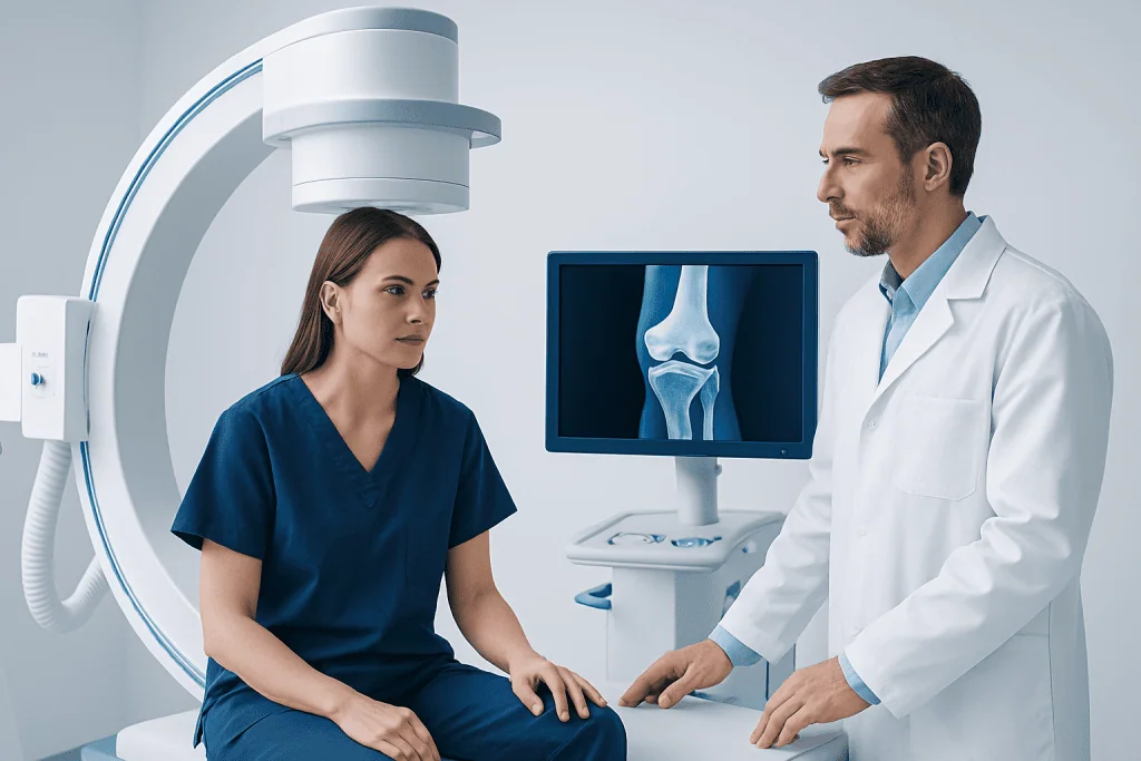
The three main knee joint bones are the femur, tibia, and patella. The femur, or thigh bone, is the upper bone of the joint. Its lower end forms the top part of the knee and meets the tibia below. The rounded ends of the femur help the knee move in a controlled way.
The tibia, or shin bone, is the larger of the two lower-leg bones. It forms the main lower part of the knee joint and carries most of the body’s weight from the femur down through the leg. The top of the tibia is shaped to support the femur and to hold the menisci, which act as cushions.
The patella, or kneecap, is a small triangular bone at the front of the knee. It sits within the quadriceps tendon and helps protect the front of the joint. It also improves the mechanical efficiency of the thigh muscles, making it easier to straighten the knee.
The fibula is often mentioned in discussions of knee anatomy because it sits next to the tibia in the lower leg. However, it is not one of the primary bones forming the main knee joint surfaces. It still matters because ligaments and muscles attach near it, and injuries around the fibula can affect knee stability and function.
How the knee bones work together
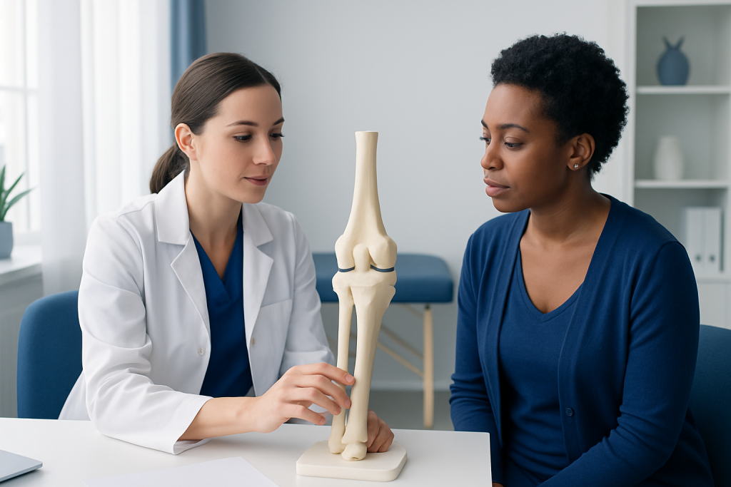
The knee includes two related joints. One is the tibiofemoral joint, where the femur meets the tibia. This is the main part of the knee and is responsible for most weight-bearing and movement. The other is the patellofemoral joint, where the patella glides along the front of the femur as the knee bends and straightens.
These bones are covered with smooth articular cartilage at the points where they touch. This cartilage reduces friction and helps the joint move comfortably. Between the femur and tibia are two pieces of fibrocartilage called the menisci, which help distribute weight, absorb shock, and improve stability.
Strong soft tissues hold the bones in place. The cruciate and collateral ligaments limit unwanted movement, while tendons connect muscles to bone. The quadriceps tendon and patellar tendon are especially important because they help extend the knee and control the position of the patella.
If any part of this system is injured or worn down, the knee may become painful, stiff, swollen, or unstable. For example, bone alignment problems can affect kneecap tracking, and cartilage loss may lead to knee osteoarthritis. This is why knee symptoms are not always caused by the bones alone, even when the pain seems deep inside the joint.
Common problems that affect knee bones and nearby structures
Several conditions can affect the bones that make up the knee joint. Fractures may involve the patella, the lower end of the femur, or the upper part of the tibia. These injuries can happen after falls, sports trauma, or direct blows. Symptoms often include pain, swelling, bruising, and difficulty bearing weight.
Degenerative changes are also common, especially with age. In osteoarthritis, the cartilage covering the ends of the bones gradually wears down. As the smooth joint surface changes, movement may become painful and stiff. Bone spurs can also form over time as part of the joint’s response to wear.
Not all knee pain comes from the bones themselves. Meniscus tears, ligament injuries, tendon irritation, bursitis, and alignment problems can also cause symptoms. A sports injury may damage the anterior cruciate ligament, while a sudden twist can affect the meniscus. Some patients may need evaluation for conditions such as meniscus tear or other internal knee injuries.
Overuse can also affect the patella and the structures around it. For example, repeated stress from running, jumping, or frequent kneeling may irritate the kneecap or the tendon below it. Pain at the front of the knee is common in these situations and may be related to patellofemoral problems rather than a major bone injury.
How doctors diagnose knee problems
Diagnosis starts with a medical history and physical examination. A doctor will ask where the pain is located, when it began, whether there was an injury, and what movements make symptoms worse. They may also ask about swelling, locking, instability, or trouble walking.
During the examination, the knee is checked for tenderness, range of motion, swelling, strength, and alignment. The doctor may gently move the joint or perform specific tests to look for meniscus or ligament injury. This hands-on assessment helps identify whether the problem is likely related to bone, cartilage, tendon, or ligament structures.
Imaging can provide more detail. X-rays are often the first test when a bone injury, arthritis, or alignment issue is suspected. They show the femur, tibia, patella, and overall joint space well. If soft tissue injury is possible, MRI may be recommended because it can show cartilage, menisci, ligaments, tendons, and bone marrow changes.
In some cases, ultrasound, CT, or lab tests may also be useful. The right test depends on the symptoms and the doctor’s findings. A careful diagnosis is important because treatment for a fracture is very different from treatment for tendon pain or a cartilage problem.
Treatment options for knee conditions
Treatment depends on which part of the knee is affected and how severe the problem is. Many mild or moderate issues improve with rest, activity changes, ice, physical therapy, supportive bracing, and anti-inflammatory treatment when appropriate. Strengthening the muscles around the knee can reduce pressure on the joint and improve stability.
Injuries involving ligaments or menisci sometimes require specialized care. For example, persistent instability after a sports injury may lead a doctor to discuss ACL surgery in selected patients. A damaged meniscus that causes locking, persistent pain, or functional limitation may be assessed for meniscus surgery when conservative care is not enough.
When knee joint surfaces become severely damaged, treatment may focus on restoring function and reducing pain. For advanced arthritis or major structural degeneration, some patients may be candidates for knee replacement after careful evaluation. Surgery is usually considered only when symptoms are significant and other treatments have not provided enough relief.
Care is individualized. Age, activity level, general health, the exact diagnosis, and personal goals all help shape the treatment plan. Near the end of the care pathway, patients seeking international evaluation may also learn that Acibadem International’s multidisciplinary specialists and JCI-accredited hospitals diagnose and treat knee conditions with coordinated orthopedic care.
Prevention and self-care for a healthy knee
Although not every knee problem can be prevented, healthy habits can lower strain on the joint. Maintaining a weight that is appropriate for the individual can reduce pressure on the femur, tibia, and patella during daily activities. Regular exercise that strengthens the thighs, hips, and core can also improve knee support.
Warm-up and technique matter, especially in sports. Proper footwear, gradual training increases, and attention to movement form can help reduce stress on the kneecap and surrounding tissues. People who kneel frequently for work or home tasks may benefit from padding and regular position changes.
After minor knee strain, self-care often includes rest, ice, compression, and elevation for a short period. Gentle range-of-motion exercises may help once pain begins to settle. However, it is usually best to avoid pushing through severe pain, sudden swelling, or a feeling that the knee may give way.
Long-term self-care also includes listening to symptoms. Repeated clicking with pain, swelling after activity, or worsening stiffness may be signs that the knee needs medical attention. Early assessment can help prevent ongoing irritation and may improve recovery.
When to see a doctor
A doctor should evaluate knee pain that is severe, does not improve, or keeps returning. Medical attention is also important if the knee becomes swollen, feels unstable, locks, or cannot fully bend or straighten. These symptoms can point to a problem involving the bones, cartilage, or internal soft tissues.
Urgent care is especially important after a major fall, sports collision, or direct blow to the knee. A fracture, dislocation, or significant ligament injury may need prompt treatment. Inability to bear weight, obvious deformity, or sudden large swelling should not be ignored.
People with fever, redness, warmth, or marked tenderness around the knee should also seek medical advice. These symptoms can have several causes, including inflammation or infection, and need proper assessment. Anyone with ongoing knee symptoms should speak with a qualified healthcare professional for an accurate diagnosis and a treatment plan tailored to their needs.
Understanding which bones make up the knee joint is helpful, but symptoms are not always caused by bone problems alone. A complete evaluation looks at the whole joint so that treatment can address the true source of discomfort and restore function as safely as possible.
Frequently asked questions
What are the three main bones of the knee?
The three main bones of the knee are the femur, tibia, and patella. The femur is the thigh bone, the tibia is the shin bone, and the patella is the kneecap. Together, they form the main structures of the knee joint.
Is the fibula part of the knee joint?
The fibula is close to the knee, but it is not one of the main bones forming the primary weight-bearing knee joint surfaces. It still plays an important supporting role because muscles and ligaments attach near it. Problems around the fibula can still affect knee function.
What does the patella do?
The patella helps protect the front of the knee and improves the leverage of the thigh muscles. This makes it easier to straighten the leg. It also glides along the femur as the knee bends and extends.
Can knee pain come from something other than the bones?
Yes, knee pain often comes from cartilage, menisci, ligaments, tendons, or bursae rather than the bones alone. For example, a meniscus tear or tendon irritation can cause pain even when the bones are intact. That is why a full medical assessment is important.
How do doctors check for knee joint problems?
Doctors usually begin with a physical exam and a discussion of symptoms, injury history, and daily activities. X-rays can show the bones and signs of arthritis or fracture, while MRI is often used to assess ligaments, menisci, and cartilage. The choice of test depends on the suspected cause.
When should someone worry about knee pain?
Knee pain should be medically assessed if it is severe, follows an injury, causes swelling, or makes walking difficult. It is also important to seek care if the knee locks, gives way, or cannot fully move. Ongoing or repeated symptoms are a good reason to see a doctor.
References
- American Academy of Orthopaedic Surgeons
- National Institute of Arthritis and Musculoskeletal and Skin Diseases
- MedlinePlus
- Mayo Clinic
- NHS
This article is for general information only and is not a substitute for professional medical advice. Please consult a qualified doctor about your individual situation.
Joint and spine care in Turkey — expert assessment & treatment
JCI-accredited · board-certified surgeons · reply within 24h
Add us as a Preferred Source to see more of our trusted health content across Google Search, AI Overviews and Discover.

More from the Health Library



Related Specialists

Dr. Salahattin Okur
Pediatrics
Dr. Tunca Cingöz
Orthopedic Surgery & Traumatology
Assoc. Prof. Dr. Alper Karakuş
Cardiology
Dilem Kadıoğlu
Physical Medicine & Rehabilitation