Peripheral Nerve Ultrasound vs EMG: How Neuromuscular Testing Is Chosen

Peripheral nerve ultrasound shows the anatomy of nerves and nearby tissues in real time. EMG and nerve conduction studies measure how nerves and muscles are working.
Key Takeaways
- Peripheral nerve ultrasound shows the anatomy of nerves and nearby tissues in real time.
- EMG and nerve conduction studies measure how nerves and muscles are working.
- Many patients benefit from both tests because they answer different clinical questions.
- The best test is chosen based on symptoms, examination findings, and the suspected condition.
- These tests can help diagnose problems such as nerve compression, neuropathy, radiculopathy, and muscle disorders.
Peripheral nerve ultrasound and EMG are complementary tests used to evaluate nerve and muscle problems. The choice depends on what the doctor needs to learn, such as whether the main question is about nerve structure, nerve function, muscle involvement, or a combination of these.
Overview: Why These Tests Are Compared
Peripheral nerve ultrasound and EMG are both important tools in neuromuscular medicine, but they do not do the same job. Peripheral nerve ultrasound uses sound waves to create images of nerves, muscles, and surrounding structures. EMG usually refers to a combined electrodiagnostic evaluation that includes nerve conduction studies and needle electromyography, which assess how well nerves transmit signals and how muscles respond.
Because these tests provide different kinds of information, the question is often not which one is better overall, but which one is better for a specific patient at a specific time. A person with numbness in the hand, weakness in the foot, muscle twitching, or pain radiating from the neck may need a tailored testing plan based on the suspected cause.
In many cases, the two methods complement one another. Ultrasound can reveal whether a nerve is enlarged, compressed, displaced, or affected by a nearby cyst or scar. EMG and nerve conduction studies can show whether the nerve is functioning abnormally, whether the problem is mild or severe, and whether it may be coming from the nerve root, peripheral nerve, neuromuscular junction, or muscle.
What Peripheral Nerve Ultrasound Shows
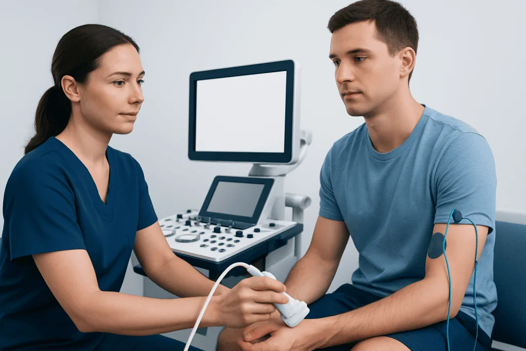
Peripheral nerve ultrasound is a noninvasive imaging test performed with a handheld probe placed on the skin. It allows the clinician to see the shape, size, and course of a nerve in real time. This can be especially useful when a focal nerve problem is suspected, such as compression at the wrist, elbow, or ankle.
Ultrasound can also show structures around the nerve that may contribute to symptoms. These may include inflamed tissue, ganglion cysts, tumors, scar tissue, anatomic variations, or swelling from injury. Dynamic scanning, which means viewing the nerve while the patient moves a joint, can help identify nerve subluxation or entrapment that may not be obvious at rest.
Another advantage is patient comfort. Unlike needle testing, ultrasound does not involve electrical stimulation or needle insertion. It is often used as part of the evaluation for conditions such as carpal tunnel syndrome or other focal neuropathies, especially when the main goal is to understand the nerve’s structure.
- Best for visualizing nerve anatomy
- Useful for focal entrapment or trauma
- Can detect masses, cysts, and scarring
- Allows dynamic assessment during movement
What EMG and Nerve Conduction Studies Show
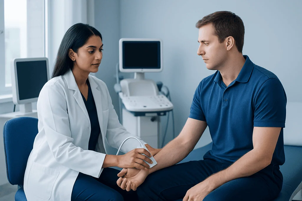
EMG testing usually includes two parts. Nerve conduction studies use small electrical stimuli on the skin to measure how fast and how strongly a nerve carries signals. Needle EMG uses a very fine needle electrode to record electrical activity in selected muscles. Together, these tests help doctors understand whether symptoms come from a nerve, the nerve root, the muscle, or the connection between the nerve and muscle.
Electrodiagnostic testing is especially valuable when the main question is about function rather than anatomy. For example, it can help confirm whether weakness is caused by a nerve injury, whether numbness is due to a generalized peripheral neuropathy, or whether symptoms reflect a pinched nerve in the spine rather than compression farther along the limb.
EMG and nerve conduction studies can also help estimate severity, distribution, and chronicity. They may show whether there is active nerve injury, whether the body is trying to reinnervate muscle, or whether multiple nerves are involved. This information often helps guide treatment decisions, rehabilitation plans, and the need for additional imaging such as MRI imaging.
How Doctors Choose Between Ultrasound and EMG
The choice between peripheral nerve ultrasound and EMG begins with the patient’s history and neurological examination. Doctors consider the pattern of symptoms, including numbness, tingling, pain, weakness, muscle wasting, balance problems, and whether symptoms are constant or brought on by movement. They also look at reflexes, sensory changes, and muscle strength.
If the concern is a localized compression or structural abnormality, ultrasound may be chosen early. If the concern is a broader disorder affecting nerve or muscle function, EMG and nerve conduction studies are often more informative. For example, widespread tingling in both feet may raise suspicion for peripheral neuropathy, while isolated hand numbness may point more strongly toward a local entrapment neuropathy.
Often, the most effective approach is to combine both tests. A clinician may use ultrasound to locate a suspicious area and then use electrodiagnostic testing to determine whether that finding is actually affecting nerve function. This combined strategy can improve diagnostic accuracy and provide a more complete picture, especially in complex or unclear cases.
- Ultrasound is often preferred for structure-focused questions
- EMG is often preferred for function-focused questions
- Both may be used when symptoms are complex or overlapping
- The testing plan may change after the physical examination
Common Conditions Where One or Both Tests Help
Focal entrapment neuropathies are among the most common reasons these tests are used. In carpal tunnel syndrome, for example, ultrasound may show enlargement of the median nerve at the wrist, while nerve conduction studies can confirm slowing across the carpal tunnel and help grade severity. Similar principles apply to ulnar neuropathy at the elbow or peroneal neuropathy near the knee.
In suspected radiculopathy, such as nerve root irritation from the neck or lower back, EMG is often more useful than ultrasound because the problem begins near the spine. Needle EMG can detect changes in muscles supplied by affected nerve roots, while ultrasound has a more limited role in evaluating deeper spinal causes. In these situations, doctors may also consider brain and nerve MRI or spine imaging depending on the symptoms.
For traumatic nerve injuries, ultrasound can be especially helpful in showing whether a nerve is swollen, partially torn, trapped in scar tissue, or displaced. EMG can then assess whether signals are still reaching muscles and whether recovery is occurring over time. In selected patients with muscle weakness or concern for a primary muscle disorder, EMG remains a core test, and further evaluation may include blood tests, imaging, or occasionally muscle biopsy.
What Patients Can Expect During Testing
Peripheral nerve ultrasound is generally quick and well tolerated. The skin is exposed over the area being examined, gel is applied, and the probe is moved along the course of the nerve. Patients may be asked to change position or move a joint so the clinician can observe the nerve during motion. There is no radiation, and discomfort is usually minimal.
Nerve conduction studies involve electrodes placed on the skin and brief electrical pulses to stimulate the nerve. These pulses can feel surprising or uncomfortable, but they are very short. Needle EMG involves inserting a fine needle into selected muscles to record electrical activity at rest and during gentle contraction. Some muscles may feel more sensitive than others, but the test is usually manageable and is performed under controlled conditions.
The exact length of testing depends on how many nerves and muscles need evaluation. In some cases only a limited study is needed, while in others a broader assessment is necessary. Patients should tell the clinician about bleeding disorders, implanted devices, blood thinners, skin infections, or severe swelling so the testing plan can be adjusted safely.
Limits, Benefits, and Why Results Need Context
Neither test is perfect on its own. Ultrasound may identify changes in a nerve’s appearance, but not every structural finding explains symptoms. Some mild enlargement or anatomic variation may be incidental. Likewise, EMG can show functional abnormalities, but very early changes may be subtle, and some small sensory fibers are not fully assessed by routine electrodiagnostic testing.
Results always need to be interpreted together with symptoms, physical examination, and sometimes additional tests. Blood tests may be needed if a metabolic, autoimmune, nutritional, or infectious cause is suspected. Imaging may be recommended if there is concern for spinal compression, masses, or more complex anatomy. This stepwise approach helps avoid overinterpreting any single test.
A multidisciplinary evaluation can be especially helpful when symptoms are persistent or the diagnosis remains unclear. Near the end of the care pathway, some patients may be referred for rehabilitation, pain management, surgery, or medical treatment depending on the cause. Acibadem International’s multidisciplinary specialists in JCI-accredited hospitals evaluate neuromuscular conditions for international patients and may use both ultrasound and electrodiagnostic testing when clinically appropriate.
When to Seek Medical Advice
A person should seek medical advice for numbness, tingling, burning pain, unexplained weakness, muscle wasting, or clumsiness that persists, recurs, or worsens. Symptoms that interfere with walking, hand function, sleep, or daily activities deserve timely assessment. Earlier evaluation can help identify treatable causes and prevent ongoing nerve irritation in some cases.
Urgent medical attention is important if weakness develops suddenly, if there is rapidly progressive loss of function, or if symptoms are accompanied by trouble breathing, swallowing, speaking, severe back or neck pain, or loss of bladder or bowel control. These signs may point to a more serious neurological problem that needs prompt care.
Even when symptoms seem mild, it is helpful to consult a qualified doctor if they are one-sided, associated with visible muscle loss, follow an injury, or occur in the setting of diabetes, autoimmune disease, cancer treatment, or known spine disease. Choosing the right test is part of good diagnosis, and a specialist can determine whether ultrasound, EMG, both, or another evaluation is most useful.
Frequently asked questions
Is peripheral nerve ultrasound better than EMG?
Neither test is universally better. Peripheral nerve ultrasound is better for showing nerve anatomy and nearby structures, while EMG and nerve conduction studies are better for evaluating how nerves and muscles function. Doctors choose based on the clinical question, and many patients benefit from both.
Can ultrasound replace EMG?
In some focused situations, ultrasound may provide enough information about a nerve compression or structural abnormality. However, it cannot fully replace EMG when the main question involves nerve function, muscle involvement, radiculopathy, or severity of nerve damage. The tests are complementary rather than interchangeable.
Which test is more uncomfortable?
Ultrasound is usually the more comfortable test because it is performed on the skin surface without needles or electrical stimulation. Nerve conduction studies involve brief electrical pulses, and needle EMG involves small needle insertions into muscles. Although EMG can be uncomfortable, most patients tolerate it well.
Why would a doctor order both ultrasound and EMG?
A doctor may want both structure and function information. For example, ultrasound may show that a nerve is enlarged or compressed, while EMG can confirm whether that change is affecting signal transmission and muscle health. Using both can improve diagnostic confidence in more complex cases.
Does EMG show nerve damage right away?
Some abnormalities can be detected early, especially on nerve conduction studies, but certain needle EMG changes may take time to appear after an injury. The timing depends on the type and location of the nerve problem. This is one reason doctors sometimes repeat testing or combine it with imaging.
How should a patient prepare for these tests?
Patients are usually advised to arrive with clean skin and avoid lotions or oils on the day of testing because they can interfere with electrodes and ultrasound contact. It is also important to mention blood thinners, pacemakers or other implanted devices, bleeding problems, and any recent infections or surgeries. The care team will explain any specific preparation needed.
References
- American Association of Neuromuscular & Electrodiagnostic Medicine
- National Institute of Neurological Disorders and Stroke
- Radiological Society of North America
- Mayo Clinic
- World Federation of Neurology
This article is for general information only and is not a substitute for professional medical advice. Please consult a qualified doctor about your individual situation.
Peripheral Nerve in Turkey — costs, top hospitals & a free quote
JCI-accredited · board-certified surgeons · reply within 24h
Add us as a Preferred Source to see more of our trusted health content across Google Search, AI Overviews and Discover.

Check-up & Preventive Medicine
Comprehensive health screenings designed for early detection and prevention.
1 specialists in this unitMore from the Health Library



Related Specialists

Assoc. Prof. Dr. Özge Begüm Comba
Ophthalmology
Dr. Cem Kara
Pediatric Surgery
Dr. Gültekin Barut
Internal Medicine
Assoc. Prof. Dr. Ceren Yıldız Eren
Gynecology & Obstetrics