

Corneal Topography: Mapping the Eye Before Vision Correction

Corneal topography maps the surface shape and curvature of the cornea. The test is quick, painless, and does not usually require touching the eye.
Key Takeaways
- Corneal topography maps the surface shape and curvature of the cornea.
- The test is quick, painless, and does not usually require touching the eye.
- It is often used before laser vision correction, cataract surgery, and specialty contact lens fitting.
- Corneal topography can help detect irregular corneal conditions such as astigmatism and keratoconus.
- Results are most useful when interpreted alongside a full eye examination.
Corneal topography is a noninvasive imaging test that creates a detailed map of the cornea, the clear front surface of the eye. It helps eye specialists plan vision correction, fit contact lenses more precisely, and detect corneal conditions early.
Overview
Corneal topography is a specialized eye imaging test that measures the shape, curvature, and surface features of the cornea. The cornea plays a major role in focusing light, so even small changes in its shape can affect vision quality. By creating a color-coded map of the cornea, this test helps eye doctors understand how the eye focuses and whether the surface is smooth, regular, or irregular.
The test is especially useful before vision correction procedures. People considering laser treatments, cataract surgery, or refractive lens procedures often have corneal topography as part of their preoperative assessment. It can also help explain blurred vision, monitor corneal changes over time, and support accurate contact lens fitting.
Unlike a standard vision chart test, corneal topography does not simply measure how clearly a person sees. Instead, it gives structural information about the eye’s front surface. This makes it an important tool in modern eye care, where treatment plans are increasingly tailored to each person’s individual eye anatomy.
How Corneal Topography Works
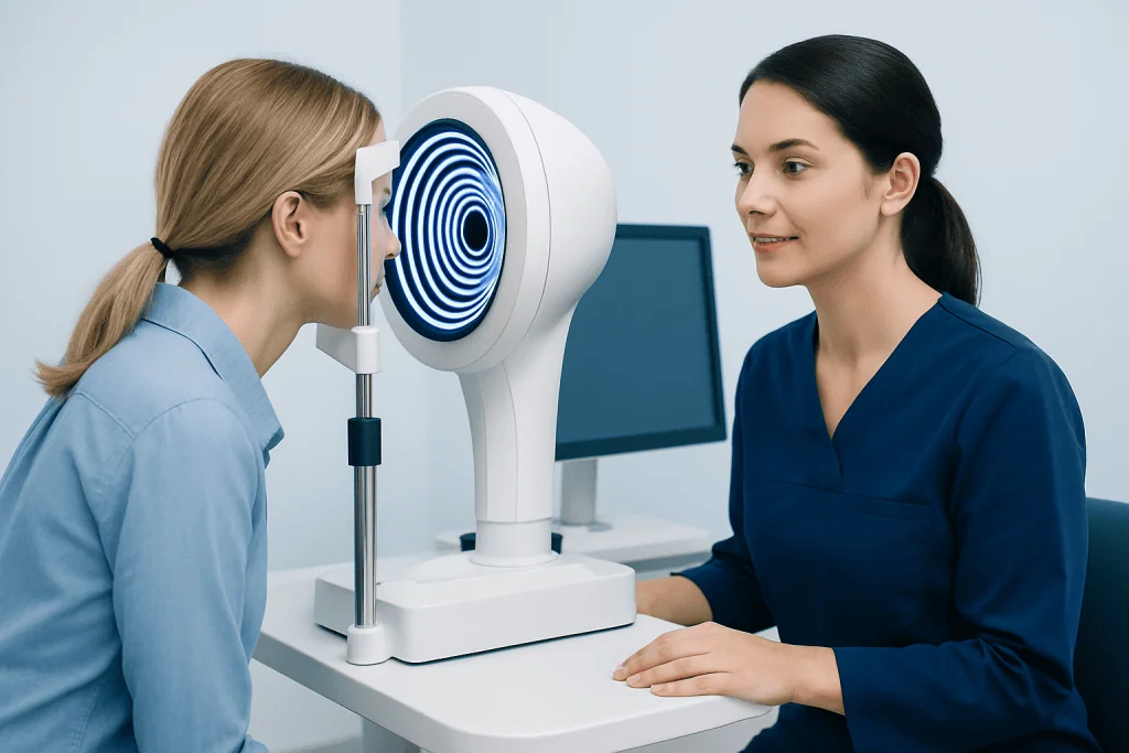
During corneal topography, the patient sits in front of a machine and looks at a target light. The device projects patterns, often rings or other structured light, onto the cornea. A computer then analyzes how those patterns reflect from the eye to build a detailed surface map.
The map usually uses colors to show different curvatures. Warmer colors may represent steeper areas, while cooler colors may show flatter areas. These patterns help specialists identify normal corneal shape, regular astigmatism, and subtle irregularities that may not be obvious during a routine eye exam.
Corneal topography is different from corneal tomography, although the terms are sometimes confused. Topography primarily looks at the front surface shape of the cornea, while tomography can provide additional information about both the front and back surfaces and corneal thickness. In many clinics, both technologies may be used together to give a more complete picture of eye health.
Why It Is Done Before Vision Correction
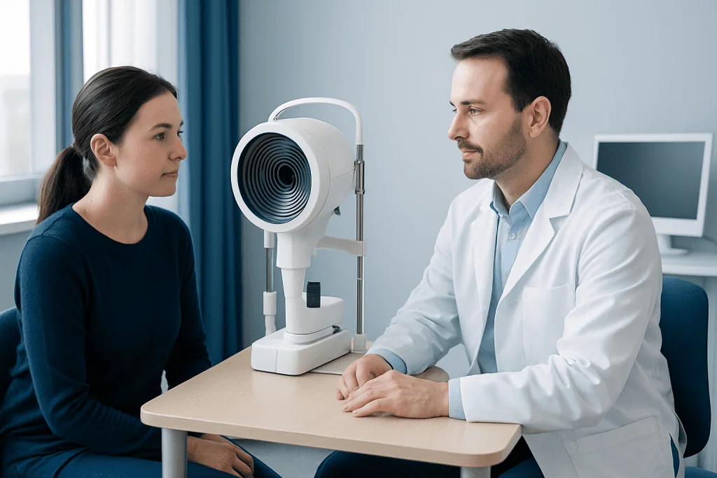
Before any procedure that changes the way the eye focuses light, doctors need a clear understanding of the cornea. Corneal topography helps determine whether the surface is suitable for treatment and whether there are any irregularities that could affect safety or results. This is one reason it is commonly included in evaluations for LASIK vision correction and other refractive procedures.
The test also helps guide treatment planning. If the cornea has a particular curvature pattern or degree of astigmatism, the surgeon can use this information to select or customize the most appropriate approach. In some cases, topography may show that a person would benefit from a different treatment than originally expected, such as no-touch laser treatment or another form of refractive care.
Corneal mapping is also valuable before cataract surgery because it improves the understanding of corneal astigmatism and optical surface quality. This can help with surgical planning and lens selection. In this way, the test supports more individualized care rather than a one-size-fits-all approach.
What Conditions It Can Help Detect
Corneal topography is often used to identify the cause of blurred or distorted vision when a standard refraction does not tell the whole story. It can reveal regular and irregular astigmatism, subtle corneal warpage, and surface changes related to prior surgery, injury, or long-term contact lens wear. These details may be important when deciding on treatment or follow-up.
One of the best-known uses of corneal topography is in screening for keratoconus, a condition in which the cornea gradually thins and bulges into an irregular shape. Early keratoconus may not cause obvious signs on a basic eye exam, but corneal mapping can sometimes detect suspicious patterns before symptoms become more advanced.
The test can also support the management of people with dry eye-related surface irregularity, corneal scars, post-surgical changes, or difficulty tolerating standard contact lenses. In specialty lens clinics, topography can help design lenses that better match the corneal surface, improving comfort and visual performance.
What to Expect During the Test
For most people, corneal topography is simple and comfortable. The patient places the chin on a support, keeps the head still, and looks straight ahead for a few seconds while images are captured. The machine does not usually touch the eye, and the test is completed quickly.
In many cases, no eye drops are needed. If the person wears contact lenses, the doctor may advise stopping lens use for a period before the test, because contact lenses can temporarily change the cornea’s shape. The required break depends on the type of lens and the purpose of the examination.
Results are available immediately in most clinics, but interpretation should be made by an eye specialist in the context of the full eye examination. Topography is highly informative, yet it is only one part of decision-making. Vision, corneal thickness, tear film quality, and general eye health also matter when planning care.
Benefits, Limits, and Accuracy
The main benefit of corneal topography is the level of detail it provides. It can detect patterns that are not visible with routine tools alone and can improve the planning of surgery or contact lens fitting. Because the test is noninvasive and repeatable, it is also helpful for monitoring changes over time.
Another advantage is patient safety. By identifying irregular or unstable corneal patterns, topography can help prevent unsuitable vision correction procedures. It can also provide reassurance when the corneal shape is normal and stable. In this way, it supports informed and individualized treatment choices.
Like any test, it has limitations. A poor tear film, blinking, difficulty keeping still, or recent contact lens use can affect image quality. Some patients may need repeat measurements to confirm findings. For the most complete assessment, specialists may combine topography with corneal thickness measurements, tomography, wavefront analysis, and a detailed clinical exam.
After the Test and Next Steps
There is no recovery period after corneal topography. People can usually return to normal daily activities right away, including work, reading, and driving, unless another eye test performed the same day causes temporary blur. The topography scan itself does not damage the eye or change vision.
What happens next depends on the reason for the test. Some people move forward with a surgical consultation, while others may be advised to update glasses, try specialized contact lenses, or undergo further testing. If the map shows irregularities, the doctor may recommend monitoring over time or additional evaluation to clarify the cause.
For patients exploring international care, Acibadem International’s multidisciplinary eye specialists and JCI-accredited hospitals evaluate and treat corneal and refractive conditions using modern diagnostic technologies. When needed, corneal topography may be part of a wider assessment that also includes imaging, refraction, and surface eye evaluation.
When to See an Eye Doctor
A person should consider an eye examination if vision becomes blurred, distorted, or less clear despite updated glasses or contact lenses. New glare, halos, ghosting, or difficulty seeing sharply at night can also justify further assessment, especially if symptoms are persistent.
Anyone considering refractive surgery should have a comprehensive preoperative evaluation, and corneal topography is often part of that process. It is also wise to seek specialist advice if there is a family history of corneal disease, previous corneal injury, or ongoing contact lens discomfort.
Prompt evaluation is particularly important when vision changes seem to worsen over time. Although many causes are manageable, the right diagnosis helps guide safer and more effective treatment decisions. An ophthalmologist or qualified eye care professional can explain whether corneal topography is appropriate and what the results mean for the individual patient.
Frequently asked questions
Is corneal topography painful?
Corneal topography is generally painless and noninvasive. In most cases, the machine does not touch the eye, and the test takes only a short time.
How long does a corneal topography test take?
The scan itself usually takes just a few minutes per eye. The full appointment may take longer if it is part of a comprehensive eye examination or surgical assessment.
Do contact lenses affect corneal topography results?
Yes, contact lenses can temporarily alter the shape of the cornea and may affect the accuracy of the map. An eye doctor may ask the patient to stop wearing lenses for a period before testing, depending on the lens type.
Can corneal topography detect keratoconus?
Corneal topography can help detect patterns that suggest keratoconus, sometimes even at an early stage. However, diagnosis is usually based on the full clinical picture, which may include other imaging and examination findings.
Is corneal topography the same as a regular eye exam?
No, it provides different information. A regular eye exam checks vision and overall eye health, while corneal topography specifically maps the shape of the cornea in detail.
Why is corneal topography important before LASIK?
It helps determine whether the cornea is regular and stable enough for treatment. It also supports surgical planning by showing curvature patterns and possible irregularities that could influence the best approach.
References
- American Academy of Ophthalmology
- National Eye Institute
- American Society of Cataract and Refractive Surgery
- Royal College of Ophthalmologists
This article is for general information only and is not a substitute for professional medical advice. Please consult a qualified doctor about your individual situation.
Explore treatments in Turkey — costs, top hospitals & a free quote
JCI-accredited · board-certified surgeons · reply within 24h
Add us as a Preferred Source to see more of our trusted health content across Google Search, AI Overviews and Discover.

More from the Health Library



Related Specialists

Asst. Prof. Dr. Ebru Kazancı
Neonatal Intensive Care Unit
Prof. Dr. Sinan Karaoğlu
Orthopedic Surgery & Traumatology
Dr. Ahmet Temizyürek
Anesthesiology
Dr. Hüseyin Çelik
Nephrology