

EOS Imaging for Spine and Joint Assessment: Lower-Dose Full-Body X-Rays Explained
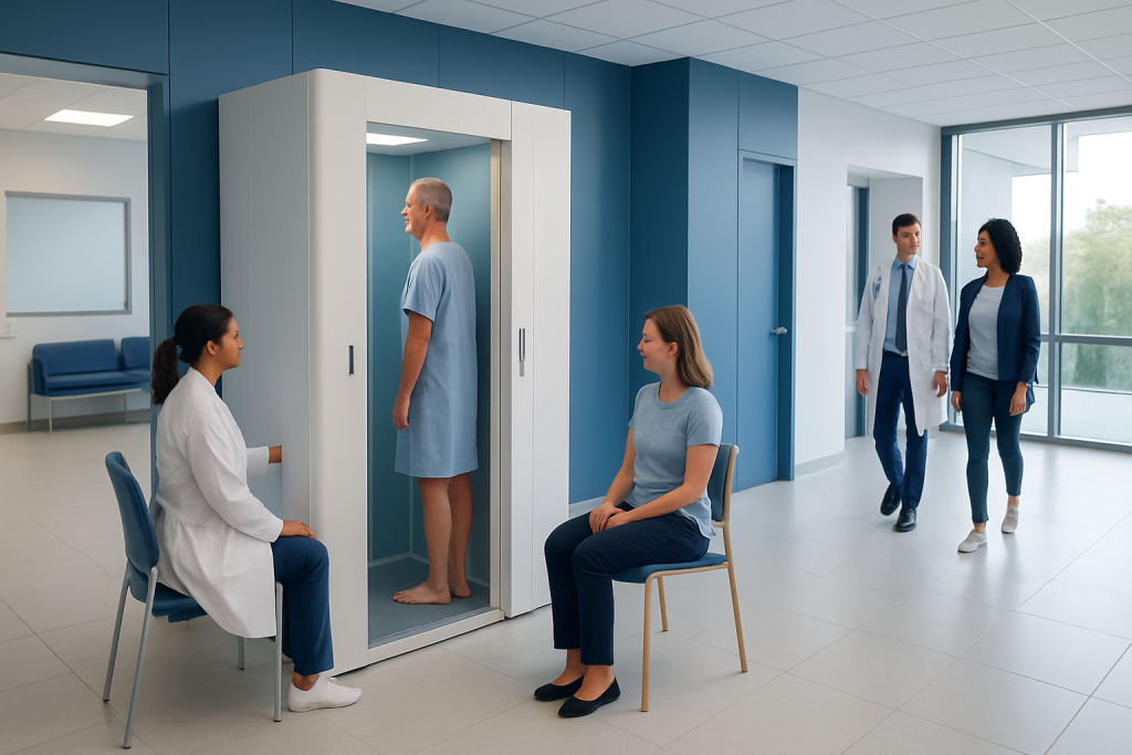
EOS imaging provides full-body images in a natural standing position. It uses a lower radiation dose than many standard full-body X-ray methods.
Key Takeaways
- EOS imaging provides full-body images in a natural standing position.
- It uses a lower radiation dose than many standard full-body X-ray methods.
- It is especially useful for spine deformities, leg length differences, and joint alignment assessment.
- The scan is quick, noninvasive, and typically does not require special preparation.
- Doctors may use EOS imaging to plan treatment and monitor changes over time.
EOS imaging is an advanced X-ray technology designed to assess the spine, hips, knees, and overall body alignment while a person is standing naturally. It uses a lower radiation dose than many conventional full-body X-ray approaches and can help doctors evaluate posture, balance, and joint relationships more precisely.
Overview of EOS imaging
EOS imaging is a specialized X-ray system used to examine the musculoskeletal system, especially the spine, pelvis, hips, knees, and lower limbs. Unlike many traditional imaging methods that focus on one body part at a time, EOS can capture full-body images in an upright, weight-bearing position. This is helpful because alignment problems often become clearer when the body is standing naturally rather than lying down.
One of the main advantages of EOS imaging is its lower radiation dose compared with many conventional full-body radiographic studies. This can be particularly important for people who need repeated imaging over time, such as children and adolescents with scoliosis or adults being followed for spine and joint alignment issues. Lower-dose imaging supports ongoing monitoring while limiting unnecessary radiation exposure.
EOS produces both front and side views at the same time, which helps clinicians understand how different parts of the body relate to each other. In selected cases, the system can also support 3D modeling of the skeleton from the X-ray images. This can give orthopedic and spine specialists a more complete picture of posture, balance, and anatomical alignment.
How EOS imaging works and what makes it different
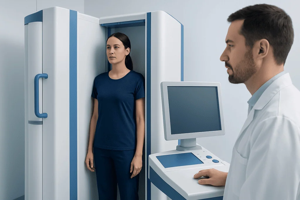
During an EOS scan, the person stands still inside an open, upright imaging cabin while two X-ray beams move vertically to capture images from the front and side simultaneously. Because the scan is performed while standing, it reflects the way the spine, pelvis, and legs carry the body under normal load. This is a key difference from standard scans performed while lying on a table.
Traditional X-rays remain very useful and widely available, but EOS imaging offers a broader view of whole-body alignment with lower radiation exposure in many situations. It is often used when doctors need to assess how the head, spine, pelvis, hips, knees, and ankles line up together. This full-body perspective can be valuable in complex orthopedic and spinal conditions.
Another important difference is the ability to reconstruct 3D models from the images in appropriate cases. These reconstructions may help clinicians evaluate rotational deformities, spinal curves, and joint positioning in more detail. For treatment planning, this can complement physical examination and other imaging tests rather than replace them entirely.
When doctors use EOS imaging for spine and joint assessment
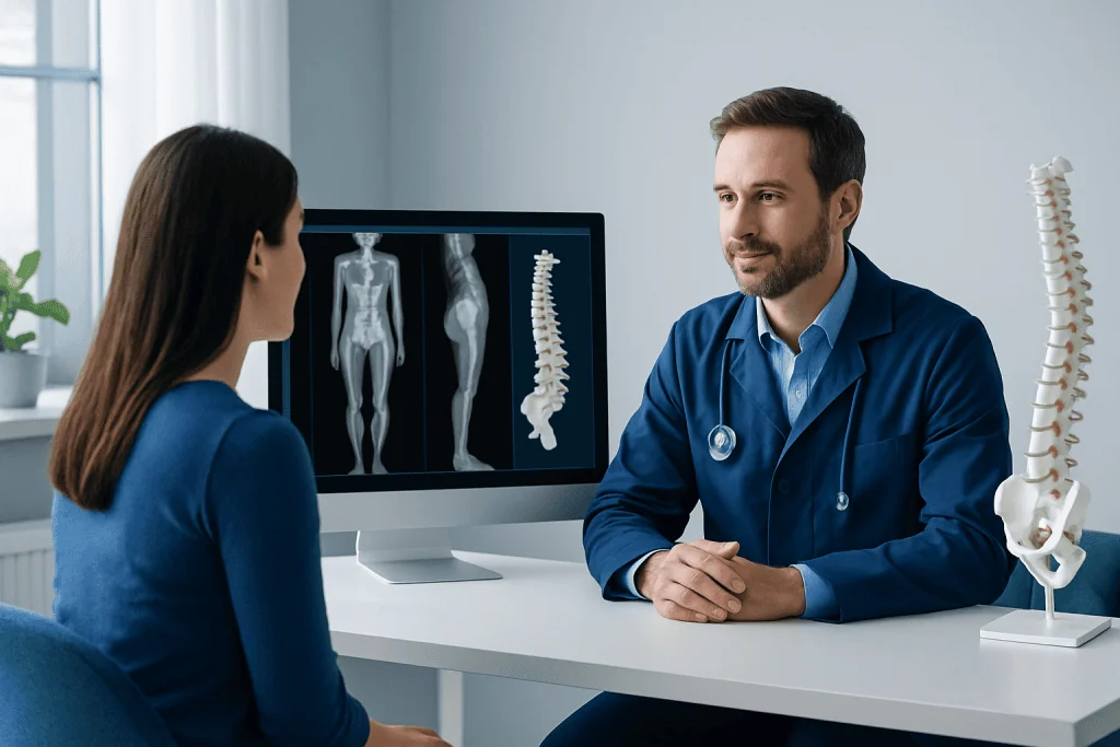
EOS imaging is often requested when a doctor needs to study body alignment as a whole. It is commonly used in children, teenagers, and adults with spinal curvature, posture concerns, limb length differences, gait problems, or joint alignment abnormalities. Because images are taken while standing, the scan can reveal changes that may not appear the same way on non-weight-bearing imaging.
In spine care, EOS may be used to assess conditions such as scoliosis and kyphosis, as well as overall sagittal and coronal balance. For joint assessment, it can help evaluate the hips, knees, and ankles together, which may be useful before or after orthopedic treatment. It is also relevant in people with degenerative changes, prior surgery, or suspected structural imbalance affecting movement.
Doctors may also use EOS imaging before procedures to improve planning and after treatment to monitor results over time. This can be especially helpful for people undergoing spine surgery, knee replacement, or hip replacement when alignment is an important part of decision-making. In some cases, it may also support assessment of complex leg length discrepancy.
What to expect during the scan
EOS imaging is a quick, noninvasive test. In most cases, the person is asked to remove metal objects such as belts, jewelry, or clothing items that could interfere with the images. Depending on the area being examined, the radiology team may ask the person to stand in a specific position and remain still for a short period while the scanner captures the images.
The scan itself is usually brief and painless. There are no injections, no enclosed tunnel, and no recovery period afterward. Patients can usually return to normal activities immediately unless their doctor has given separate instructions related to another test or procedure.
Children may need reassurance and simple coaching to stand still, but sedation is generally not required. The radiology team provides clear guidance throughout the process. If a person has difficulty standing independently, the healthcare team will decide whether EOS is appropriate or whether another imaging method would be more suitable.
Benefits and limitations of EOS imaging
The main benefits of EOS imaging include lower radiation exposure, full-body weight-bearing views, and the ability to assess alignment from head to toe in a single examination. This can improve understanding of how one part of the body influences another, such as how pelvic tilt may affect the spine or how knee alignment may change overall posture. For many orthopedic questions, this broader view adds clinically useful information.
Another advantage is follow-up monitoring. People with chronic spine or joint conditions may need repeated imaging over months or years. In these cases, lower-dose techniques are especially appealing. EOS may also help surgeons and rehabilitation teams track changes after treatment and compare images over time using a consistent imaging approach.
Even so, EOS imaging has limitations. It does not replace MRI or CT when doctors need detailed information about soft tissues, nerves, discs, ligaments, or internal structures. It may also be less suitable for patients who cannot safely stand. The best imaging test depends on the clinical question, and many people need more than one type of study for a complete evaluation.
- Best for: alignment, posture, spinal curves, leg length, and weight-bearing assessment
- Not ideal for: detailed soft tissue injuries, nerve compression, or emergency internal findings
- May be combined with: standard X-rays, MRI, CT, or physical examination findings
Safety, radiation exposure, and who may benefit most
All X-ray tests use ionizing radiation, so imaging should always be chosen thoughtfully. EOS technology was developed to reduce exposure while still producing useful diagnostic images. For patients who require periodic monitoring, especially younger patients with developing bones, reducing cumulative radiation can be an important part of safe long-term care.
People who may benefit most include children and adolescents with spinal deformities, adults with posture and balance concerns, and patients preparing for orthopedic procedures where precise alignment matters. It may also help athletes or active individuals being assessed for biomechanical issues that affect performance or comfort. However, the decision to use EOS should be based on medical need rather than convenience alone.
As with other X-ray examinations, patients should tell the radiology team if they are pregnant or might be pregnant. The healthcare team will weigh risks and benefits and may recommend postponing the scan or choosing another test if appropriate. When used appropriately, EOS imaging is considered a valuable and reassuring diagnostic tool within modern musculoskeletal care.
Understanding the results and next steps
After the scan, a radiologist and the referring specialist review the images. They look at spinal curvature, pelvic position, joint spacing, limb alignment, and the overall relationship between the body segments. In some cases, software-assisted measurements or 3D reconstructions are used to support a more detailed analysis.
The results are then interpreted in the context of symptoms, physical examination, and any prior imaging. A scan may show findings such as scoliosis progression, uneven leg length, altered posture, or imbalance in the hips and knees. These findings do not always mean surgery is needed; often they help guide conservative care, follow-up intervals, rehabilitation, or more targeted testing.
Next steps may include observation, physical therapy, bracing, medication for symptom relief, or referral to an orthopedic or spine specialist. When treatment planning is complex, a multidisciplinary approach can be helpful. Acibadem International’s multidisciplinary specialists and JCI-accredited hospitals diagnose and treat spine and joint conditions for international patients, using advanced imaging when clinically appropriate.
Frequently asked questions
Is EOS imaging the same as a regular X-ray?
EOS imaging is a type of X-ray, but it is designed specifically for lower-dose, full-body, weight-bearing assessment. It captures front and side images at the same time while the person is standing, which can provide more useful alignment information for certain spine and joint conditions.
Why is standing position important in EOS imaging?
Standing shows how the spine, pelvis, hips, knees, and ankles align under the body's natural weight. This can reveal posture and balance problems more clearly than images taken while lying down, especially when doctors are assessing the whole musculoskeletal system.
Who commonly needs EOS imaging?
It is often used for children and adults with scoliosis, posture problems, leg length differences, and orthopedic alignment concerns. Doctors may also request it before or after certain spine or joint procedures to help with treatment planning and follow-up.
Does EOS imaging replace MRI or CT scans?
No. EOS is excellent for assessing bone alignment and weight-bearing posture, but it does not provide the same level of soft tissue detail as MRI or the same cross-sectional detail as CT. Doctors choose the imaging method based on the question they need to answer.
How should a patient prepare for an EOS scan?
Preparation is usually simple. Patients may be asked to wear comfortable clothing and remove metal objects such as jewelry, belts, or items that could affect image quality. The imaging team will explain exactly how to stand and what to do during the scan.
Is EOS imaging safe for children?
When medically indicated, EOS imaging is generally considered a useful option for children because it uses a lower radiation dose than many standard full-body radiographic methods. This can be especially helpful when repeat imaging is needed over time, such as in monitoring scoliosis.
References
- World Health Organization
- International Society of Radiology
- Radiological Society of North America
- American Academy of Orthopaedic Surgeons
- Scoliosis Research Society
This article is for general information only and is not a substitute for professional medical advice. Please consult a qualified doctor about your individual situation.
Joint and spine care in Turkey — expert assessment & treatment
JCI-accredited · board-certified surgeons · reply within 24h
Add us as a Preferred Source to see more of our trusted health content across Google Search, AI Overviews and Discover.

More from the Health Library



Related Specialists
Dr. Mehmet Tayfun Osmanağaoğlu
Gynecology & Obstetrics
Assoc. Prof. Dr. Umut Karabulut
Cardiology
Prof. Dr. Sinan Karaoğlu
Orthopedic Surgery & Traumatology
Prof. Dr. Kaan Erler
Orthopedic Surgery & Traumatology