What Is Neuroradiology? Imaging of the Brain, Spine, and Nerves Explained
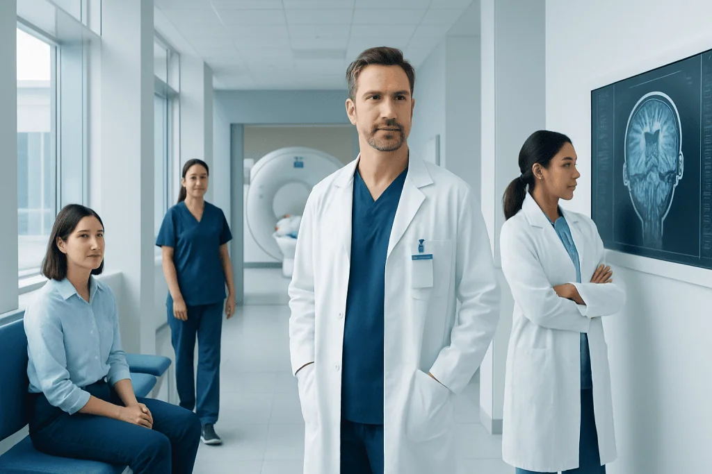
Neuroradiology focuses on imaging of the brain, spine, head, neck, and nerves. Common tests include MRI, CT, angiography, ultrasound, and sometimes PET imaging.
Key Takeaways
- Neuroradiology focuses on imaging of the brain, spine, head, neck, and nerves.
- Common tests include MRI, CT, angiography, ultrasound, and sometimes PET imaging.
- These scans help diagnose stroke, tumors, bleeding, multiple sclerosis, disc disease, and many other neurological conditions.
- Some neuroradiologists also perform image-guided procedures, such as treatments for blood vessel abnormalities.
- Most neuroradiology exams are noninvasive, and the right test depends on the symptoms and clinical question.
Neuroradiology is a medical specialty that uses advanced imaging to examine the brain, spine, spinal cord, head, neck, and nervous system. It helps doctors diagnose many conditions and, in some cases, guide minimally invasive treatments.
Overview of Neuroradiology
Neuroradiology is a subspecialty of radiology focused on imaging the nervous system. This includes the brain, spinal cord, spine, head, neck, and the blood vessels and nerves connected to these areas. By producing detailed images, neuroradiology helps doctors understand what may be causing symptoms such as headache, weakness, numbness, seizures, balance problems, back pain, or changes in vision and speech.
Neuroradiologists are doctors trained to interpret these scans and, in some settings, perform image-guided procedures. They work closely with neurologists, neurosurgeons, spine specialists, oncologists, emergency physicians, and rehabilitation teams. Their role is to help answer important questions: Is there a stroke? Is there a tumor, infection, inflammation, or bleeding? Is a nerve being compressed? Is a blood vessel narrowed, blocked, or abnormally formed?
For patients, neuroradiology is often a key part of diagnosis. It can provide information that a physical examination alone cannot show. In emergencies, it may help guide urgent treatment decisions. In long-term conditions, it can help monitor changes over time and assess how well treatment is working.
What Parts of the Body Does It Assess?
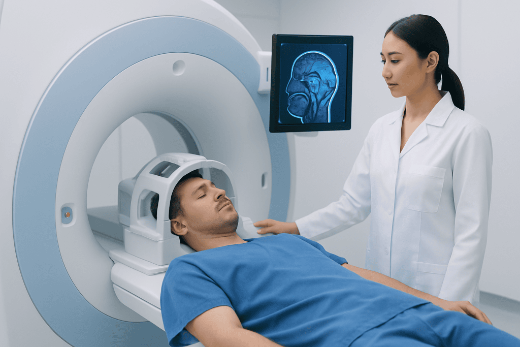
Although the name suggests a focus only on the brain, neuroradiology covers a wider area. It includes imaging of the brain and skull, the spine and spinal cord, the nerves that travel from the brain and spinal cord, and the structures of the head and neck. This broad view is important because symptoms can arise from many different points within the nervous system.
Neuroradiology may be used to assess:
- The brain, including areas involved in movement, language, memory, and vision
- The spinal cord and spinal canal
- The bones, discs, and joints of the spine
- Blood vessels in the brain, neck, and spine
- Cranial nerves and peripheral nerve pathways in selected cases
- The pituitary gland, inner ear region, eye sockets, and facial structures
This imaging is useful for both sudden symptoms and chronic concerns. For example, it may be used after head trauma, during stroke evaluation, or when looking into ongoing symptoms such as chronic neck pain, dizziness, hearing changes, or suspected nerve compression.
Common Imaging Tests Used in Neuroradiology

Several types of scans are used in neuroradiology, and each has strengths. Magnetic resonance imaging, or MRI, is especially useful for looking at the brain, spinal cord, discs, nerves, and soft tissues. It does not use ionizing radiation and can show very fine details, making it important in conditions such as multiple sclerosis, tumors, infections, or spinal cord problems. A specialized exam called MRI is often one of the most informative neuroradiology tools.
Computed tomography, or CT, uses X-rays to create cross-sectional images. It is fast and widely used in emergencies, especially when doctors need to check for bleeding, fractures, stroke-related changes, or severe head injury. A CT scan may also be combined with contrast dye to study blood vessels or detect tumors and inflammation more clearly.
Other tests may include angiography, which looks closely at blood vessels; ultrasound, which is useful for some neck vessels; and, in selected cases, PET-CT to help evaluate certain tumors or complex neurological conditions. Some imaging studies use contrast material to highlight tissues or vessels. Before contrast is given, the care team may ask about allergies, kidney function, pregnancy, implants, or devices such as pacemakers to make sure the exam is appropriate and safe.
Why Neuroradiology Is Performed
Doctors request neuroradiology tests when symptoms suggest a problem in the nervous system or when a known condition needs monitoring. Imaging can help confirm a diagnosis, narrow down possible causes, plan treatment, and check for complications. The right test depends on the symptoms, how quickly the problem developed, the person’s age and medical history, and whether an emergency is suspected.
Common reasons for neuroradiology imaging include severe or unusual headaches, seizures, weakness on one side of the body, numbness, confusion, fainting, memory changes, speech difficulty, dizziness, back or neck pain, and problems with walking or coordination. Imaging is also used after trauma and when doctors are concerned about stroke, infection, hydrocephalus, spinal cord compression, or spread of cancer to the brain or spine.
Neuroradiology can help identify or support the diagnosis of conditions such as brain tumors, disc herniation, spinal stenosis, aneurysms, vascular malformations, demyelinating diseases such as multiple sclerosis, and stroke. It may also help evaluate unexplained hearing loss, facial pain, pituitary problems, and symptoms related to nerve inflammation or compression.
How Doctors Interpret the Results
A neuroradiologist reviews the images in detail and prepares a report for the referring doctor. The report describes any abnormal findings, such as swelling, bleeding, tissue damage, masses, narrowing of blood vessels, disc changes, fractures, or signs of infection or inflammation. It also comments on what the findings may mean in the context of the symptoms and whether further imaging or follow-up is needed.
Imaging results are most useful when interpreted together with the clinical picture. A scan may show changes that explain symptoms clearly, but sometimes findings are nonspecific or incidental. For example, age-related spine changes can appear on imaging even in people without pain. That is why doctors combine scan results with a medical history, neurological examination, and sometimes blood tests or other procedures before making a final diagnosis.
If the scan identifies an urgent problem, treatment decisions may follow quickly. In other cases, doctors may compare the images with earlier studies to see whether a condition is stable, improving, or progressing. This can be important for monitoring diseases such as tumors, inflammatory disorders, or long-term spine problems.
Interventional Neuroradiology and Treatment Planning
Neuroradiology is not only diagnostic. In some centers, specially trained doctors perform interventional neuroradiology procedures. These are minimally invasive treatments guided by imaging, usually through a thin catheter placed in a blood vessel. They are used for selected problems involving the blood vessels of the brain, head, neck, or spine.
Examples include treating aneurysms, some types of stroke, and abnormal connections between arteries and veins. Image-guided care may be considered for conditions such as brain aneurysm or vessel blockages, depending on the individual case. In emergency situations, some patients may be assessed for thrombectomy to remove a clot, or for embolization to block abnormal bleeding or blood flow to a lesion.
Even when no procedure is needed, neuroradiology helps treatment planning. Surgeons may use imaging to map anatomy before an operation. Neurologists may use MRI findings to choose medical treatment. Oncologists may rely on scan results to monitor response to therapy. Near the end of the care pathway, rehabilitation teams may also use imaging information to better understand recovery needs. For international patients, Acibadem International’s multidisciplinary specialists and JCI-accredited hospitals diagnose and treat neurological and vascular conditions with coordinated imaging and specialist care.
Preparing for a Neuroradiology Exam and What to Expect
Preparation depends on the type of scan. Many neuroradiology tests require little or no special preparation. Patients may be asked to remove metal items before MRI, avoid food for a few hours before some contrast studies, or bring information about implants, prior scans, medications, allergies, and kidney problems. If claustrophobia is a concern, it is helpful to mention this in advance so the care team can discuss options.
During MRI, the patient lies still on a table that moves into the scanner. The machine can be noisy, and ear protection is usually provided. CT scans are generally quicker and more open. If contrast material is used, it may be given through a vein, and some people notice a brief warm sensation or metallic taste. Imaging staff monitor the patient throughout the exam and provide instructions to help the process go smoothly.
After the test, most people can return to normal activities unless they have received sedation or have been told otherwise. Results may be available quickly in urgent cases, while routine reports may take longer. Patients should ask when and how they will receive their results and who will explain the findings in relation to their symptoms.
Limitations, Safety, and When to Seek Medical Advice
Neuroradiology is highly valuable, but no single scan can answer every question. Some conditions may not be visible early on, and some findings can overlap between different disorders. At times, follow-up imaging, a different scan type, or other tests are needed for clarity. This is a normal part of careful diagnosis rather than a sign that something has gone wrong.
Safety is an important part of imaging. CT uses ionizing radiation, so doctors weigh the benefit of the test against the exposure, especially in children and during pregnancy. MRI does not use radiation, but certain implants, metal fragments, or devices may require extra precautions. Contrast agents are often used safely, though they may not be suitable for everyone. Sharing full medical information with the imaging team helps reduce risk.
Medical advice should be sought promptly for sudden neurological symptoms such as facial droop, arm weakness, severe confusion, loss of consciousness, sudden severe headache, seizures, or sudden trouble speaking, seeing, or walking. These symptoms may need urgent imaging and emergency care. For non-emergency symptoms such as ongoing back pain, recurring headaches, numbness, or balance problems, a doctor can decide whether neuroradiology is appropriate and which test would be most useful.
Frequently asked questions
What is neuroradiology in simple terms?
Neuroradiology is the branch of medical imaging that looks at the brain, spine, spinal cord, head, neck, and related nerves and blood vessels. It helps doctors diagnose conditions affecting the nervous system and sometimes guide treatment.
Is neuroradiology the same as neurology?
No. Neurology is the medical specialty that diagnoses and treats disorders of the nervous system, while neuroradiology focuses on imaging those areas. The two specialties often work closely together to understand symptoms and plan care.
Which scans are most commonly used in neuroradiology?
MRI and CT are the most common neuroradiology scans. Depending on the situation, doctors may also use angiography, ultrasound, or other specialized imaging to look at blood vessels, nerves, or tumors.
Does a neuroradiology scan hurt?
Most neuroradiology scans are painless. The main challenge is usually lying still, and some people may feel uncomfortable in a confined MRI space or notice a brief sensation if contrast dye is used.
Why would someone be referred for neuroradiology?
A doctor may refer someone for neuroradiology because of symptoms such as headache, weakness, numbness, seizures, dizziness, memory changes, or neck and back pain. It is also commonly used after injury or when conditions like stroke, tumors, or spinal problems are suspected.
Is neuroradiology only for serious conditions?
No. It is used for a wide range of concerns, from relatively common spine pain and chronic headaches to more urgent conditions such as stroke or bleeding. The purpose is to gather clear information so care can be better tailored to the patient.
References
- Radiological Society of North America
- American College of Radiology
- National Institute of Neurological Disorders and Stroke
- World Health Organization
- National Health Service
This article is for general information only and is not a substitute for professional medical advice. Please consult a qualified doctor about your individual situation.
Neuroradiology in Turkey — costs, top hospitals & a free quote
JCI-accredited · board-certified surgeons · reply within 24h
Add us as a Preferred Source to see more of our trusted health content across Google Search, AI Overviews and Discover.