Transcranial Doppler Ultrasound: How It Evaluates Brain Blood Flow

Transcranial Doppler ultrasound uses sound waves to measure blood flow in brain arteries. The test does not use radiation and is usually quick, safe, and painless.
Key Takeaways
- Transcranial Doppler ultrasound uses sound waves to measure blood flow in brain arteries.
- The test does not use radiation and is usually quick, safe, and painless.
- It can help evaluate stroke risk, vessel narrowing, vasospasm, and other circulation-related concerns.
- Results are interpreted together with symptoms, examination findings, and sometimes other imaging tests.
- Preparation is usually simple, though a doctor may give specific instructions based on the reason for testing.
Transcranial Doppler ultrasound is a painless, noninvasive test that evaluates how blood moves through the brain’s major arteries. It is often used to help assess stroke risk, monitor circulation, and support diagnosis in certain neurological and vascular conditions.
Overview
Transcranial Doppler ultrasound, often called TCD, is a specialized ultrasound test that measures blood flow through the brain’s main arteries. It works by sending high-frequency sound waves through thin areas of the skull and analyzing how those waves reflect off moving red blood cells. This helps clinicians understand how fast blood is flowing and whether circulation appears normal.
Unlike imaging tests that mainly show the structure of the brain, transcranial Doppler focuses on blood flow dynamics. In other words, it gives functional information about circulation rather than detailed pictures of brain tissue. This can be especially useful when a doctor wants to monitor changes in real time or evaluate how blood vessels are behaving during illness or recovery.
The test is noninvasive, does not involve radiation, and is usually performed at the bedside, in an outpatient imaging area, or in the hospital. Because it is relatively quick and repeatable, it is commonly used for follow-up and monitoring. In some situations, it may be used alongside other tests such as a standard ultrasound examination, CT, MRI, or vascular studies.
What the Test Evaluates
Transcranial Doppler ultrasound evaluates blood flow in major arteries inside the skull, including vessels that supply important parts of the brain. By measuring flow velocity and wave patterns, clinicians can look for signs that blood is moving too slowly, too quickly, or unevenly. These patterns may suggest narrowing, blockage, spasm, or changes in pressure and circulation.
One of the main uses of TCD is stroke-related evaluation. It may help detect changes associated with reduced blood supply, monitor blood flow after a stroke, or identify abnormalities that increase the chance of future events. It can also support the assessment of conditions such as stroke and transient ischemic attack when blood vessel function is an important concern.
TCD is also used in people with subarachnoid hemorrhage to check for vasospasm, a narrowing of blood vessels that can develop after bleeding around the brain. In some patients, it helps detect tiny emboli, monitor cerebral circulation during procedures, or assess sickle cell disease in children who are at higher risk of stroke. Because it can be repeated easily, it is often valuable for tracking changes over time rather than relying on a single snapshot.
When Doctors Recommend Transcranial Doppler

A doctor may recommend transcranial Doppler ultrasound when symptoms or medical history suggest a possible problem with blood flow to the brain. This can include sudden weakness, speech changes, dizziness, temporary vision loss, fainting episodes, or symptoms that raise concern about reduced circulation. The test may also be used after a known neurological event to monitor recovery or watch for complications.
In hospital settings, TCD is frequently used in patients who have had bleeding around the brain, vascular surgery, or intensive care monitoring. It can help clinicians follow blood flow changes without moving a critically ill patient repeatedly for more complex imaging. For some people, the goal is diagnosis; for others, it is ongoing surveillance.
Doctors may also use TCD in selected people with known blood vessel disorders, heart-related embolic risk, or inherited conditions such as sickle cell disease. In certain cases, it is part of a broader evaluation that may include MRI or CT scanning to compare blood flow findings with structural images of the brain and blood vessels.
How the Procedure Is Performed
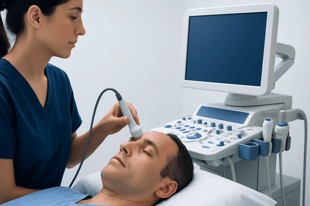
During the test, the patient usually lies comfortably on an examination bed or sits in a relaxed position. A trained healthcare professional places a small handheld probe, called a transducer, against specific areas of the head where the skull is thinner. These areas may include the temples, around the eyes in selected cases, the back of the head, or the upper neck.
A gel is applied to help sound waves travel effectively between the probe and the skin. The transducer sends sound waves into the skull, and a computer analyzes the returning signals. As blood cells move through the arteries, they create measurable changes in the reflected sound, known as the Doppler effect. These signals are displayed as waveforms and flow velocities for interpretation.
The examination is usually painless. Some people notice mild pressure from the probe or hear pulsing sounds from the machine, but discomfort is generally minimal. The test often takes between 30 and 60 minutes, depending on the reason for the study and whether both sides of the brain or repeated measurements are needed.
In some situations, an additional maneuver may be performed, such as breath-holding, a change in body position, or contrast-enhanced monitoring if the doctor is investigating specific circulation questions. The healthcare team explains these steps in advance so the patient knows what to expect.
What the Results Can Show
Transcranial Doppler results are based on the speed and pattern of blood flow in the arteries being examined. Higher-than-expected flow velocities can suggest vessel narrowing or vasospasm, while lower flow may point to reduced circulation, blockage, or other hemodynamic changes. The exact meaning depends on which artery is involved, the patient’s age, medical condition, and the reason the test was ordered.
Abnormal findings do not always mean there is permanent damage to the brain. Instead, they often show that circulation needs closer assessment. A doctor may compare TCD findings with symptoms, neurological examination, blood tests, and other imaging. If needed, further studies such as vascular ultrasound, angiographic imaging, or carotid artery disease evaluation may be recommended.
In monitoring situations, trends can be just as important as one measurement. For example, a series of TCD studies may show whether vasospasm is improving, whether treatment is helping maintain flow, or whether new changes are developing. This makes the test especially useful in hospital care, where early detection of circulation changes can guide timely decisions.
Benefits, Limits, and Safety
One of the biggest benefits of transcranial Doppler ultrasound is that it is safe, noninvasive, and free of ionizing radiation. It can be repeated as often as needed, which is valuable when a patient requires close monitoring. It is also portable in many clinical settings, making it useful for bedside assessment.
Another advantage is that TCD provides real-time information about blood flow behavior. Structural scans can show whether a blood vessel looks narrowed, but TCD can help reveal how that narrowing is affecting circulation at that moment. This functional information can support treatment planning and follow-up care.
Still, the test has limitations. It does not provide the same anatomical detail as CT angiography or MR angiography, and image quality can vary depending on skull thickness and the quality of acoustic windows. In some adults, especially older adults, it may be difficult to obtain strong signals through the skull. A normal TCD also does not rule out every possible brain or vascular problem, so it is best viewed as one part of a complete evaluation.
Overall, transcranial Doppler is considered very safe when performed by trained professionals. Doctors choose it carefully based on the clinical question, often together with other diagnostic tools to build the clearest picture of brain circulation.
Preparing for the Test and What Happens Afterward
Preparation for transcranial Doppler ultrasound is usually simple. Many patients do not need special fasting or major changes to their routine before the test. However, the healthcare team may ask about current medications, recent neurological symptoms, smoking, caffeine use, or prior imaging results, since these factors can sometimes influence circulation or test planning.
It is often helpful to wear comfortable clothing and arrive with a list of medications and medical history. If the test is being performed to investigate a specific condition, the doctor may provide more personalized instructions. Patients should follow those directions closely and ask questions if anything is unclear.
After the procedure, most people can return to normal activities right away unless they are in the hospital for another reason or undergoing additional tests. A specialist reviews the waveforms and measurements, then sends a report to the referring doctor. The doctor explains what the results mean and whether any further evaluation or treatment is needed.
Near the end of the care pathway, some patients may benefit from multidisciplinary assessment if circulation abnormalities are found. Acibadem International’s multidisciplinary specialists and JCI-accredited hospitals evaluate and treat neurological and vascular conditions for international patients when more detailed assessment is needed.
When to See a Doctor
A person should seek medical attention promptly if they develop symptoms that could suggest reduced blood flow to the brain. These may include sudden weakness or numbness on one side, difficulty speaking, sudden confusion, loss of balance, severe dizziness, or changes in vision. These symptoms need urgent medical evaluation, even if they improve quickly.
Transcranial Doppler ultrasound is not a substitute for emergency care. Instead, it is a diagnostic tool that may be used as part of the evaluation once a clinician has assessed the situation. People with a history of stroke, transient neurological symptoms, vascular disease, or conditions that increase clotting risk should discuss ongoing monitoring with their doctor.
Even when symptoms are mild or intermittent, it is wise to arrange medical review if they are new, recurring, or worsening. Early assessment can help identify whether further imaging, blood vessel studies, or specialist care is appropriate and can support timely treatment when needed.
Frequently asked questions
What is transcranial Doppler ultrasound used for?
Transcranial Doppler ultrasound is used to measure blood flow in the brain’s major arteries. Doctors use it to help assess stroke risk, monitor circulation after certain brain events, and look for changes such as vessel narrowing or vasospasm.
Is transcranial Doppler ultrasound painful?
The test is usually painless. Most people only feel light pressure from the probe on the skin and may hear pulsing sounds during the examination.
Does transcranial Doppler use radiation?
No, transcranial Doppler ultrasound does not use radiation. It uses sound waves, which makes it a noninvasive and repeatable test for monitoring blood flow.
How long does a transcranial Doppler test take?
The test commonly takes about 30 to 60 minutes. The exact time depends on why it is being done and how many blood vessels need to be assessed.
Can transcranial Doppler diagnose a stroke by itself?
Transcranial Doppler can provide important information about blood flow, but it does not usually diagnose a stroke on its own. Doctors interpret the results together with symptoms, physical examination, and other imaging tests such as CT or MRI.
Do patients need to prepare for a transcranial Doppler ultrasound?
Preparation is often minimal, and many people do not need to fast or make major changes beforehand. The healthcare team may still give specific instructions based on the reason for the test and the patient’s medical history.
References
- World Health Organization
- American Stroke Association
- National Institute of Neurological Disorders and Stroke
- Radiological Society of North America
- Society of NeuroInterventional Surgery
This article is for general information only and is not a substitute for professional medical advice. Please consult a qualified doctor about your individual situation.
Ultrasound in Turkey — costs, top hospitals & a free quote
JCI-accredited · board-certified surgeons · reply within 24h
Add us as a Preferred Source to see more of our trusted health content across Google Search, AI Overviews and Discover.

Check-up & Preventive Medicine
Comprehensive health screenings designed for early detection and prevention.
1 specialists in this unit