Ultrasound-Guided vs CT-Guided Procedures: What Is the Difference?

Ultrasound-guided procedures use real-time sound-wave imaging and do not involve ionizing radiation. CT-guided procedures use detailed cross-sectional X-ray images and can be especially helpful for deeper or harder-to-see targets.
Key Takeaways
- Ultrasound-guided procedures use real-time sound-wave imaging and do not involve ionizing radiation.
- CT-guided procedures use detailed cross-sectional X-ray images and can be especially helpful for deeper or harder-to-see targets.
- The choice between ultrasound and CT depends on visibility, precision needs, body location, patient factors, and the type of procedure.
- Both methods are commonly used for biopsies, drainages, injections, and other minimally invasive procedures.
- Doctors also consider comfort, speed, safety, and the need to avoid radiation when selecting the guidance method.
Ultrasound-guided and CT-guided procedures both help doctors place needles or instruments accurately, but they use different imaging methods and each has distinct advantages. The best choice depends on the body area, the target tissue, and the patient’s individual needs.
Overview: What image-guided procedures do
Image-guided procedures help doctors perform biopsies, drain fluid collections, place tubes, or deliver treatment with greater accuracy. Instead of relying only on physical examination or surface landmarks, the doctor uses live or near-live imaging to see internal structures and guide a needle or instrument to the right place.
Two common guidance methods are ultrasound and computed tomography, often called CT. In ultrasound-guided procedures, sound waves create real-time images on a screen. In CT-guided procedures, a series of X-ray images produces detailed cross-sectional views of the body. Both approaches are widely used and can make procedures safer and more precise.
Neither method is automatically better in every case. The right option depends on what part of the body is being treated, how easy the target is to see, whether the doctor needs continuous real-time imaging, and whether reducing radiation exposure is especially important. The goal in both cases is the same: accurate treatment with as little disruption to the body as possible.
How ultrasound-guided procedures work
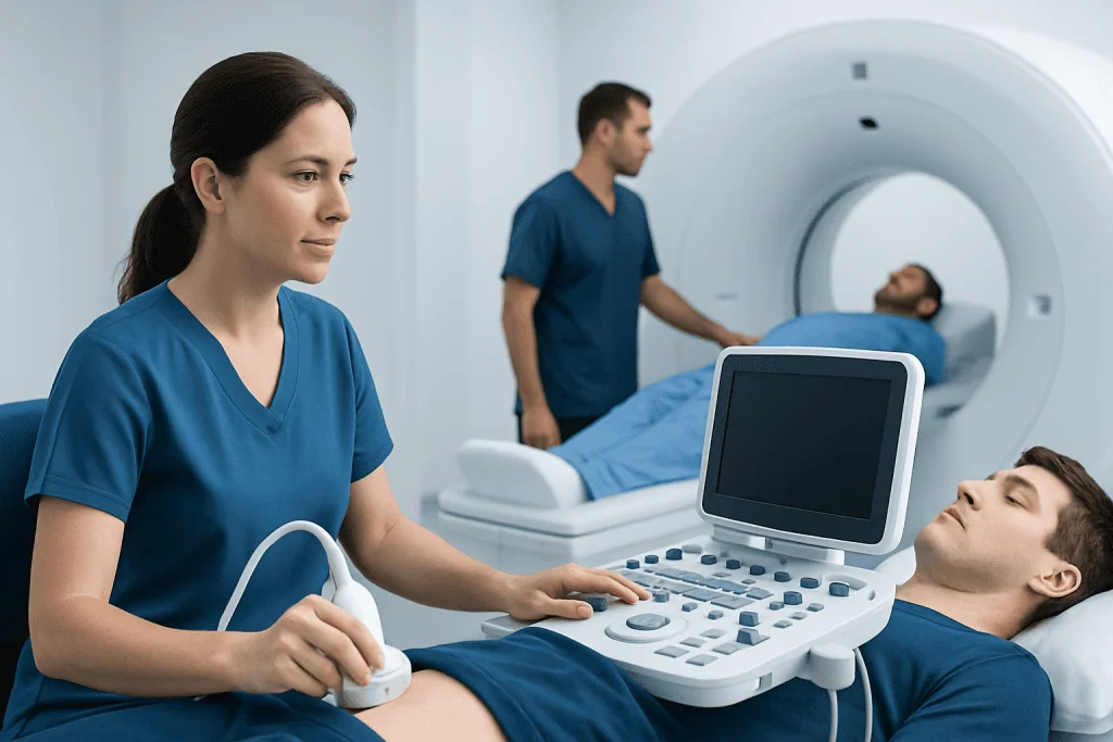
Ultrasound-guided procedures use a handheld device called a transducer, which is placed on the skin. The transducer sends sound waves into the body and receives returning echoes, creating images of organs, soft tissues, blood vessels, or fluid collections. Because the image appears in real time, the doctor can often watch the needle move as it advances.
This method is especially helpful for structures that are easy to see with ultrasound, such as fluid pockets, superficial soft tissue lesions, thyroid nodules, breast findings, lymph nodes, joints, tendons, and many abdominal or pelvic targets. It is also commonly used for vascular access and for guiding injections into specific joints or soft tissue spaces.
A major advantage of ultrasound is that it does not use ionizing radiation. It is often portable, relatively quick to set up, and can allow the patient to change position easily during the procedure. In many situations, it can improve comfort because the doctor can make adjustments instantly while viewing the target on the screen.
However, ultrasound has limits. Some targets are difficult to see because of overlying bone, bowel gas, body habitus, or depth. The quality of the examination can also depend on the operator’s experience and the acoustic window available. If the target cannot be seen clearly, another imaging method may be more suitable.
How CT-guided procedures work
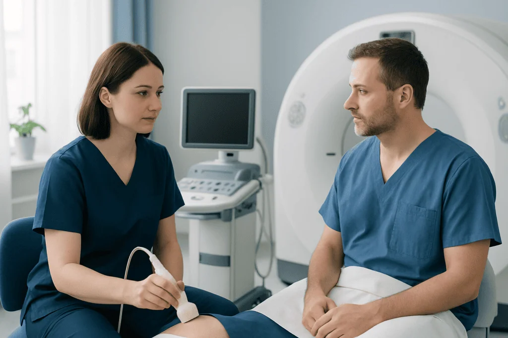
CT-guided procedures use a CT scanner to create detailed images of internal anatomy. The patient lies on a table that moves through the scanner, and the doctor uses these images to plan the safest path to the target. In many cases, the needle is advanced in small steps, with repeat CT images confirming its position.
CT is often chosen when the area is deep in the body or difficult to visualize with ultrasound. It can be particularly useful for procedures involving the lungs, deep abdominal structures, retroperitoneal areas, certain bones, or lesions partly hidden by gas or bone. The detailed anatomic information can help the doctor avoid nearby organs, blood vessels, or other sensitive structures.
One key strength of CT is clarity. It provides excellent cross-sectional detail and can show relationships between structures very clearly. This makes it valuable for lesions that are small, deep, or in complex locations where precision is critical.
The main limitation is that CT uses ionizing radiation. While doses are managed carefully and the expected benefit generally outweighs the risk when CT guidance is chosen, reducing unnecessary radiation remains an important consideration. CT guidance may also be less flexible than ultrasound for continuous real-time viewing, although newer techniques can improve workflow in selected settings.
Main differences between ultrasound-guided and CT-guided procedures
The biggest difference is the imaging technology. Ultrasound uses sound waves and produces live images without radiation. CT uses X-rays to create detailed cross-sectional images and does involve radiation exposure. This difference often shapes the decision, especially for younger patients, repeat procedures, or situations where avoiding radiation is preferred.
Another major difference is visibility. Ultrasound is excellent for many soft tissues and fluid-filled areas, but it may be limited by bone, air, or very deep targets. CT usually provides a broader and clearer map of deeper anatomy and can better show structures hidden from ultrasound. For this reason, CT may be favored when the doctor needs a more complete view of surrounding tissues.
Real-time guidance is also important. Ultrasound allows the doctor to watch movement as it happens, which is very useful when a target shifts with breathing or when precise needle control is needed throughout the procedure. CT often relies on step-by-step image checks rather than continuous live imaging, although this can still provide excellent accuracy.
Practical factors matter as well. Ultrasound-guided procedures may be faster, more portable, and more convenient at the bedside. CT-guided procedures may require scheduling in a CT suite and can take longer because images are obtained in stages. The doctor balances all of these factors to choose the safest and most effective approach for each patient.
- Ultrasound: no radiation, live imaging, portable, best for many soft tissue and fluid targets
- CT: detailed anatomy, useful for deep or complex targets, involves radiation, often stepwise image confirmation
- Both: improve procedural accuracy and help doctors reach a target with fewer unnecessary passes
When one method may be preferred over the other
Ultrasound is often preferred when the target is clearly visible and accessible with sound-wave imaging. This can include many thyroid biopsies, breast biopsies, liver lesion sampling, joint injections, abscess drainages, and procedures involving superficial lymph nodes or soft tissue masses. It is also commonly chosen when radiation avoidance is desirable.
CT may be preferred when the target is deep, very small, close to critical structures, or difficult to identify with ultrasound. Examples can include some lung biopsies, deep pelvic or abdominal collections, certain bone procedures, and lesions obscured by bowel gas or bone. In these situations, CT’s detailed cross-sectional view can make the access route clearer.
Sometimes the decision is influenced by patient-specific factors such as body habitus, breathing pattern, ability to lie still, pregnancy status, prior surgeries, implanted devices, or the need for sedation. The type of procedure also matters. A simple fluid drainage may be ideal for ultrasound, while a complex deep biopsy may be more suitable for CT.
Doctors may also consider whether another guidance option is appropriate, depending on the condition and anatomy. The choice is individualized rather than one-size-fits-all, and the imaging team explains why a specific method is recommended for that situation.
Safety, preparation, and what patients can expect
Both ultrasound-guided and CT-guided procedures are generally considered safe when performed by trained teams. As with any procedure involving a needle or instrument, possible risks can include bleeding, infection, discomfort, or injury to nearby structures. The exact risks depend on the body area and the reason for the procedure.
Before the procedure, the medical team reviews the patient’s symptoms, medications, allergies, and relevant test results. Blood-thinning medicines may need special instructions, and some patients may be asked not to eat or drink for a period beforehand, especially if sedation could be used. The team also explains the benefits, possible risks, and what will happen during recovery.
During an ultrasound-guided procedure, the skin is cleaned, gel is applied, and local anesthetic is often used to numb the area. During a CT-guided procedure, the patient is positioned in the scanner, the area is marked using imaging, and the needle is advanced with repeat scans as needed. In both cases, patients may feel pressure, but the team works to keep them as comfortable as possible.
Afterward, patients are usually observed for a short time, although some procedures require longer monitoring. Instructions may include rest, wound care, and watching for symptoms such as increasing pain, fever, shortness of breath, or significant swelling. If concerns arise, the patient should contact the care team promptly.
Questions to ask the doctor and making the right choice
Patients often feel more confident when they understand why one imaging method has been selected. Helpful questions include whether the target can be seen clearly with ultrasound, whether CT would provide more precision, whether radiation is a concern, and what risks are specific to the planned procedure. Asking about recovery time and the possibility of sedation can also be useful.
It is reasonable to ask whether there are alternatives and what the benefits of each option are. In many situations, the doctor’s recommendation is based on the safest path to the target and the highest chance of obtaining a clear sample or successful treatment in a single session. A tailored plan is usually more important than a general preference for one technology over another.
For patients having related imaging or procedures, broader radiology and minimally invasive care may also be part of the discussion, such as interventional radiology, ultrasound imaging, or CT scanning. Acibadem International’s multidisciplinary specialists and JCI-accredited hospitals diagnose and treat conditions requiring image-guided procedures for international patients.
The key point is that both ultrasound-guided and CT-guided procedures are established tools in modern medicine. The best method is the one that allows the doctor to see the target clearly, avoid important nearby structures, and perform the procedure as accurately and safely as possible.
Frequently asked questions
Is ultrasound-guided or CT-guided better?
Neither is universally better. Ultrasound-guided and CT-guided procedures each have strengths, and the better choice depends on the location of the target, how clearly it can be seen, and the goals of the procedure. The doctor chooses the method that offers the safest and most accurate approach.
Do ultrasound-guided procedures use radiation?
No. Ultrasound uses sound waves rather than X-rays, so it does not expose the patient to ionizing radiation. This is one reason it is often preferred when the target can be seen clearly with ultrasound.
Why would a doctor choose CT guidance instead of ultrasound?
A doctor may choose CT guidance when the target is deep, small, difficult to visualize, or partly hidden by bone or bowel gas. CT can provide more detailed cross-sectional anatomy, which may help in planning a safe needle path. It is often helpful for certain lung, bone, or deep abdominal procedures.
Are these procedures painful?
Most patients feel pressure or brief discomfort rather than significant pain. Local anesthetic is commonly used to numb the area, and some procedures may involve sedation depending on the body area and complexity. The medical team will explain what sensations to expect beforehand.
How long does recovery take after an image-guided procedure?
Recovery depends on the type of procedure and the area treated. Many patients go home the same day after a short observation period, while some procedures require longer monitoring. The care team provides specific instructions about activity, wound care, and warning signs to watch for.
Can patients ask for one method instead of the other?
Patients can always discuss preferences and concerns with their doctor. However, the final decision is usually based on which imaging method can show the target most clearly and allow the safest access. Shared decision-making is important, especially if radiation exposure or comfort is a concern.
This article is for general information only and is not a substitute for professional medical advice. Please consult a qualified doctor about your individual situation.
Ultrasound in Turkey — costs, top hospitals & a free quote
JCI-accredited · board-certified surgeons · reply within 24h
Add us as a Preferred Source to see more of our trusted health content across Google Search, AI Overviews and Discover.

Check-up & Preventive Medicine
Comprehensive health screenings designed for early detection and prevention.
1 specialists in this unitMore from the Health Library

Quest Diagnostics Appointment: How to Book, Prepare, and Check In

What Happens After an Abnormal Imaging Result? Next Tests, Follow-Up, and Timing

Biopsy vs Imaging: When a Scan Is Enough and When Tissue Testing Is Needed

Can You Get Lab Testing Without an Appointment?

What Is a Diagnostic Test? A Patient Guide to Lab and Imaging Results