Biopsy vs Imaging: When a Scan Is Enough and When Tissue Testing Is Needed
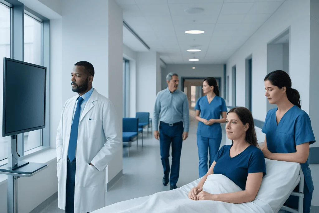
Imaging shows size, shape, location, and behavior of an abnormality, but it cannot always identify the exact cell type. A biopsy removes or samples tissue so it can be examined under a microscope for a definitive diagnosis in many cases.
Key Takeaways
- Imaging shows size, shape, location, and behavior of an abnormality, but it cannot always identify the exact cell type.
- A biopsy removes or samples tissue so it can be examined under a microscope for a definitive diagnosis in many cases.
- Some findings on ultrasound, CT, MRI, or mammography are so typical that biopsy may not be necessary right away.
- Biopsy is often recommended when cancer, infection, inflammation, or an unclear mass must be confirmed.
- Doctors decide between scan follow-up and tissue testing based on symptoms, imaging features, risk factors, and the likely impact on treatment.
Biopsy and imaging are complementary diagnostic tools rather than competing ones. In many situations, scans provide enough information to guide care, while in others a tissue sample is needed to confirm exactly what a lesion or abnormality is.
Overview: How Biopsy and Imaging Work Together
When doctors investigate a symptom, lump, pain, or unexpected test result, they often use imaging first. Imaging tests such as ultrasound, X-ray, computed tomography (CT), magnetic resonance imaging (MRI), and mammography create pictures of structures inside the body. These pictures can reveal whether an abnormality is present, where it is located, how large it is, and whether it has features that look harmless or concerning.
A biopsy is different. Instead of creating an image, it takes a sample of cells or tissue for laboratory examination. A pathologist then studies that sample under a microscope and may use additional tests to identify infection, inflammation, benign growths, or cancer. For many conditions, especially suspected tumors, biopsy provides the most definite answer about what the tissue actually is.
Because they answer different questions, biopsy and imaging are often used together rather than one replacing the other. Imaging may detect an abnormality and help decide whether a biopsy is needed. It can also guide the biopsy needle to the safest and most accurate area. In other situations, imaging findings are so typical that doctors may recommend observation, repeat imaging, or treatment without tissue sampling.
When a Scan May Be Enough

In some cases, imaging findings are highly characteristic and can provide enough confidence for a doctor to recommend monitoring or treatment without immediate biopsy. This is most common when an abnormality has clear benign features. Examples may include a simple cyst filled with fluid, a classic benign-appearing liver hemangioma, or a fracture or kidney stone that is well seen on imaging and matches the person’s symptoms.
Scans may also be enough when biopsy would not change management. For example, if a person has a straightforward muscle strain, imaging may confirm the injury but tissue sampling would offer no additional benefit. Similarly, if a tiny incidental finding appears stable over time and has no suspicious features, doctors may advise periodic follow-up imaging rather than an invasive procedure.
Imaging can also be sufficient when the goal is to assess anatomy, extent, or response rather than establish cell type. In conditions such as lung cancer, imaging is essential for locating a lesion, estimating spread, and planning care, even though a biopsy is often still needed for confirmation. In other words, a scan may be enough for some decisions, but not always for the final diagnosis.
- Clearly benign imaging appearance
- Findings that match a common non-cancerous condition
- Stable lesions on repeated imaging
- Situations where biopsy would not alter treatment
- Cases where biopsy risk outweighs likely benefit
When Tissue Testing Is Needed
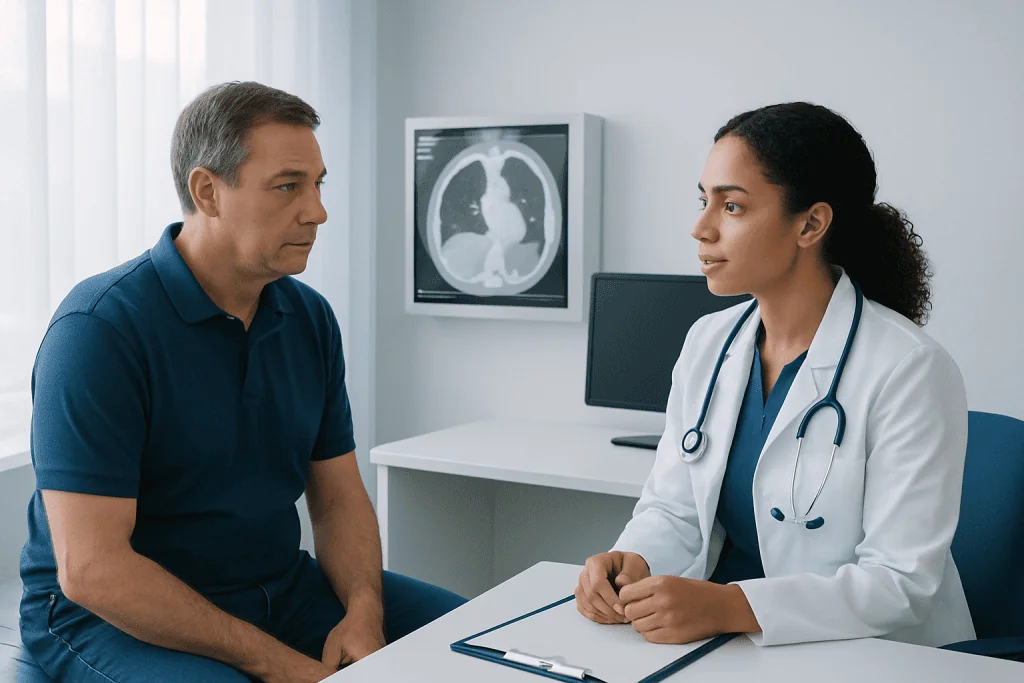
A biopsy is usually recommended when imaging alone cannot confidently distinguish between different possibilities. A scan can show that a mass exists, but several conditions may look similar on images. A tumor, scar, infection, inflammatory lesion, or area of tissue damage can sometimes overlap in appearance. If treatment depends on knowing exactly which one is present, tissue testing becomes important.
Biopsy is especially valuable when cancer is suspected. Imaging can strongly suggest malignancy, but many cancers require tissue confirmation before major treatment begins. This is because the pathology report can identify the cancer type, grade, and sometimes molecular features that guide therapy. In breast, lung, prostate, liver, lymph node, skin, and many other abnormalities, the biopsy result may directly shape the next step.
Tissue testing may also be needed when an infection or inflammatory disease is unclear, when a lesion is growing, when symptoms are significant despite uncertain imaging, or when doctors need to rule out recurrence after previous treatment. In oncology, for example, a suspicious area on PET-CT scanning may show increased activity, but that activity alone does not always prove cancer. A biopsy may still be necessary to make the diagnosis with confidence.
How Doctors Decide Between Follow-Up and Biopsy
Doctors do not base this decision on one image alone. They consider the whole clinical picture, including the person’s age, symptoms, medical history, family history, laboratory results, and known risk factors. A finding that may simply be monitored in one person could reasonably be biopsied in another if the overall risk is higher.
The appearance of the lesion matters greatly. Radiologists look at size, shape, borders, density or signal, blood flow, calcifications, and how the area behaves with contrast enhancement. Smooth borders and simple fluid content may suggest a benign process, while irregular margins, rapid growth, invasion of nearby tissue, or abnormal uptake on functional imaging may raise concern.
Doctors also weigh practicality and safety. Some lesions are easy to access with ultrasound or CT guidance, while others are in sensitive or deep locations where biopsy carries more risk. If an abnormality has a very typical benign appearance and a biopsy would expose the patient to unnecessary harm, surveillance may be more appropriate. If the result would meaningfully change treatment, however, tissue diagnosis often becomes worthwhile.
Common Tests Used Before or During Biopsy
Several imaging methods help determine whether a biopsy is needed and how it should be performed. Ultrasound is often used for thyroid nodules, breast lumps, lymph nodes, and soft-tissue masses because it can distinguish fluid from solid tissue and guide needles in real time. CT is useful for deeper lesions in the chest, abdomen, pelvis, and bones. MRI offers detailed soft-tissue contrast and can better characterize some liver, brain, prostate, and musculoskeletal findings.
Screening and diagnostic breast imaging are a common example of this stepwise approach. A mammogram may show a suspicious area, additional ultrasound or MRI may further clarify it, and biopsy is then performed only if the appearance remains concerning. In some settings, advanced imaging such as MRI or mammography helps avoid unnecessary biopsies by identifying features that are very likely benign.
When a biopsy is needed, imaging often guides it. Ultrasound-guided, CT-guided, stereotactic, or MRI-guided biopsies can improve precision and reduce trauma to surrounding tissue. This approach helps doctors sample the most informative area of a lesion while making the procedure safer and more efficient.
Benefits, Limits, and Risks of Each Approach
Imaging has important advantages. It is generally noninvasive, can assess large areas of the body, and can be repeated over time to monitor change. It is very helpful for detecting abnormalities early, evaluating treatment response, and guiding procedures. However, imaging has limits: it may not always tell benign from malignant with certainty, and results can occasionally be indeterminate or lead to incidental findings that need further assessment.
Biopsy offers more specific information because it looks directly at cells and tissue architecture. In many cases, it provides the confirmation needed before starting surgery, chemotherapy, radiation therapy, or other targeted treatment. But biopsy is invasive, even when minimally invasive. Depending on the site, possible risks may include bleeding, infection, pain, bruising, or a small chance of sampling error if the tissue taken does not fully represent the lesion.
Neither method is perfect on its own. A scan may strongly suggest a diagnosis but still need confirmation. A biopsy may give a definitive answer but can miss the abnormal area if the lesion is difficult to sample. That is why the best results usually come from combining radiology, pathology, and clinical judgment rather than relying on a single test in isolation.
What Patients Can Expect During the Decision Process
For patients, being told that a biopsy might be needed can feel stressful, but it does not automatically mean cancer or a serious illness. Often, it simply means the medical team wants a clearer answer before recommending the next step. Many biopsies ultimately show benign or treatable conditions. Asking why the biopsy is being recommended and what information it is expected to provide can help make the process feel more manageable.
Patients may also be offered short-interval follow-up imaging instead of immediate biopsy. This means repeating the scan after a set period to see whether the finding changes. If it remains stable or disappears, biopsy may be avoided. If it grows or develops more suspicious features, tissue sampling may then be advised.
It is reasonable for patients to ask which imaging findings are reassuring, what the alternatives are, what risks the biopsy carries, and whether the result would change treatment. Near the end of the evaluation pathway, some people benefit from care in centers with radiology, pathology, surgery, and medical specialists working together. Acibadem International’s multidisciplinary specialists and JCI-accredited hospitals diagnose and treat patients who need advanced imaging and biopsy-based evaluation, including image-guided procedures such as biopsy.
When to Seek Medical Advice Promptly
Most imaging findings are not emergencies, but some situations deserve prompt medical review. A person should contact a doctor sooner if there is a new or enlarging lump, unexplained bleeding, persistent pain, unexplained weight loss, ongoing fever, shortness of breath, or neurological symptoms such as weakness or new severe headaches. These symptoms do not necessarily mean a serious disease, but they should not be ignored.
Medical advice is also important when a scan report mentions an indeterminate, suspicious, or recommended follow-up finding. Patients should not feel they need to interpret complex imaging language on their own. The radiology report is only one part of the decision; the treating doctor connects it with symptoms, examination, and test results to decide whether watchful waiting, more imaging, or biopsy is best.
If there is uncertainty, a second opinion can be helpful, especially before an invasive procedure or major treatment. Informed decisions are usually made by balancing the chance of missing an important diagnosis against the risks of unnecessary testing. A clear discussion with a qualified doctor remains the safest and most reassuring way to decide when a scan is enough and when tissue testing is needed.
Frequently asked questions
Can imaging replace a biopsy?
Sometimes, but not always. Imaging can be enough when a finding looks clearly benign or when tissue results would not change treatment. A biopsy is usually needed when the exact diagnosis must be confirmed, especially if cancer is a concern.
Why would a doctor order a biopsy after a scan already found the problem?
A scan can show that an abnormality is present, but it may not reveal exactly what the cells are. A biopsy helps distinguish between benign growths, cancer, infection, inflammation, and other causes. This information often guides the safest and most effective treatment plan.
Does a recommendation for biopsy mean the lesion is cancer?
No. A biopsy is a diagnostic tool, not a diagnosis by itself. Many biopsied lesions turn out to be benign, but tissue testing is sometimes the best way to rule out more serious causes with confidence.
What types of imaging commonly guide biopsy decisions?
Ultrasound, CT, MRI, mammography, and sometimes PET-CT are commonly used. These tests help doctors assess the lesion’s appearance, location, and level of concern. They can also help choose the safest and most accurate biopsy approach.
Is it safe to wait and repeat the scan instead of having an immediate biopsy?
In selected cases, yes. If the imaging features suggest a low-risk finding, a doctor may recommend follow-up imaging after a period of time to check for changes. This approach should always be based on medical advice, because not all findings are suitable for watchful waiting.
What questions should patients ask before agreeing to a biopsy?
Helpful questions include why the biopsy is needed, what diagnosis is being considered, whether the result will change treatment, and what risks or alternatives exist. Patients can also ask whether repeat imaging is a reasonable option and how the procedure will be guided.
This article is for general information only and is not a substitute for professional medical advice. Please consult a qualified doctor about your individual situation.
Oncology care in Turkey — second opinion and treatment plan
JCI-accredited · board-certified surgeons · reply within 24h
Add us as a Preferred Source to see more of our trusted health content across Google Search, AI Overviews and Discover.

More from the Health Library
Diagnostic Medical Ultrasound: Who May Need It and What It Can Detect
Lab Test Results: What Abnormal Values Mean and When to Follow Up
Bone Density Test: DEXA Scan for Osteoporosis Risk
What Is a Diagnostic Test? Common Types and Why Doctors Order Them
What Happens During a Blood Test Visit?