

Doppler Ultrasound: Checking Blood Flow in Veins and Arteries

Doppler ultrasound measures the direction and speed of blood flow in veins and arteries without radiation. It is commonly used to assess blood clots, narrowed arteries, varicose veins, aneurysms, and circulation problems.
Key Takeaways
- Doppler ultrasound measures the direction and speed of blood flow in veins and arteries without radiation.
- It is commonly used to assess blood clots, narrowed arteries, varicose veins, aneurysms, and circulation problems.
- The test is usually painless, does not require needles or contrast dye, and can often be completed in less than an hour.
- Different Doppler techniques, such as color Doppler and duplex ultrasound, provide both images and blood-flow information.
- Results should always be interpreted by qualified healthcare professionals in the context of symptoms, medical history, and other tests.
Doppler ultrasound is a noninvasive imaging test that uses sound waves to assess how blood moves through veins and arteries. It helps doctors evaluate circulation, detect blockages or clots, and guide treatment decisions safely and comfortably.
Overview
Doppler ultrasound is a medical imaging test that shows how blood flows through the body’s veins and arteries. It uses high-frequency sound waves, not X-rays, to create real-time information about circulation. The test can help doctors see whether blood is moving normally, slowing down, flowing in the wrong direction, or being obstructed by a clot or narrowing.
A standard ultrasound creates images of organs and tissues. Doppler ultrasound adds information about motion, especially the movement of red blood cells inside blood vessels. This makes it particularly useful for evaluating the vascular system, including arteries that carry blood away from the heart and veins that return blood to the heart.
The examination may be requested for many parts of the body, including the legs, arms, neck, abdomen, kidneys, liver, and during pregnancy when fetal or placental blood flow needs assessment. It is widely used because it is noninvasive, generally painless, repeatable, and does not expose the patient to ionizing radiation.
How Doppler Ultrasound Works
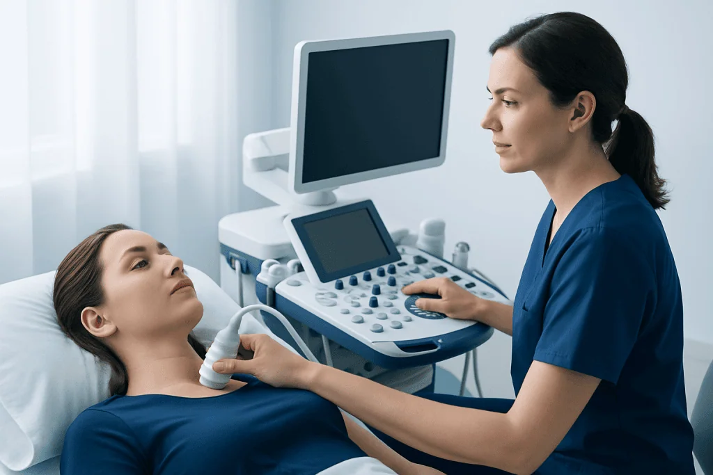
Doppler ultrasound is based on the Doppler effect, a change in sound-wave frequency that occurs when sound reflects off moving objects. During the test, a small handheld device called a transducer sends sound waves into the body. When these waves bounce back from moving blood cells, the ultrasound system analyzes the changes and displays information about blood-flow speed and direction.
Several types of Doppler ultrasound may be used depending on the clinical question. Color Doppler shows blood flow as color over a grayscale ultrasound image, helping the examiner identify vessels and flow direction. Spectral Doppler displays flow patterns as waveforms, which can show whether blood flow is normal, reduced, turbulent, or blocked. Duplex ultrasound combines standard imaging with Doppler measurements, giving both anatomic and functional information.
In some situations, power Doppler may be used to detect very small or slow blood flow, although it does not usually show direction as clearly as color Doppler. The choice of technique depends on the area being examined, the suspected condition, and the information the doctor needs to guide care.
When Doppler Ultrasound Is Recommended

A doctor may recommend Doppler ultrasound when symptoms suggest a circulation problem. In the legs, it is often used to look for deep vein thrombosis, a blood clot in a deep vein, especially when there is swelling, pain, warmth, or redness. It may also be used to evaluate chronic venous insufficiency, varicose veins, or valve problems that allow blood to pool in the legs.
Arterial Doppler ultrasound can help assess reduced blood flow caused by narrowed or blocked arteries. This may be considered when a person has leg pain while walking, cold feet, slow-healing wounds, or weak pulses. In the neck, carotid Doppler ultrasound evaluates the carotid arteries, which supply blood to the brain, and may be used when there is concern about narrowing that could increase stroke risk.
Doppler ultrasound may also be used in abdominal and organ imaging. Examples include checking blood flow to the kidneys, assessing liver blood vessels, evaluating an abdominal aortic aneurysm, or monitoring transplanted organs. In obstetric care, Doppler measurements may be used when clinically indicated to assess blood flow in the umbilical cord, placenta, or fetal vessels.
What to Expect During the Test
The procedure is usually straightforward. The patient may be asked to lie on an examination table, and the area being studied is exposed while the rest of the body is covered for comfort and privacy. A clear gel is applied to the skin to help sound waves pass between the transducer and the body. The examiner then moves the transducer gently over the skin while watching images and flow signals on a monitor.
The test may involve light to moderate pressure, especially when examining leg veins for clots. This pressure helps determine whether a vein compresses normally. Some patients may hear whooshing or pulsing sounds from the ultrasound machine; these sounds represent blood-flow signals and are a normal part of the examination.
Most Doppler ultrasound studies take about 20 to 60 minutes, depending on the area examined and the complexity of the findings. The patient can usually return to normal activities immediately afterward. Because the test does not use radiation and usually does not require injections, recovery time is not expected.
Preparation and Safety
Preparation depends on the part of the body being examined. Many Doppler ultrasound studies, such as leg vein or carotid artery scans, require little or no special preparation. Patients may be advised to wear loose, comfortable clothing and to remove jewelry or clothing that covers the area being scanned.
For some abdominal Doppler examinations, fasting for several hours may be requested so that intestinal gas is reduced and blood vessels can be seen more clearly. Patients should follow the instructions provided by the imaging center or doctor. Regular medications should generally be continued unless a healthcare professional gives different instructions.
Doppler ultrasound is considered a safe diagnostic tool when performed by trained professionals for appropriate medical reasons. It does not use ionizing radiation and is commonly used across many age groups, including children and pregnant patients when indicated. As with any test, its value depends on choosing the right examination and interpreting the results in the context of the patient’s overall health.
Understanding Results
Doppler ultrasound results may describe whether blood vessels are open, narrowed, blocked, enlarged, or leaking backward through valves. In veins, the report may mention whether a clot is present, whether the vein compresses normally, and whether blood flow changes appropriately with breathing or gentle compression. In arteries, the report may include blood-flow velocities and waveform patterns that suggest normal flow, narrowing, or obstruction.
It is important to understand that Doppler ultrasound findings are not interpreted in isolation. A doctor considers the patient’s symptoms, physical examination, risk factors, and sometimes other tests such as blood tests, CT angiography, MR angiography, or conventional angiography. In some cases, follow-up ultrasound is recommended to monitor a known condition or to check how treatment is working.
Results may be available soon after the examination, but timing varies by facility. A radiologist, vascular specialist, or other qualified physician typically reviews the images and prepares a formal report for the referring doctor. Patients should ask their healthcare provider to explain what the findings mean and what steps, if any, are recommended next.
Benefits, Limitations, and Alternatives
Doppler ultrasound has several important benefits. It is noninvasive, widely available, and does not expose the patient to radiation. It can show blood flow in real time, which is valuable when evaluating circulation problems. It can also be repeated when needed, making it useful for monitoring certain vascular conditions over time.
However, Doppler ultrasound also has limitations. Image quality may be affected by body habitus, swelling, open wounds, heavy calcification in blood vessels, bowel gas, or the depth and location of the vessel. Some vessels may be difficult to see clearly, and very small or complex abnormalities may require additional imaging. The accuracy of the test also depends on the training and experience of the examiner.
Alternatives may include CT angiography, MR angiography, venography, or catheter-based angiography, depending on the medical question. These tests can provide different types of detail, but they may involve contrast dye, radiation, or more invasive techniques. The best test is chosen based on the patient’s condition, kidney function, allergies, urgency, and the information needed for treatment planning.
When to See a Doctor
A person should seek medical advice if they develop symptoms that may suggest a circulation problem, such as persistent leg swelling, calf pain, unexplained redness or warmth, new varicose vein symptoms, coldness in a limb, weak pulses, or wounds that do not heal as expected. Sudden neurological symptoms, chest pain, severe shortness of breath, or sudden severe limb pain require urgent medical assessment.
People with known vascular disease, diabetes, high blood pressure, high cholesterol, smoking history, kidney disease, or a history of blood clots may need evaluation if new symptoms appear. A doctor can decide whether Doppler ultrasound is appropriate or whether another test is needed.
At Acibadem International, multidisciplinary specialists and JCI-accredited hospitals diagnose and treat vascular and circulatory conditions for international patients, including those who may need Doppler ultrasound as part of their assessment. Patients should bring previous imaging reports, medication lists, and relevant medical records to help the care team interpret results accurately.
Frequently asked questions
Is Doppler ultrasound painful?
Doppler ultrasound is usually not painful. The examiner may apply gentle pressure with the transducer, and leg vein studies may require firmer pressure to check whether veins compress normally. Any discomfort is typically brief, but patients should tell the examiner if an area is very tender.
Does Doppler ultrasound use radiation?
No. Doppler ultrasound uses sound waves rather than ionizing radiation. This is one reason it is commonly used for evaluating blood flow in many areas of the body, including in pregnancy when clinically indicated.
Can Doppler ultrasound detect a blood clot?
Yes, Doppler ultrasound is commonly used to evaluate suspected blood clots in deep veins, especially in the legs. The test can show whether a vein compresses normally and whether blood flow is present or obstructed. If symptoms are strong but results are unclear, a doctor may recommend repeat ultrasound or another test.
How should a patient prepare for a Doppler ultrasound?
Preparation depends on the body area being examined. Many vascular Doppler tests need no special preparation, but some abdominal studies may require fasting for several hours. Patients should follow the instructions from their doctor or imaging center and ask whether they should take regular medications as usual.
How long does it take to get results?
The scan itself often takes 20 to 60 minutes. A qualified physician usually reviews the images and sends a report to the referring doctor. The timing of results varies, and urgent findings may be communicated more quickly according to the facility’s clinical process.
What is the difference between venous and arterial Doppler?
Venous Doppler evaluates veins, which return blood to the heart, and is often used to look for clots or valve problems. Arterial Doppler evaluates arteries, which carry blood away from the heart, and is commonly used to assess narrowing, blockage, or reduced circulation. The scanning technique and measurements differ depending on the type of vessel.
Are there any risks or side effects?
Doppler ultrasound is considered very safe when used appropriately by trained professionals. It does not involve radiation, needles, or contrast dye in most routine vascular examinations. Patients can usually resume normal activities immediately after the test.
References
- World Health Organization
- American College of Radiology
- Radiological Society of North America
- Society for Vascular Surgery
- National Institute for Health and Care Excellence
This article is for general information only and is not a substitute for professional medical advice. Please consult a qualified doctor about your individual situation.
Ultrasound in Turkey — costs, top hospitals & a free quote
JCI-accredited · board-certified surgeons · reply within 24h
Add us as a Preferred Source to see more of our trusted health content across Google Search, AI Overviews and Discover.

Check-up & Preventive Medicine
Comprehensive health screenings designed for early detection and prevention.
3 specialists in this unitMore from the Health Library



Related Specialists

Prof. Dr. Ercan Karaarslan
Radiology
Prof. Dr. Bora Peynircioğlu
Interventional Radiology
Prof. Dr. Koray Güven
Interventional Radiology
Assoc. Prof. Dr. Deniz Can Aliş
Radiology