8 week ultrasound: A Complete Medical Guide for Patients

An 8 week ultrasound is commonly used to confirm pregnancy location, dating, and early development. Most scans at this stage are done transvaginally because it gives clearer images in early pregnancy.
Key Takeaways
- An 8 week ultrasound is commonly used to confirm pregnancy location, dating, and early development.
- Most scans at this stage are done transvaginally because it gives clearer images in early pregnancy.
- It is normal if a person cannot see much detail yet; the scan is mainly for medical confirmation rather than appearance.
- Sometimes dates are off because ovulation happened later than expected, so follow-up imaging may be recommended.
- Urgent medical review is important for heavy bleeding, severe pain, dizziness, or fainting in early pregnancy.
An 8 week ultrasound is an early pregnancy scan used to confirm that the pregnancy is in the uterus, estimate gestational age, and check for early fetal cardiac activity. It can also help explain symptoms such as pain or bleeding and guide next steps in prenatal care.
Overview: what an 8 week ultrasound is
An 8 week ultrasound is an imaging test performed in early pregnancy to look at the uterus and the developing embryo. At this stage, the main goals are to confirm that the pregnancy is inside the uterus, estimate how far along the pregnancy is, and look for expected early findings such as a gestational sac, yolk sac, embryo, and often cardiac activity.
For many patients, this scan is the first visual confirmation of pregnancy. It can be reassuring, but it is also a medical assessment. The information from the scan helps clinicians compare the ultrasound findings with the last menstrual period, decide whether the pregnancy appears to be developing as expected, and plan any needed follow-up.
An ultrasound at 8 weeks may be done routinely or because of symptoms such as pelvic pain, spotting, bleeding, or uncertainty about dates. It may also be recommended after fertility treatment, after a previous pregnancy loss, or when there is concern for an early pregnancy problem such as ectopic pregnancy or miscarriage.
Why this scan is done and what it can show
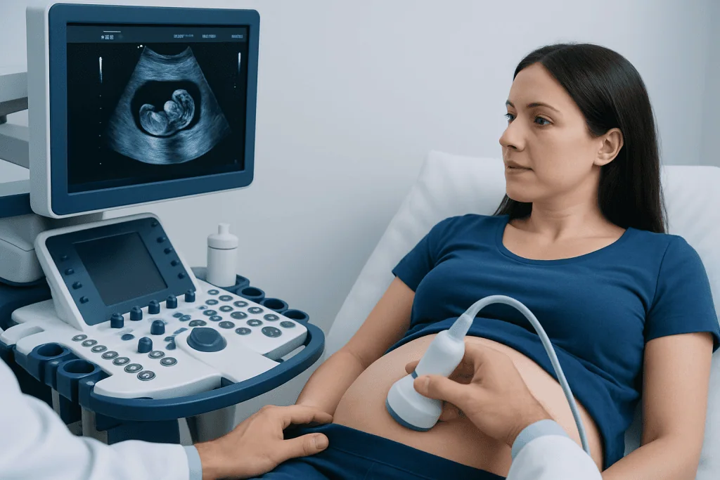
The value of an 8 week ultrasound is that it answers several important questions at once. First, it can confirm whether the pregnancy is located in the uterus. This matters because a pregnancy outside the uterus needs urgent medical attention. Second, it can measure the embryo, usually by crown-rump length, which is one of the most accurate ways to estimate gestational age in the first trimester.
The scan can also assess whether there is one embryo or more than one. In twin or multiple pregnancies, early ultrasound helps identify how many embryos are present and whether they appear to be developing in the expected way. This information is useful for later prenatal care.
Another important purpose is checking for early cardiac activity. At around 8 weeks, this is often visible if the dates are correct. However, ultrasound results must always be interpreted carefully in relation to cycle length, ovulation timing, and previous test results. A follow-up scan may be needed if the findings are uncertain rather than abnormal.
- Confirms pregnancy location
- Estimates due date more accurately
- Checks for single or multiple pregnancy
- Looks for early cardiac activity
- Helps investigate pain or bleeding
What to expect during the appointment

At 8 weeks, the clearest images are often obtained with a transvaginal ultrasound. During this test, a slim covered probe is gently inserted into the vagina to view the uterus and pregnancy from close range. This approach is commonly used in early pregnancy because the embryo is still very small, and an abdominal scan may not provide enough detail.
An abdominal ultrasound may sometimes be performed as well, especially later in the first trimester or when anatomy and body shape allow a good view. The clinician or sonographer will explain which method is best and why. Most patients find the transvaginal scan uncomfortable rather than painful, and it is usually brief.
Before the appointment, patients may be advised whether they need a full bladder. This depends on the type of scan planned. During the examination, the imaging specialist records the structures seen, takes measurements, and prepares a report for the clinician. If symptoms such as bleeding or cramping are present, these should be mentioned before the scan begins.
What is usually seen at 8 weeks
By about 8 weeks of pregnancy, ultrasound often shows a gestational sac within the uterus, a yolk sac, and an embryo. The embryo is still very small, and the image may not look like the later pregnancy scans that many people expect. This is normal. Early ultrasound is focused on confirmation and measurement rather than detailed anatomy.
Cardiac activity is often visible at this stage if the pregnancy dates are accurate. The clinician may discuss whether it was seen and whether the embryo’s size matches the expected timing. If a patient is certain they are 8 weeks pregnant but the scan appears earlier, one common explanation is that ovulation or implantation happened later than assumed.
Sometimes the scan does not give a final answer on the first visit. This does not always mean something is wrong. If the embryo is measuring smaller than expected or if structures are not yet clearly visible, the healthcare team may recommend repeating the scan after several days to one or two weeks. Follow-up is often the safest way to distinguish a normal early pregnancy from a pregnancy that is not developing as expected.
Reasons results may differ from expectations
Patients often expect the scan findings to match the date calculated from the last menstrual period, but early pregnancy dating is not always exact. Menstrual cycles vary, ovulation may happen later than average, and implantation timing differs from person to person. Because of this, an ultrasound that seems to show 6 or 7 weeks instead of 8 weeks may reflect different timing rather than a problem.
There are also medical reasons why an early scan may be unclear or concerning. These include a pregnancy that has stopped developing, a pregnancy of uncertain viability, or a pregnancy outside the uterus. In some cases, the uterus may show early changes before the embryo is visible. Blood tests that measure pregnancy hormone levels may be used alongside ultrasound to help clarify the situation.
When symptoms or findings raise concern, clinicians may evaluate for conditions such as ovarian cysts that can also cause pelvic pain in early pregnancy, or they may arrange further review. If treatment or procedural care becomes necessary, options may involve imaging follow-up, medication, or specialist support, depending on the diagnosis.
How doctors use the results
The ultrasound report helps the clinical team decide what should happen next. If the pregnancy is in the uterus, measurements match the expected stage, and cardiac activity is seen, routine prenatal follow-up is usually recommended. The estimated due date may be based on the first-trimester ultrasound if this is more accurate than menstrual dating.
If the results are uncertain, the next step is often repeat imaging rather than immediate conclusions. A second scan allows time for normal early growth, which can make the picture much clearer. Blood tests may also be used to track the pattern of pregnancy hormone changes when clinicians need more information.
If a complication is diagnosed, management depends on the specific cause and the patient’s symptoms. For example, suspected pregnancy loss may be assessed further and managed expectantly, medically, or with a procedure such as dilation and curettage when clinically appropriate. If a pregnancy outside the uterus is suspected, prompt evaluation is essential and may require urgent treatment, including laparoscopy in selected cases.
Preparation, emotional impact, and self-care
There is usually little preparation needed for an 8 week ultrasound beyond following the clinic’s instructions about bladder filling. It may help to bring previous test results, know the date of the last menstrual period, and make a note of any symptoms such as pain, spotting, or one-sided discomfort. Patients who have had fertility treatment should tell the sonographer or clinician, as this can affect how results are interpreted.
Early pregnancy scans can bring relief, uncertainty, or anxiety. These feelings are common, especially after previous pregnancy loss or when symptoms are present. It can be helpful to attend with a trusted support person if permitted and to ask the clinician to explain findings in simple terms. If follow-up is needed, asking what is known now and what the next scan is expected to clarify may make the waiting period easier.
General self-care in early pregnancy includes rest when needed, staying hydrated, avoiding smoking and alcohol, taking prenatal supplements as advised, and seeking medical advice before starting or stopping medicines. When broader gynecologic assessment is needed, specialist care such as gynecology evaluation can help coordinate early pregnancy and women’s health concerns.
When to seek medical care
Medical care should be sought promptly if there is heavy vaginal bleeding, severe or worsening pelvic pain, shoulder pain, dizziness, fainting, or marked weakness in early pregnancy. These symptoms do not always mean a serious problem, but they need timely assessment because they can occur with miscarriage or ectopic pregnancy.
Patients should also contact a healthcare professional if they have ongoing spotting with cramps, fever, foul-smelling discharge, or if pregnancy symptoms change suddenly and they are worried. New symptoms after an ultrasound should not be ignored simply because a scan was already done.
For international patients who need coordinated assessment, Acibadem International’s multidisciplinary specialists and JCI-accredited hospitals diagnose and treat early pregnancy conditions using modern imaging and gynecologic care pathways.
Frequently asked questions
Is an 8 week ultrasound necessary for every pregnancy?
Not every pregnancy requires an ultrasound exactly at 8 weeks, but many clinicians recommend an early scan based on symptoms, medical history, or uncertainty about dates. It is especially useful when there is bleeding, pain, fertility treatment, or a need to confirm pregnancy location and viability.
Is a transvaginal ultrasound safe at 8 weeks?
Yes. A transvaginal ultrasound is widely used in early pregnancy and is considered safe when performed by trained professionals. It does not cause miscarriage and often provides the clearest view at this stage.
Can an 8 week ultrasound be wrong about how far along the pregnancy is?
Ultrasound dating in the first trimester is usually very reliable, but differences can happen if ovulation occurred later than expected. This is why doctors compare scan findings with menstrual dates and may repeat the scan if the timing is uncertain.
What if no heartbeat is seen at the 8 week ultrasound?
This can be concerning, but it does not always mean the pregnancy is not developing. If the dates may be off or the findings are borderline, a repeat scan is often recommended after a short interval to confirm what is happening.
Can an 8 week ultrasound detect twins?
Yes, twins or other multiple pregnancies are often visible at this stage. Early ultrasound can help determine how many embryos are present and support planning for prenatal follow-up.
Will the scan show if everything is normal with the baby?
An 8 week ultrasound can confirm early development, but it is too early to assess all anatomy in detail. Later scans in pregnancy are used to look more closely at fetal structure and growth.
References
- World Health Organization
- American College of Obstetricians and Gynecologists
- National Institute for Health and Care Excellence
- Royal College of Obstetricians and Gynaecologists
- Society of Obstetricians and Gynaecologists of Canada
This article is for general information only and is not a substitute for professional medical advice. Please consult a qualified doctor about your individual situation.
Ultrasound in Turkey — costs, top hospitals & a free quote
JCI-accredited · board-certified surgeons · reply within 24h
Add us as a Preferred Source to see more of our trusted health content across Google Search, AI Overviews and Discover.
Check your numbers in seconds
BMI, calories, due date, blood pressure and 30+ more clinical calculators — free, instant, doctor-reviewed ranges.

Check-up & Preventive Medicine
Comprehensive health screenings designed for early detection and prevention.
3 specialists in this unitMore from the Health Library



Related Specialists

Asst. Prof. Dr. Mustafa Sungur
Orthopedic Surgery & Traumatology
Dr. Didem Çevik Gündoğan
Pediatrics
Dr. İlkay Kisa Özdemir
Intensive Care
Dt. Anil Ayvat
Oral & Dental Health