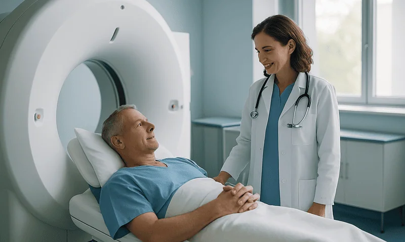
How Long CT Scan Results Take: Typical Timelines, Urgent Cases and What Delays Them
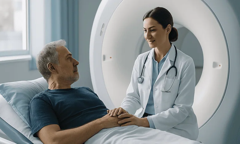
Most routine outpatient CT scan results reach the ordering clinician within a few days to a couple of weeks, while emergency department scans are…
Read article

Articles on medical imaging: MRI, CT, ultrasound, PET-CT and X-ray, covering preparation, contrast agents, radiation and how to read the wording of a report. The report is interpreted together with your symptoms by your doctor.
Most routine outpatient CT scan results reach the ordering clinician within a few days to a couple of weeks, while emergency department scans are…
Doctors usually choose ultrasound first when the question involves soft tissue near the surface, fluid, the pelvis, the pregnant uterus or moving blood, because…
EKG and ECG are two abbreviations for the same test: the electrocardiogram, a painless recording of the heart's electrical activity taken through sticky electrodes…
An ultrasound scan is a painless imaging test that uses high-frequency sound waves, not radiation, to build live pictures of organs, blood vessels and…
An echocardiogram is a painless ultrasound scan that shows the heart's size, pumping strength, valve function, and blood flow in real time. Doctors order…
Cardiac stress tests show how your heart performs under demand. A treadmill test records the heart's electrical activity during exercise; a stress echocardiogram adds…
A full-body MRI scan can detect some tumors, aneurysms, cysts, and structural abnormalities without radiation, but no major medical organization recommends it for screening…
Most MRI scans take between 15 and 90 minutes, depending on the body part, the number of imaging sequences, and whether contrast dye is…
A CT scan can detect many tumors, especially solid masses in the lungs, liver, kidneys, pancreas, bones and lymph nodes, often when they are…
A Holter monitor is a small, portable device that records every heartbeat for 24 to 48 hours through adhesive chest electrodes. It helps doctors…
An echocardiogram is an ultrasound scan of the heart that shows its size, the thickness of its walls, how strongly it pumps, and whether…
An abdominal ultrasound uses sound waves to examine the liver, gallbladder, pancreas, spleen, kidneys, and abdominal aorta, detecting problems such as gallstones, cysts, fluid…
An MRCP (magnetic resonance cholangiopancreatography) test is a non-invasive MRI scan that creates detailed pictures of the bile ducts, gallbladder, and pancreatic duct without…
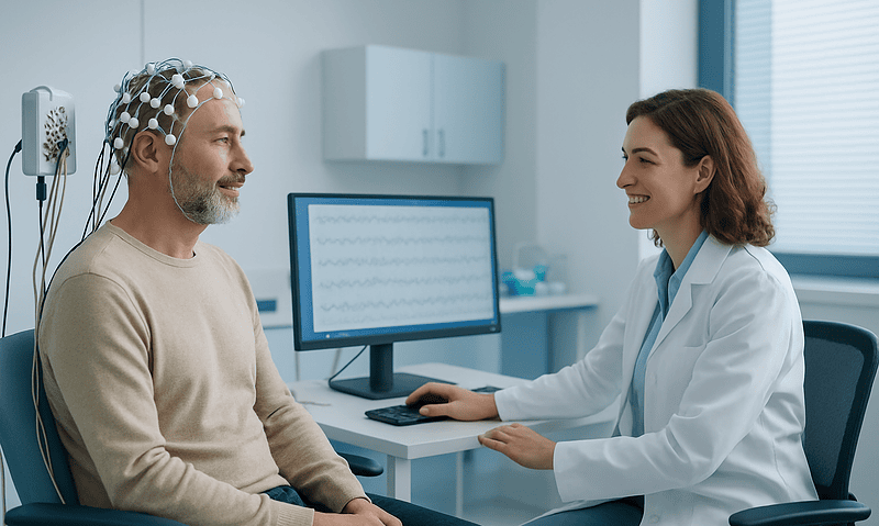
Neurophysiology tests measure the electrical activity of nerves, muscles, and the brain. An EMG records signals inside muscle, a nerve conduction study times impulses…
A renal CT scan is an X-ray-based imaging test that creates detailed cross-sectional pictures of the kidneys, ureters, and bladder. Doctors order it to…
A chest X-ray uses a small dose of radiation to picture the lungs, heart, airways, blood vessels, and bones of the chest. It can…
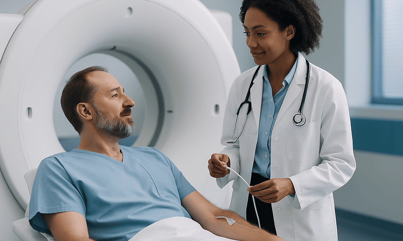
A CT scan with contrast uses an iodine-based dye, swallowed or given through an IV, to make blood vessels, organs, and abnormal tissue stand…
A CT angiogram is a noninvasive imaging test that uses X-rays and an injected contrast dye to create detailed pictures of blood vessels, …
Choose which cookies you allow. Essential cookies are always on because the site cannot work without them.