

How Laser Treatment Corrects Myopia: Flattening the Cornea to Refocus Distant Objects

Key Takeaways
- The cornea provides roughly two-thirds of the eye's focusing power, which is why flattening its center by a few microns can shift the focal point back onto the retina.
- An excimer laser removes corneal tissue by breaking molecular bonds with ultraviolet light rather than heat, so the procedure sculpts rather than burns.
- The NHS advises that a glasses prescription should ideally have been stable for about 2 years before laser correction, because an eye that is still changing will drift off target.
- The Mayo Clinic notes that full healing and stable vision can take 2 to 3 months, with dryness and night glare common during that window.
- Laser treatment corrects the blur of myopia but not the elongated shape of the eye, so the retinal and glaucoma risks the NEI links to high myopia remain and regular dilated exams continue.
- Presbyopia, the age-related loss of near focus, arrives in the forties whether or not you had laser surgery, so reading glasses later in life should be expected.
Laser treatment corrects myopia by reshaping the cornea, the clear front window of the eye. In a nearsighted eye, light focuses in front of the retina instead of on it. An excimer laser removes a microscopically thin, precisely mapped layer of central corneal tissue, flattening its curve and reducing its focusing power so distant light lands on the retina. Suitability depends on prescription stability, corneal thickness and overall eye health, all assessed by an eye specialist.
The glasses fog the moment she steps in from the cold. Her contact lens slides off a fingertip and vanishes into the sink drain, again. She has read that a laser can fix all this in minutes, and she has also heard a colleague say the laser “burns your eye.” Neither picture is quite right, and the gap between them is where most of the anxiety lives.
Understanding how laser treatment corrects myopia turns out to be a lesson in geometry rather than drama. The eye is a small camera with two lenses. Nearsightedness means the camera focuses a fraction of a millimeter too early. The surgeon’s job is not to shorten the eye, which is impossible, but to weaken its front lens by a calculated amount so the focal point drifts back onto the retina.
This explainer walks through that geometry, what happens in the room, who is usually a candidate, what the first weeks feel like, and which warning signs deserve a same-day call.
How laser treatment corrects myopia: why the cornea is the target
Two structures bend light inside the eye. The cornea, the clear dome at the very front, does most of the work, contributing roughly two-thirds of the eye’s total focusing power because it sits at the boundary between air and fluid, where light bends most sharply. The crystalline lens behind the pupil adds the rest and fine-tunes focus for near and far.
In myopia, this system is slightly too strong for the length of the eye. Either the eyeball has grown a little too long from front to back, or the cornea is a little too steeply curved, or both. Light from a distant street sign converges before it reaches the retina, the light-sensing layer at the back, then spreads out again by the time it arrives. The result is a blurred sign and a sharp phone screen, since near objects naturally focus further back.
A laser cannot make the eye shorter. What it can do is change the shape of the cornea with extraordinary precision. By removing a thin layer of tissue from the center and progressively less toward the edge, the surgeon flattens the central curve. A flatter surface bends light less. Less bending pushes the focal point backward, ideally onto the retina itself.
Glasses achieve the same optical result from outside the eye, using a concave lens that diverges light slightly before it enters. Laser correction, in effect, carves that same corrective lens into the front of the eye so it travels with you. The Mayo Clinic describes this reshaping as the shared principle behind all corneal refractive procedures for nearsightedness, whatever the technique used to reach the tissue.
The laser itself is an excimer laser, a source of ultraviolet light that breaks the molecular bonds in tissue and removes it layer by layer with almost no heat. That is why “burning” is the wrong word; the process is closer to sculpting.
What myopia actually is, and why glasses only mask it
Myopia is a refractive error, meaning a focusing mismatch, not a disease of the eye’s tissues. The National Eye Institute (part of the NIH) explains that nearsightedness usually appears in childhood, tends to progress through the school years as the eye grows, and typically settles in early adulthood once growth stops. This matters for surgery timing, as later sections explain.

Glasses and contact lenses do not change the eye at all. They intercept light before it enters and adjust its path so the eye’s own optics land it correctly. Take them off and the mismatch is exactly as it was. Laser correction changes the eye’s own optics, which is why the effect persists without a device.
One point deserves plain statement because it shapes how people should think about the procedure. The NEI notes that higher degrees of myopia carry an increased lifetime risk of certain eye conditions, including retinal detachment, glaucoma and damage to the macula at the center of the retina. Those risks are linked mainly to the elongated shape of a myopic eye. Flattening the cornea corrects the focus but leaves the eye’s length untouched, so a person who was highly nearsighted before laser treatment still has the anatomy of a highly nearsighted eye afterward.
In practice this means the procedure changes what you see, not what your retina needs. Regular dilated eye examinations remain part of long-term care for anyone with significant myopia, corrected or not. Eye specialists generally frame laser surgery as a way to reduce dependence on lenses, not as a way to make the eye “normal” in every respect.
Holding both ideas at once, better focus and unchanged anatomy, is the most useful mental model a patient can bring into the consultation.
How laser treatment corrects myopia in numbers: diopters and microns
Two units run through every conversation about laser vision correction. A diopter is the unit of lens power; a nearsighted prescription is written as a negative number, and a larger negative number means a stronger correction is needed. A micron is one thousandth of a millimeter, roughly one-hundredth the width of a human hair.
The central cornea is a little over half a millimeter thick, thinner than a bank card. The laser works within the front portion of that thickness. The more diopters of myopia to correct, the more tissue must be removed from the center to achieve the required flattening, with the removal tapering smoothly toward the edge of a circular treatment zone that roughly matches the pupil. Surgeons calculate the depth from a standard optical formula, and the exact figure for any individual depends on prescription and the chosen zone size.
This arithmetic explains three things patients often hear in consultation. First, why corneal thickness is measured so carefully: enough tissue must remain underneath to keep the cornea structurally stable for life. Second, why very high prescriptions may be steered toward other options; the NHS notes that lens-based surgery may be more suitable for people with a high spectacle prescription. Third, why a larger pupil, which needs a wider treatment zone, can influence the plan.
Modern systems add a second layer of numbers. Corneal topography, a detailed map of the surface curvature, and wavefront analysis, a measurement of how light is distorted through the whole eye, feed into a customized treatment profile rather than a single average curve. Eye-tracking in the laser adjusts for tiny movements many times per second.
None of these figures should be treated as promises about the outcome. They describe the plan. Healing, which the next sections cover, introduces variation the laser cannot fully control.
What actually happens in the laser room
Most people are surprised by how ordinary the setting feels. You lie on a reclining bed beneath the laser. The Mayo Clinic notes that the procedure for both eyes generally takes around 30 minutes or less, and that you remain awake throughout.
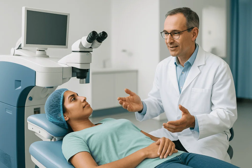
The sequence for the most widely known technique, LASIK (laser-assisted in situ keratomileusis), runs roughly as follows. Numbing eye drops are applied, so the eye feels pressure and touch but not pain. A small device holds the eyelids open, and a suction ring steadies the eye; this is usually the least pleasant moment, described by many as firm pressure with brief dimming of vision.
Next, a thin hinged flap is created in the outer cornea, either with a femtosecond laser, which uses very short pulses to separate tissue, or with a fine mechanical blade. The flap is folded back like the cover of a book. You may be asked to stare at a blinking target light while the excimer laser works on the exposed tissue for a matter of seconds per eye. A faint clicking sound and a smell sometimes described as similar to singed hair are normal; the smell comes from the tissue removal process, not from heat damage.
The flap is then laid back into position, where it adheres naturally without stitches within minutes. The Cleveland Clinic and MedlinePlus both describe the same core steps, differing only in wording.
Afterward you sit up, vision typically hazy as though looking through a steamed window, and someone else drives you home. Protective shields, prescribed drops and a follow-up appointment are arranged before you leave. Surface techniques such as PRK skip the flap and remove the thin outer layer instead, which changes the recovery story, as the comparison below shows.
LASIK vs PRK vs SMILE for nearsightedness: how each reaches the cornea
All three mainstream laser techniques flatten the central cornea by the same optical logic. They differ in how they get to the tissue, and that difference drives comfort, recovery pace and suitability. The NHS lists LASIK, surface procedures (PRK and LASEK) and SMILE as the laser options generally offered for refractive error.
| Technique | How the tissue is reached | Typical recovery pattern | Often considered when |
|---|---|---|---|
| LASIK | Thin hinged flap lifted, excimer laser reshapes beneath, flap replaced | Vision often clears within days; flap-related risks exist | Adequate corneal thickness, no flap concerns |
| PRK / LASEK | Outer surface layer removed or moved aside, laser applied directly | More discomfort in the first days; vision clears more slowly as the surface regrows | Thinner corneas, higher risk of eye trauma, some occupations |
| SMILE | Femtosecond laser cuts a lens-shaped disc inside the cornea, removed through a small keyhole | No flap; recovery generally comparable to LASIK in early reports | Moderate myopia; availability varies |
PRK stands for photorefractive keratectomy; the outer cell layer, called the epithelium, is removed and regrows over several days, which is why the early period feels grittier. LASEK is a variant that preserves and repositions that layer. SMILE, small incision lenticule extraction, removes a pre-cut internal disc rather than ablating the surface, leaving the outer cornea largely intact.
No technique is universally superior. The Mayo Clinic frames the choice as depending on corneal measurements, prescription, lifestyle and the surgeon’s assessment, and that neutral framing is the honest one. Someone who plays contact sports may be steered away from a flap. Someone with a thinner cornea may be steered toward a surface procedure. The right question is not which method is best, but which method fits this pair of eyes.
Who is not suitable for laser eye surgery, and who is asked to wait
Suitability rests on three pillars: a stable eye, a healthy eye and a cornea with enough tissue to spare. The NHS advises that laser eye surgery is generally offered to adults, and that ideally the glasses prescription should have stayed roughly the same for about 2 years beforehand. A prescription still drifting means the eye is still changing, and a correction set today may be off target within a few years.
The Mayo Clinic lists circumstances in which surgeons commonly advise against, or defer, laser correction. These include:
- Pregnancy or breastfeeding, when hormonal shifts can temporarily alter the cornea and prescription.
- Keratoconus or other conditions that thin or weaken the cornea, or a family history suggesting them.
- Autoimmune or immune-related conditions that can impair healing.
- Poorly controlled diabetes, which can affect wound healing and vision stability.
- Significant dry eye disease, which laser treatment can worsen.
- Existing cataract, glaucoma, or other eye disease that would limit the benefit.
- A prescription outside the range the cornea can safely accommodate.
Some categories are about waiting rather than a permanent no. A person in their late teens with a changing prescription may be asked to return in a year or two. Someone recovering from an eye infection or inflammation is usually asked to wait until the eye has been quiet for a period set by the specialist.
Occupation and hobbies count too. People at high risk of blows to the face may be steered away from flap procedures. Those whose jobs demand exceptional night vision are counseled carefully about glare and halos.
The decision is made jointly with the surgeon after examination, and it is never a matter of persuasion. A specialist who suggests waiting is protecting the result, not withholding it.
The tests before surgery: what your care team measures and why
The pre-operative assessment is usually longer than the surgery itself, and the thoroughness is deliberate. Each measurement answers a specific safety question.
A full refraction establishes the exact prescription, often repeated with dilating drops that relax the focusing muscle so the true error is measured without the eye compensating. Corneal topography maps the surface curvature point by point, screening for subtle irregularities that might indicate a cornea prone to weakening. Pachymetry measures corneal thickness, the figure that determines how much tissue can be removed while leaving a safe reserve.
Pupil size is measured in dim light because a large pupil may extend beyond the treated zone at night, which is one contributor to halos around headlights. Tear film quality is assessed, since existing dryness tends to worsen in the months after surgery. A dilated examination of the retina checks for thinning, tears or other changes, which is particularly relevant in higher myopia.
Contact lens wearers are asked to stop wearing lenses for a period before the assessment and again before surgery. The Mayo Clinic explains that contact lenses temporarily alter the shape of the cornea, and measurements taken too soon would not reflect the eye’s true form. The exact interval depends on lens type and is set by the clinic team; rigid lenses generally require a longer break than soft ones.
You will also be asked about medicines, medical conditions and past eye problems. Some medicines affect healing or tear production, so bring a current list rather than relying on memory.
Expect the assessment to end in one of three places: a recommendation to proceed with a specified technique, a recommendation to wait and re-measure, or a recommendation toward an alternative. All three are legitimate outcomes of a careful workup.
The first days and weeks after laser treatment for myopia
The first evening is usually spent with eyes closed. The Mayo Clinic describes the common early sensations after LASIK: itching, a gritty or burning feeling, watering, light sensitivity and blurred vision. Pain is typically mild and short-lived after LASIK, more pronounced for a few days after surface procedures such as PRK while the outer layer regrows.
Most people notice clearer distance vision by the next morning after LASIK, though it fluctuates. A follow-up visit is generally scheduled within the first day or two so the surgeon can check the flap or surface and confirm healing is on track. Prescribed drops, usually an antibiotic to reduce infection risk and an anti-inflammatory to calm the healing response, are used on the schedule the team provides; lubricating drops help with dryness.
A few practical rules dominate the early weeks:
- Do not rub the eyes, since the flap can be displaced in the first days.
- Wear the protective shields provided at night for as long as advised.
- Avoid swimming pools, hot tubs and open water for the period your team specifies, because of infection risk.
- Hold off on eye makeup and creams around the eyes until cleared.
- Return to driving only when the surgeon confirms your vision meets the legal standard.
Vision continues to settle for longer than people expect. The Mayo Clinic notes that it can take 2 to 3 months for the eye to heal fully and vision to stabilize. Night glare and dryness are common during this window and tend to ease, though not for everyone.
Follow-up appointments typically continue over the first several months. Attending them is not a formality; they are when under- or over-correction is identified and when any need for additional treatment is discussed.
Is laser eye surgery safe for myopia? Risks explained without spin
Safety is best discussed in terms of specific, known risks rather than reassurance. The Mayo Clinic and the NHS describe a consistent set, and every candidate should hear them plainly.
Dry eyes are the most common complaint. Cutting through corneal nerves reduces the signals that drive tear production, and the effect can persist for months. The Mayo Clinic notes dryness generally improves over time, but a minority experience it long-term. Glare, halos and double images, especially around lights at night, are frequent early on and usually diminish as the cornea settles; for some they remain noticeable.
Under-correction and over-correction happen because healing varies between individuals. Too little tissue removed leaves residual myopia; too much tips the eye toward farsightedness. The surgeon may discuss a further procedure once the eye has stabilized, but that decision depends on thickness and healing. New or altered astigmatism can result from uneven tissue removal.
Flap complications are specific to LASIK: the flap may fold, wrinkle or become displaced, and cells can grow beneath it. Most are treatable if caught early, which is one reason follow-up matters. Corneal ectasia, a progressive weakening and bulging, is uncommon but serious; careful pre-operative screening exists largely to avoid it.
Infection and inflammation are rare but real. Very rarely, vision loss can occur that lenses cannot fully restore. The NHS describes serious complications as uncommon but notes that no surgery is risk-free.
What the evidence supports, then, is a nuanced answer. For well-selected candidates who are screened thoroughly, laser correction has a long track record and a well-characterized risk profile. “Safe” is not the same as “without risk,” and an honest surgeon will spend as long on this list as on the benefits.
Does LASIK permanently fix myopia?
The tissue the laser removes does not grow back, so the change to corneal shape is permanent in that literal sense. Whether your vision stays where it was set is a different question, and the honest answer is: usually largely yes, with several well-understood exceptions.
The first is regression. In some eyes, particularly those that started with higher prescriptions, a small amount of myopia returns over months or years as the cornea remodels during healing. The Mayo Clinic lists this among recognized outcomes. It is not a failure of the laser; it is biology adjusting to a new shape. Where enough corneal thickness remains and the eye has stabilized, an enhancement procedure may be considered, but that call rests with the treating surgeon.
The second is that the eye itself can still change. If surgery is done before the prescription has settled, the underlying myopia may continue to progress, which is why stability for about 2 years is the usual expectation described by the NHS.
The third is presbyopia, the age-related stiffening of the lens that makes near focus difficult, typically beginning in the forties. Laser correction of distance vision does nothing to prevent it. Many people who had LASIK in their twenties find themselves reaching for reading glasses two decades later, exactly as they would have without surgery. Some clinics discuss deliberately leaving one eye slightly nearsighted to soften this, a strategy with trade-offs that deserves a frank conversation.
Finally, cataract develops with age in corrected and uncorrected eyes alike. Prior laser surgery does not prevent cataract surgery later, though it does make the calculations for the replacement lens more complex, so keep records of your pre-operative measurements for life.
“Permanent” therefore describes the corneal change accurately and the whole visual future only approximately.
Alternatives when the cornea is not the right place to work
A recommendation against corneal laser surgery is not the end of the conversation. Several other routes exist, each with its own logic.
Glasses and contact lenses remain the most widely used correction and carry no surgical risk. Modern contact lens materials and daily disposables have reduced, though not eliminated, the infection risk associated with lens wear, and the Cleveland Clinic notes that proper hygiene remains essential.
Orthokeratology uses rigid lenses worn overnight to temporarily flatten the cornea, producing clear daytime vision without lenses. The effect reverses if wear stops, and the same infection precautions apply.
For higher prescriptions or thinner corneas, surgeons may discuss lens-based surgery. An implantable collamer lens, sometimes called a phakic intraocular lens, is a thin corrective lens placed inside the eye in front of the natural lens, which is left in place. The correction is reversible in principle because the implant can be removed. Refractive lens exchange replaces the natural lens with an artificial one, the same operation used for cataract; the NHS notes lens surgery may suit people with a high prescription or those in later life for whom cataract is on the horizon.
For children, whose eyes are still growing, laser surgery is not offered. The focus is on slowing progression. Options studied for myopia control include specially designed spectacle and contact lenses and low-concentration atropine eye drops, which appear to act on signaling in the eye that influences growth. Evidence for these approaches is still developing, and any decision about a child’s treatment sits with the pediatric eye specialist.
Each alternative trades one set of risks and limits for another. The specialist’s task is to match the option to the eye, not to the person’s preference for a particular procedure.
What people often get wrong about laser treatment for myopia
Myths cluster around this procedure, partly because it is common and partly because it involves a laser near the eye. A few corrections, grounded in what the major medical sources actually say.
“The laser burns the eye.” The excimer laser removes tissue by breaking molecular bonds with ultraviolet light, producing negligible heat. The smell some notice comes from the removal process itself.
“You could blink or move and ruin it.” A lid holder prevents blinking, and the laser tracks eye movement many times per second, pausing if the eye moves beyond a set range.
“It fixes your eyes for life.” The corneal change is permanent, but presbyopia arrives regardless, regression can occur, and cataract develops with age. Reading glasses in the forties are the rule, not the exception.
“Everyone ends up with perfect vision.” Outcomes vary with the starting prescription and healing. The Mayo Clinic is explicit that results are less predictable for higher degrees of myopia and that some people need lenses for certain tasks afterward.
“If I’m very nearsighted, I need it more.” High myopia is exactly where corneal surgery is most likely to be limited by tissue thickness and where lens-based options enter the discussion. Need and suitability are different things.
“Laser surgery removes the risks of being nearsighted.” It removes the blur. The elongated eye, and the retinal and glaucoma risks the NEI links to high myopia, remain and still warrant regular dilated examinations.
“Dry eye is a rare side effect.” It is the most common one, and anyone with existing dryness should expect a careful conversation about it beforehand.
Correcting these misconceptions tends to produce a calmer, better-prepared patient, which is worth more than any reassurance.
Questions to ask your care team before deciding
A good consultation should leave you able to explain the plan in your own words. These questions help get there, and none of them should be unwelcome.
- Which technique are you recommending for my eyes, and what in my measurements led you there rather than to another method?
- How thick are my corneas, and how much tissue would remain after treatment?
- Has my prescription been stable long enough, by your assessment, to proceed now?
- How large are my pupils in dim light, and does that affect the expected night vision result?
- What did my tear film assessment show, and how do you expect dryness to behave in my case?
- What does my retinal examination show, given my level of myopia?
- What range of visual outcome is realistic for a prescription like mine, and what would make you consider a further procedure?
- Which side effects should I expect in the first weeks, and which would be unusual?
- What is the follow-up schedule, and who do I contact out of hours if something feels wrong?
- How will presbyopia affect me later, and is there a reason to consider leaving one eye slightly nearsighted?
- Are there reasons in my medical history, medicines or occupation that make you hesitate?
- If laser surgery is not ideal for me, which alternative would you discuss and why?
Bring your current glasses and contact lens details, a list of medicines, and any records of past eye problems. Take notes or bring someone with you; the amount of information is considerable, and drops may blur your vision for a few hours after the assessment.
A specialist who welcomes these questions, and who is comfortable saying “not yet” or “not this method,” is demonstrating exactly the judgment the procedure depends on.
When to call your doctor: red-flag signs after laser eye surgery
Most of what people feel in the first days is expected: grittiness, watering, light sensitivity, fluctuating blur. Some signs are not, and they warrant a same-day call to the surgical team or, if that is not possible, urgent eye care. Do not wait for the next scheduled appointment.
Contact your care team urgently if you notice any of the following:
- Severe or worsening eye pain, particularly pain that increases rather than eases after the first day, or pain out of proportion to what you were told to expect.
- A sudden drop in vision, or vision that was clearing and then becomes markedly worse.
- New flashes of light, a shower of new floaters, or a shadow or curtain across part of your vision. These can signal a retinal tear or detachment, a risk that is higher in nearsighted eyes regardless of surgery and needs urgent assessment.
- Increasing redness, thick discharge, or a white or gray spot on the cornea, which may indicate infection.
- Any blow to the eye, or a sense that something has shifted after rubbing, in the weeks when the flap is still settling.
- Sudden severe headache with eye pain, nausea and halos, which can indicate a dangerous rise in eye pressure.
The Mayo Clinic and the NHS both emphasize that early treatment of infection, flap problems or pressure changes improves the chance of preserving vision, which is why the threshold for calling should be low.
For the longer term, keep the schedule of dilated eye examinations your specialist recommends. A corrected myopic eye still has the anatomy of a myopic eye, and the same retinal warning signs apply years after the laser has done its work. Every treatment decision, including whether any further procedure is appropriate, rests with the team that examined you.
Frequently asked questions
Is laser eye surgery safe for myopia?
For carefully screened adults with stable prescriptions and healthy corneas, laser correction has a long track record and well-described risks rather than unknown ones. The most common side effects are dry eyes and night glare or halos, which the Mayo Clinic notes usually ease over months but can persist. Rare complications include infection, flap problems and corneal weakening. Thorough pre-operative testing exists to identify eyes that should not proceed.
Who is not suitable for laser eye surgery?
People with a changing prescription, thin or irregular corneas, keratoconus, significant dry eye, cataract, glaucoma, uncontrolled diabetes or immune conditions that impair healing are commonly advised against it, according to the Mayo Clinic. Pregnancy and breastfeeding are usual reasons to defer. Very high prescriptions may exceed what the cornea can safely accommodate, in which case lens-based options are discussed. The final judgment rests with the examining specialist.
LASIK vs PRK for nearsightedness: which heals faster?
LASIK typically gives clearer vision within a day or two because the surface layer is preserved as a flap, while PRK removes that layer and vision clears more gradually as it regrows over several days. PRK involves more discomfort early on but avoids flap-related risks and may be preferred for thinner corneas or people at risk of eye trauma. Both flatten the cornea by the same principle.
Does LASIK permanently fix myopia?
The change to corneal shape is permanent, but vision can still shift. Some regression of myopia can occur as the cornea heals, particularly with higher prescriptions, and the underlying eye can change if surgery happens before the prescription has settled. Presbyopia in the forties and cataract in later life develop regardless. Any further procedure depends on remaining corneal thickness and is decided by the surgeon.
How long does laser treatment for myopia take on the day?
The Mayo Clinic notes that LASIK for both eyes generally takes about 30 minutes or less, with the laser itself active for only seconds per eye. Most of that time goes on positioning, numbing drops, steadying the eye and creating and replacing the flap. You go home the same day, but vision is hazy for the first hours so someone else needs to drive.
Why does the surgeon flatten the cornea instead of fixing the eye's length?
The eyeball’s length cannot be surgically shortened, but its front lens can be weakened. Because the cornea does most of the eye’s focusing, removing a thin central layer flattens its curve, reduces how sharply it bends light and moves the focal point backward onto the retina. This produces the same optical effect as a concave spectacle lens, built into the eye itself.
Can very high myopia be treated with laser?
It depends on how much corneal tissue would need to be removed and how much would remain. Higher prescriptions require deeper removal, and the surgeon must leave a safe reserve for structural stability. The NHS notes that lens-based surgery, such as an implantable lens or lens exchange, may be more suitable for high prescriptions. Results for high myopia are also less predictable, so expectations are set carefully.
What does recovery look like in the first week after laser eye surgery?
Expect grittiness, watering, light sensitivity and fluctuating blur, with a follow-up visit within the first day or two. Prescribed drops reduce infection and inflammation, and lubricating drops help dryness. You will be asked not to rub the eyes, to wear shields at night, and to avoid swimming and eye makeup for a period set by your team. Driving resumes only when the surgeon confirms your vision meets the legal standard.
Will I still need glasses after laser correction for myopia?
Many people rely far less on lenses for distance tasks, but outcomes vary with the starting prescription and healing, and the Mayo Clinic notes some people still use glasses for certain activities. Reading glasses become likely in the forties as presbyopia develops, regardless of surgery. Your surgeon can give a realistic range for your own measurements rather than a general figure.
Does laser surgery reduce the eye risks that come with being nearsighted?
No. The National Eye Institute links higher myopia to increased risk of retinal detachment, glaucoma and macular damage, and those risks come mainly from the elongated shape of the eye, which laser treatment does not change. Corrected eyes still need the regular dilated examinations recommended for the original level of myopia, and new flashes, floaters or a shadow in vision remain urgent warning signs for life.
References
- NHS: Laser eye surgery and lens surgery
- MedlinePlus: LASIK eye surgery
- Cleveland Clinic: LASIK eye surgery
- National Eye Institute (NIH): Nearsightedness (myopia)
This article is for general information only and is not a substitute for professional medical advice. Please consult a qualified doctor about your individual situation.

More from the Blog

Questions to Ask Before Laser Treatment for Farsightedness (and What the Answers Mean)
Before laser treatment for farsightedness, ask your care team which procedure they propose and why, how high and how stable your prescription is, how…
How Eye Muscle Surgery for Strabismus Works: Loosening, Tightening and Adjustable Sutures
Strabismus surgery realigns the eyes by repositioning one or more of the six small muscles attached to the outside of each eyeball. A surgeon…
Is Super LASIK Only for Complex Prescriptions? Who Actually Benefits From Custom Treatment
No. Custom or wavefront-guided LASIK, often marketed as Super LASIK, is not reserved for complex prescriptions. It uses a detailed optical map of each…
When Does a Drooping Eyelid Need Oculoplastic Surgery? Vision, Comfort and Timing
A drooping eyelid usually needs surgery when the lid margin sits low enough to block the upper or central field of vision, when it…
Who Is Steered Toward LASEK PRK? Thin Corneas, Contact Sports and Dry Eye Explained
LASEK and PRK are surface laser treatments that reshape the cornea without cutting a flap, so eye surgeons often suggest them for people whose…
Preparing for Vitrectomy: Fasting, Medication Review and Arranging Help for the First Days
Preparing for vitrectomy usually means attending a pre-operative assessment, following the fasting instructions your surgical team gives you, reviewing every medicine and supplement with…