

ICL Surgery: The Implantable Lens Alternative to LASIK

Key Takeaways
- An ICL is a phakic lens, meaning it is added behind the iris while your natural lens stays in place, so the cornea is never reshaped.
- FDA labeling covers adults aged 21 to 45 with myopia roughly between -3 and -20 diopters and a prescription stable within about half a diopter for a year.
- The central-port lens approved in 2022 removed the need for a preparatory laser iridotomy, but its long-term data are shorter than those of older designs.
- Most patients see clearly within a day, yet swimming, eye rubbing and contact sports are typically off-limits for one to two weeks while the incision seals.
- Cataract, raised eye pressure, endothelial cell loss and night halos are the main downsides, and roughly 1 to 3 percent of patients need the lens resized or repositioned.
- The implant has no expiry date, but it does not prevent presbyopia or age-related cataract, both of which arrive on their usual timetable.
ICL surgery places a thin, flexible implantable collamer lens inside the eye, between the iris and the natural lens, to correct nearsightedness and astigmatism without reshaping the cornea. It suits adults roughly aged 21 to 45 with stable prescriptions, including many people turned away from LASIK because of thin corneas or high myopia. The lens is removable, recovery is usually quick, and risks include cataract, pressure rises and night halos.
The reel is nine seconds long. A woman in her late twenties holds up a pair of thick glasses, blinks at the camera, and mouths the caption: “LASIK said no. ICL said yes.” It has millions of views, and the comments are a flood of the same question: what is this, and why has nobody told me?
Plenty of people are asking. As of early 2026, searches for ICL surgery have climbed steadily since the US Food and Drug Administration approved a redesigned version of the lens in 2022, one with a tiny central port that removed a separate laser step and made the procedure simpler. Athletes and influencers followed, and the phrase “implantable contact lens” started circulating as if it were brand new. It is not. Surgeons have been implanting these lenses for more than two decades.
What is new is the audience. Many of the people now curious about it have thin corneas, very high prescriptions or dry eyes, the very things that make laser surgery a poor fit. This piece walks through what the procedure does, what can go wrong, and what the evidence actually supports.
What is ICL surgery and how does it work?
ICL stands for implantable collamer lens. Collamer is a soft, biocompatible material made from collagen and a plastic called HEMA, chosen because the eye tends to tolerate it without inflammation. The lens itself is a transparent, flexible disc, thinner than a contact lens, with four small footplates that let it rest gently in the eye.
The surgery is a form of phakic intraocular lens implantation. “Phakic” simply means the natural lens stays in place; nothing is removed. Instead, the surgeon slides the folded ICL through a small opening at the edge of the cornea and positions it in the narrow space between the colored iris and the natural lens, a region called the posterior chamber. Once unfolded, it sits behind the pupil, invisible to anyone looking at you.
The optical principle is the same one your glasses use. A refractive error is a mismatch between the eye’s focusing power and its length. In nearsightedness (myopia), the eye is slightly too long, so light focuses in front of the retina and distant objects blur. A minus-powered lens bends incoming light outward just enough to push the focus back onto the retina. The ICL does this from inside the eye rather than from a frame on your nose.
Laser procedures such as LASIK achieve the same correction by permanently thinning and reshaping the cornea, the clear dome at the front of the eye. ICL surgery leaves the cornea essentially untouched apart from the entry incision, which is why it is often described as additive rather than subtractive. That distinction matters most for people with high prescriptions, where the laser would need to remove a lot of tissue, and for people whose corneas are too thin to spare any.
Current lenses correct myopia across a wide range and, in a toric version, astigmatism as well. They do not correct ordinary farsightedness in the United States, and they do not treat presbyopia, the age-related loss of near focus that sends most people to reading glasses after 45.
What changed recently and why ICL surgery is trending
Three things pushed a twenty-year-old procedure into the feed. The first was regulatory. In March 2022, the FDA approved a redesigned implantable collamer lens with a 360-micron hole at its center. Earlier models required the surgeon to make one or two small openings in the iris beforehand, usually with a laser, so that fluid inside the eye could keep circulating. The central port lets the eye’s natural fluid pass straight through the lens, so that preparatory step disappeared. Fewer visits, less inflammation, a simpler story to tell.
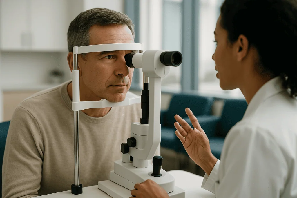
The second shift was cultural. Through 2024 and 2025, professional athletes, musicians and a long tail of creators posted their own experiences, often framed as “the thing you get when LASIK rejects you.” Social video rewards a clean narrative, and this one has it: no cutting of the cornea, reversible, done in minutes.
The third driver is demographic. Myopia is rising worldwide. The World Health Organization has flagged uncorrected refractive error as a leading cause of vision impairment, and in many urban populations more than half of young adults are nearsighted. A growing share of them have prescriptions high enough that laser surgery is marginal or off the table. For that group, an implantable lens is not an exotic alternative; it is often the only surgical option a careful clinic will offer.
What has not changed is the underlying science. Long-term outcome data, including studies that followed patients for 10 years or more, come mostly from the earlier lens designs. The central-port version has shorter follow-up by definition, though the published results so far track closely with its predecessors. When you read that the new lens has “solved” the old complications, treat that as a hopeful extrapolation rather than a settled finding. The NHS and Mayo Clinic resources listed at the end describe lens surgery in neutral terms, and that is the tone the data deserve.
Who is a good candidate for ICL surgery?
The ideal patient, in the words of most refractive surgeons, is someone in their twenties or thirties whose prescription has barely budged in a year and who is nearsighted enough that glasses are a daily nuisance. The FDA labeling covers adults aged 21 to 45 with moderate to severe myopia, with or without astigmatism, whose refraction has been stable within about half a diopter for the past 12 months. A diopter is the unit of lens power; the more negative the number, the stronger the correction.
Several measurements decide the matter more than age does. The surgeon needs enough room in the front of the eye, specifically an anterior chamber depth of roughly 3 millimeters or more between the back of the cornea and the front of the natural lens. Too shallow, and the implant could crowd the structures around it and raise pressure. The inner lining of the cornea, a single layer of cells called the endothelium, must also be healthy and reasonably dense, because those cells do not regenerate and the lens sits in their neighborhood.
People who are commonly steered toward ICL rather than laser include those with:
- Thin corneas, where LASIK would leave too little tissue for long-term stability.
- Prescriptions beyond about minus 8 diopters, where laser reshaping becomes less predictable.
- Significant dry eye, which laser surgery tends to worsen for months.
- Keratoconus suspects or irregular corneal maps, where any corneal reshaping is unwise.
Others are generally advised against it: anyone with glaucoma or a family history that raises the risk, early cataract, a history of uveitis (inflammation inside the eye), unstable or progressing myopia, or pregnancy, since hormonal changes shift refraction temporarily. Diabetes with eye involvement and certain retinal conditions also call for caution. None of these are tick-box rules; the decision rests on a dilated examination, a corneal map and the surgeon’s judgment.
ICL vs LASIK: what is the real difference?
Picture two ways to fix a blurry projector. You can grind the lens inside the projector into a new shape, or you can slip a corrective filter in front of the bulb. LASIK is the first approach; ICL is the second.

In LASIK, a surgeon creates a thin hinged flap in the cornea, folds it back, and uses an excimer laser to vaporize microscopic layers of tissue so that the cornea’s curve matches the prescription. The flap is laid back down and heals without stitches. PRK, an older cousin, skips the flap and lasers the surface directly, which means a slower, more uncomfortable recovery but no flap-related risks. SMILE uses a femtosecond laser to carve a small lenticule of tissue that is removed through a keyhole incision.
Every laser option shares one fact: the cornea is permanently thinner afterward. That is fine when the correction is modest and the cornea thick. It becomes a problem when the prescription is high, because more tissue must go, and when the cornea is already thin or irregular. Removing too much tissue weakens the dome and can lead to a bulging condition called ectasia years later.
ICL surgery sidesteps that trade-off. The cornea keeps its thickness and natural shape, which is one reason studies consistently report better contrast sensitivity and fewer induced optical aberrations in high myopes treated with phakic lenses compared with laser. It is also the reason the procedure is reversible in a way laser surgery is not. The lens can be removed or swapped if the prescription changes or a problem develops.
The price of that flexibility is that something is now living inside the eye. Laser surgery’s risks are mostly corneal and front-of-eye: dry eye, flap problems, haze, regression. ICL’s risks are intraocular: cataract, pressure spikes, endothelial cell loss and, rarely, infection inside the eye. Neither is risk-free, and neither is universally better. The right question is which risk profile fits your particular eye.
What happens on the day of ICL surgery?
The procedure is shorter than most people expect. From the moment you lie down to the moment you sit up, 15 to 30 minutes is typical for one eye, and many surgeons treat both eyes the same day or a week apart.
Preparation starts earlier. Your pupil is dilated with drops so the surgeon can see the full working space, and anesthetic drops numb the surface of the eye. Some clinics add mild sedation by mouth or vein for comfort; most patients remain awake and aware of light and gentle pressure but feel no pain. A small device holds the eyelids open.
Inside, the steps follow a set sequence. The surgeon makes a self-sealing incision at the edge of the cornea, usually about 3 millimeters wide, and fills the front of the eye with a protective gel called a viscoelastic. The folded ICL is loaded into a cartridge and injected through the incision; it unfolds slowly as it meets the eye’s warmth. Using a fine instrument, the surgeon tucks each of the four footplates behind the iris so the lens settles into the posterior chamber. The gel is rinsed out, the incision is checked for leaks, and the pupil is often constricted with a drop to help the lens seat.
Many surgeons also measure the vault before you leave. Vault is the gap between the back of the implant and the front of the natural lens; it needs to be neither too tight nor too loose, and lens sizing is largely about getting this right.
Afterward you rest briefly, have your eye pressure checked once or twice over the next hours, and go home with a protective shield, a set of prescribed drops and a schedule for follow-up. You cannot drive yourself. Vision is typically hazy for the rest of the day, then noticeably clearer by the next morning.
ICL surgery recovery time: the first week and beyond
One of the reasons ICL spreads so easily on social media is the speed of visible results. Most people can read a clock across the room the day after surgery. Still, “back to normal” is a longer arc than “can see,” and it helps to know both timelines.
In the first 24 hours, expect light sensitivity, a gritty or scratchy feeling, mild aching and some tearing. Halos around lights are common while the pupil is still dilated. Pressure checks in this window matter because retained viscoelastic gel or an inflammatory response can push eye pressure up temporarily.
Over the first week, vision steadies as the cornea settles and the pupil returns to its normal size. You will be using prescribed anti-inflammatory and antibiotic drops on the schedule your surgeon sets. Most people return to desk work within two to three days, and many feel comfortable driving once their surgeon confirms their vision meets the legal standard.
A few restrictions typically run for one to two weeks: no rubbing the eyes, no eye makeup, no swimming or hot tubs, and no heavy lifting or contact sports, since the incision is still sealing and a blow to the eye could shift the lens. Showering is fine as long as water is kept out of the eyes. Screens are allowed; they do not harm the implant, though dryness may make long sessions tiring.
By one month, the picture is usually stable and the final vision check happens. Compared with laser surgery, where dry eye and fluctuating vision can linger for three to six months, the ICL recovery window is often shorter and less bumpy, mainly because the corneal nerves are not disrupted. Follow-up then thins out to a visit at around three to six months and annual checks after that, with pressure, cell counts and vault measured each time.
What is the downside of ICL surgery? The common side effects
Honest answers beat enthusiastic ones here, so start with what is ordinary rather than rare. Nearly everyone sees some halos or starbursts around lights at night in the early weeks. The central port in the current lens is a tiny aperture, and light passing through it can scatter into a faint ring, particularly when the pupil is large in dim conditions. For most people this fades or becomes unnoticeable over a few months; for a small minority it persists and is bothersome.
Dryness is milder than after LASIK but not absent, because the incision and drops still irritate the surface. Glare in bright sun, a feeling of pressure and transient blurry patches are also common early on.
Two measurable changes deserve attention. The first is endothelial cell loss. Everyone loses a small percentage of these corneal cells with age, and surgery of any kind inside the eye accelerates that slightly. Long-term ICL studies report an additional loss in the low single digits over the first years, which is generally well tolerated in a healthy cornea but is the reason surgeons insist on a good starting count and annual monitoring.
The second is vault. If the lens sits too close to the natural lens, it can disturb its nutrition and encourage a cataract, a clouding of that natural lens. If it sits too far forward, it can narrow the drainage angle and raise eye pressure. Modern sizing has reduced both problems, but roughly 1 to 3 percent of patients still need the lens repositioned or exchanged for a different size.
Finally, residual refractive error is a possibility. A lens is chosen from a set of powers, and healing varies, so some people end up slightly under- or over-corrected and may need thin glasses for night driving or a later touch-up. Expectation-setting at the consultation prevents most of the disappointment that follows.
Can ICL surgery go wrong? Rare but serious complications
Yes, and the fact that most people do well does not make the serious outcomes imaginary. Four deserve plain description.
Cataract is the one most discussed. In the earlier generations of the lens, anterior subcapsular opacities, small clouds on the front surface of the natural lens, appeared in a few percent of eyes over a decade, more often in older patients and those with very high myopia. Many were visually insignificant; some progressed and required removal of both the ICL and the natural lens, with a standard cataract implant placed instead. Reports with the central-port design show lower rates so far, though follow-up is shorter.
Raised eye pressure comes in two flavors. The early, temporary kind usually responds to drops and resolves within days. The rarer sustained kind, when the lens crowds the drainage angle or pigment from the iris rubs off and clogs it, can lead to glaucoma, the progressive optic nerve damage that steals peripheral vision. This is why candidates with narrow angles or existing glaucoma are generally declined.
Endophthalmitis, an infection inside the eye, is the complication every intraocular surgeon fears. It is very rare, estimated at well under 1 in 1,000 for procedures of this type, but it is an emergency that can cost sight within days. Sudden pain, rapidly worsening vision and a red eye in the first week are the signals.
Retinal detachment is the fourth. Highly myopic eyes already carry a higher lifetime risk because the retina is stretched thin, and any intraocular procedure may add a small increment. New floaters, flashes of light or a curtain across the vision warrant same-day assessment.
Lens rotation (in toric models), lens dislocation after trauma, and persistent inflammation round out the list. Every one of these is treatable if caught early, which is the strongest argument for keeping every scheduled follow-up even when vision feels perfect.
How long does ICL last, and what about cataracts later?
The lens itself does not wear out. Collamer is stable in the eye for decades, and there is no built-in expiry or replacement interval. The more useful question is how long the correction remains the right one for your eye, and here the answer depends on biology rather than engineering.
Your prescription can still drift. ICL corrects the refractive error you have at the time of surgery; it does not stop the eye from lengthening further if your myopia has not fully stabilized, which is one reason surgeons wait for a year of stable readings. Published cohorts followed for 10 years show that the great majority of eyes stay within about a diopter of the target, with small average regression that is often clinically trivial.
Presbyopia arrives on schedule regardless. Sometime after 45, the natural lens stiffens and near focus fades. The ICL does nothing to prevent this, and many patients add reading glasses in their late forties exactly as they would have without surgery.
Then there is the cataract chapter. Everyone who lives long enough develops some clouding of the natural lens, usually from the sixties onward. When that day comes, the ICL is removed through a small incision, the cloudy natural lens is taken out, and a conventional artificial lens is implanted. The procedure is routine, and the earlier implant does not prevent it. Some surgeons describe ICL as a bridge: it covers the decades between stable young-adult myopia and the age at which cataract surgery would correct the refractive error anyway.
Removal for other reasons is possible at any point. If a complication develops or you simply want it out, the lens can be explanted and the eye generally returns to roughly its pre-operative prescription, because the cornea was never reshaped. That reversibility is real, but it is not the same as consequence-free; each additional surgery carries its own small risks.
What the evidence actually says about ICL surgery
Grading the evidence matters more than counting the testimonials, so here is where the studies sit on the ladder of certainty.
At the top are randomized trials, and there are few. A Cochrane systematic review comparing phakic intraocular lenses with excimer laser surgery for moderate to high myopia pooled a small number of randomized studies and found that phakic lenses produced better contrast sensitivity, less loss of best-corrected vision and higher patient satisfaction at one year, with the authors cautioning that follow-up was too short to judge long-term safety, particularly cataract. That is moderate-quality evidence for short-term visual outcomes and essentially no randomized evidence beyond a year.
Next come the pivotal regulatory trials. The FDA approvals rested on prospective, multicenter studies of several hundred eyes, followed for one to three years, with structured reporting of safety events. These are rigorous but single-arm, meaning no comparison group, and they reflect carefully selected patients treated by experienced surgeons.
The bulk of what we know about durability comes from observational cohorts, some tracking patients for 10 years or longer. They consistently report stable refraction, high rates of 20/20 or better uncorrected vision in previously highly myopic eyes, low but non-zero cataract rates, and modest endothelial cell loss. Observational data can overstate benefit, since patients who do poorly sometimes drop out, but the consistency across countries and decades is reassuring.
Claims about the central-port lens being safer rest mostly on medium-term registries and comparisons with historical data for older designs. That is reasonable, promising and not yet definitive.
Where the evidence is thinnest: outcomes in people over 45, in eyes with borderline anterior chamber depth, and in the subgroup who report persistent halos. Here guidance leans on expert consensus. A good surgeon will tell you which category your eye falls into and how confident the numbers allow them to be.
ICL, LASIK, PRK and SMILE at a glance
A table cannot pick a procedure for you, but it can show where each one sits. The figures below are typical ranges from published outcome studies and labeling; your own suitability depends on measurements that only an examination can supply.
| Feature | ICL surgery | LASIK | PRK | SMILE |
|---|---|---|---|---|
| What is changed | Lens added inside eye; cornea untouched | Cornea reshaped under a flap | Cornea surface reshaped, no flap | Tissue lenticule removed through keyhole |
| Typical myopia range | About -3 to -20 D | Up to about -10 D | Up to about -10 D | About -1 to -10 D |
| Astigmatism | Toric version, up to about 4 D | Yes | Yes | Limited |
| Thin or irregular cornea | Often suitable | Usually unsuitable | Sometimes suitable | Usually unsuitable |
| Reversible | Yes, lens can be removed | No | No | No |
| Functional vision | Often next day | Often next day | Several days to weeks | One to a few days |
| Dry eye after surgery | Mild, short-lived | Common for months | Common for months | Less than LASIK |
| Main long-term concerns | Cataract, pressure, cell loss | Ectasia, flap issues, regression | Haze, regression | Regression, limited retreatment |
| Presbyopia correction | No | No | No | No |
Two patterns stand out. The implantable lens wins on range and reversibility and loses on the fact that it introduces intraocular risk. Laser procedures win on decades of enormous patient numbers and lose when the cornea is thin or the prescription is high. Patients in the middle, with moderate myopia and healthy, thick corneas, often qualify for both, and for them the choice usually comes down to dry-eye history and personal attitude toward reversibility.
Common myths about ICL surgery, corrected
Viral posts compress a complex decision into a few confident lines. Several of those lines deserve unpacking.
“ICL is just a permanent contact lens.” The nickname “implantable contact lens” is marketing shorthand. A contact lens sits on the tear film and comes out at night. An ICL is a surgical implant placed inside the eye, behind the iris, and it carries intraocular risks that no contact lens does. The comparison undersells both the benefit and the seriousness.
“It is completely reversible, so there is no risk.” Reversible is accurate. Risk-free is not. Removing the lens is another operation, and complications such as cataract or endothelial loss that have already occurred do not reverse when the implant comes out.
“The new lens has no cataract risk.” Rates with the central-port design appear lower in the studies published so far, which cover roughly a decade of use. Lower is not zero, and the longest follow-up still belongs to the older designs.
“Anyone rejected for LASIK can have ICL.” Many can; many cannot. Thin corneas are one reason for a LASIK refusal, and ICL handles that well. Shallow anterior chambers, low endothelial counts, glaucoma, early cataract or unstable prescriptions rule out ICL too.
“It fixes your vision forever.” It corrects the refractive error you have at the time of surgery. It does not halt presbyopia, prevent cataract or guarantee that your myopia will never shift again.
“You will feel the lens.” You will not. It sits behind the iris, away from any nerve endings that register touch, and patients cannot see or sense it.
“ICL is experimental.” Phakic collamer lenses have been in clinical use since the 1990s and FDA-approved in the United States since 2005. The design has evolved; the concept is mature.
What a thorough ICL consultation should include
A consultation that lasts ten minutes and ends in a booking is a warning sign. The measurements that make ICL surgery safe are specific, and you are entitled to know they were taken.
Expect the appointment to begin with a full refraction, repeated if you have been wearing soft contact lenses recently, since lenses can temporarily warp the cornea. Soft lens wearers are usually asked to go without them for one to two weeks beforehand, longer for rigid lenses, so the cornea returns to its true shape.
The core of the visit is imaging. Corneal topography or tomography maps the surface and thickness of the cornea and screens for keratoconus. Anterior segment imaging, often with optical coherence tomography or ultrasound, measures the depth of the anterior chamber and the width of the drainage angle, and estimates the sulcus-to-sulcus distance that governs lens sizing. Specular microscopy counts the endothelial cells. A dilated examination checks the natural lens for early cataract and the retina for thinning or tears, which are more common in high myopia and sometimes need laser treatment before any refractive surgery.
The questions worth asking are concrete rather than reassuring:
- What are my anterior chamber depth and endothelial count, and how do they compare with the minimums you use?
- How many ICL procedures have you performed, and what is your personal rate of lens exchange or removal?
- How do you size the lens, and how often do your patients end up with a vault outside the target range?
- What is your plan if I develop raised pressure or a cataract in the years ahead?
- Would you also consider me for a laser procedure, and why or why not?
You should leave with written information about risks, a clear follow-up schedule and time to think. A surgeon who answers numerically and welcomes a second opinion is showing you exactly the temperament you want operating inside your eye.
When to see a doctor after ICL surgery
Scheduled visits catch most problems before you notice them, which is why they are not optional. Between those visits, certain symptoms should trigger a same-day call to your surgeon or, if the practice is closed, a trip to an emergency department with eye cover.
Seek urgent care if you notice any of the following:
- Sudden or rapidly worsening blurred vision, especially in the first week.
- Deep, increasing eye pain or pain accompanied by nausea and vomiting, which can signal a sharp rise in eye pressure.
- A red eye with discharge, swollen lids or a cloudy appearance to the front of the eye.
- New floaters, flashes of light, or a shadow or curtain creeping across your vision, the classic signs of a retinal tear or detachment.
- Severe light sensitivity that is getting worse rather than better after the first day or two.
- Any direct blow or injury to the eye, even if vision seems fine, since the lens can shift.
Non-urgent but worth reporting at your next visit: halos that are not improving by three months, noticeably different vision between the two eyes, intermittent blurring that comes and goes with the time of day, or a sense that your distance vision is drifting back toward blurry over months. Each of these can be assessed with a quick pressure check, a vault measurement and a refraction.
Beyond the first year, treat your annual eye examination as a standing appointment for life. The checks that matter, eye pressure, endothelial cell density and the clarity of the natural lens, change slowly and silently. An implant that is doing its job is easy to forget; it still needs someone to look at it.
None of the information here replaces that examination. Whether ICL surgery is appropriate for you, which lens size and power are chosen, and what to do if a complication arises are decisions that belong to the ophthalmologist who has measured your eye.
Frequently asked questions
What is the downside of ICL surgery?
The main downsides are that a surgical implant now sits inside the eye and carries intraocular risks: cataract formation, raised eye pressure, gradual loss of corneal endothelial cells and, very rarely, infection. Night halos from the lens’s central port are common early on. A small percentage of patients need the lens exchanged or repositioned, and the procedure does nothing for presbyopia.
How long does ICL eye surgery last?
The lens itself is designed to remain in the eye indefinitely and does not degrade. Published cohorts followed for 10 years or more show stable correction in most eyes. What changes is the eye around it: your prescription may drift slightly, reading glasses arrive after 45, and when age-related cataract develops the ICL is removed during routine cataract surgery.
Can ICL surgery go wrong?
Yes. Serious problems are uncommon but include cataract, sustained pressure rises that can lead to glaucoma, endophthalmitis (infection inside the eye) and retinal detachment, to which highly myopic eyes are already prone. Most are treatable when caught early, which is why follow-up visits and urgent attention to pain, sudden blur or flashes matter.
What are the most common ICL surgery side effects?
Halos and starbursts around lights at night, light sensitivity, a gritty feeling and mild dryness are the typical early effects, and most fade over weeks to months. Temporary pressure rises in the first days are managed with drops. Longer-term, a small additional loss of corneal endothelial cells is expected and monitored at annual checks.
How long does it take to recover from laser eye surgery compared with ICL?
Both LASIK and ICL usually give functional vision by the next day. LASIK patients more often report dry eye and fluctuating vision for three to six months because corneal nerves are cut. ICL recovery tends to be smoother on the surface, with restrictions on rubbing, swimming and contact sports for one to two weeks and a final stable check at about a month.
Is ICL better than LASIK for high myopia?
For prescriptions above roughly -8 diopters or in thin corneas, evidence from a Cochrane review and long-term cohorts favors phakic lenses for contrast sensitivity and preservation of best-corrected vision, because no corneal tissue is removed. The trade-off is intraocular risk. For moderate myopia with a healthy cornea, both are reasonable and the choice depends on individual measurements.
Does ICL surgery hurt?
Most patients describe pressure and brightness rather than pain. Numbing drops anesthetize the eye, and some clinics add light sedation. Afterward, a scratchy or aching feeling for a day is common and usually eased by the prescribed drops. Severe or escalating pain is not expected and should be reported to your surgeon immediately.
Can an ICL be removed if I do not like it?
Yes. Because the cornea was never reshaped, the lens can be explanted through a small incision and the eye generally returns close to its previous prescription. Removal is a second operation with its own small risks, and any complication that already occurred, such as cataract or cell loss, does not reverse. Surgeons also remove the ICL at the time of future cataract surgery.
Who should not have ICL surgery?
People with a shallow anterior chamber, a low endothelial cell count, glaucoma or narrow drainage angles, existing cataract, a history of inflammation inside the eye, an unstable prescription, or who are pregnant are generally advised against it. Age over 45 and certain retinal or diabetic eye conditions also call for caution. A dilated examination and imaging settle the question for each eye.
Will I still need reading glasses after ICL?
Eventually, yes, for most people. The ICL corrects distance vision and astigmatism but does not alter presbyopia, the stiffening of the natural lens that reduces near focus after about 45. Patients in their twenties and thirties typically enjoy glasses-free vision for years, then add readers on the same timeline they would have without surgery.
References
- NHS: Laser eye surgery and lens surgery
- MedlinePlus: LASIK eye surgery
- National Eye Institute (NIH): Nearsightedness (Myopia)
This article is for general information only and is not a substitute for professional medical advice. Please consult a qualified doctor about your individual situation.

More from the Blog
Dental Anxiety: Why It Is Common and the Options From Sedation to CBT
Dental anxiety is common because the mouth is a sensitive, personal space and dental care combines needles, noise, loss of control and past bad…

Charcoal Toothpaste: The Whitening Claim vs the Enamel Risk
Charcoal toothpaste can lift some surface stains because activated charcoal is abrasive, but reviews in dental journals have found no good evidence that it…
Sensitive Teeth and Enamel Erosion: Causes and What Actually Helps
Sensitive teeth usually happen when protective enamel wears thin or gums recede, exposing dentin, the porous layer that carries sensations toward the tooth's nerve.…

BPPV: The Most Common Cause of Vertigo and the Maneuver That Fixes It
BPPV (benign paroxysmal positional vertigo) is the most common cause of vertigo. Loose calcium crystals drift into an inner-ear canal and send false motion…

Bad Breath: The Causes Beyond Brushing, From Tonsil Stones to Reflux
Most bad breath, roughly nine in ten cases by dental estimates, begins in the mouth: sulfur-producing bacteria on the back of the tongue, gum…

TMJ Exercises: The Jaw Routine Physiotherapists Use
TMJ exercises are gentle jaw movements physiotherapists teach to relax the chewing muscles, restore smooth opening and retrain jaw posture. Small randomized trials and…