

Robotic Lung Surgery vs VATS vs Open Thoracotomy: How Surgeons Choose the Approach

Key Takeaways
- Robotic and VATS lung surgery are both keyhole approaches through small incisions without spreading the ribs; the robot adds wristed instruments and a three-dimensional view but performs no movement on its own.
- The large difference in pain and recovery is between minimally invasive and open surgery, not between robotic and VATS, because thoracotomy stretches the intercostal nerves beneath the ribs.
- Comparative studies of robotic versus VATS lobectomy show broadly similar complication rates, hospital stays and cancer outcomes, with longer operating times for the robotic approach.
- Open thoracotomy is reserved rather than retired: it is chosen for tumors invading nearby structures, dense scarring from prior surgery or infection, and emergencies during a keyhole operation.
- Conversion from a minimally invasive to an open approach is a planned safety option discussed at consent, and it changes your recovery timeline rather than signaling a mistake.
- The extent of lung removed and the thoroughness of lymph node assessment depend on the tumor and the surgeon's training, not on which platform is used.
Robotic surgery, VATS and open thoracotomy are three ways to reach the same lung. Robotic and VATS both use small incisions and a camera; robotic adds wristed instruments controlled from a console. Open thoracotomy uses a longer incision between the ribs. Surgeons choose based on tumor size and position, prior chest surgery, lung function, team experience and what gives the safest, most complete operation for that patient.
The pulmonologist slides a laminated card across the desk. On it are three small drawings of a chest: one with a single long curve along the ribs, one with three dots, one with four dots and a boxy machine beside the table. “The surgeon will walk you through which of these makes sense for you,” she says. The patient nods, then spends the drive home trying to remember what VATS stands for.
That card captures a question thousands of people now face after a lung nodule or an early cancer is found: robotic vs VATS lung surgery, or the older open thoracotomy? The internet offers strong opinions, often from people selling one of the three. The honest answer is quieter and more interesting.
All three approaches can remove the same piece of lung. What differs is how the surgeon gets there, how the ribs are treated along the way, and how your body is likely to feel during the first weeks afterward. This explainer sets out what each approach involves, what the evidence shows, and the questions worth asking before anyone picks up a scalpel.
What are the three approaches to lung surgery, in plain language?
Start with the destination, not the route. Most lung operations for a nodule or early cancer remove a defined piece of lung: a wedge, a segment or a whole lobe. The right lung has three lobes and the left has two, so a lobectomy, the removal of one lobe, takes out roughly a fifth to a third of your total lung tissue. The approach is simply the pathway the surgeon uses to reach that tissue and take it out.
Open thoracotomy is the traditional pathway. The surgeon makes an incision that runs several inches along the side of the chest, parts the muscles and spreads two ribs to create a working window. Everything is done under direct vision with handheld instruments. According to MedlinePlus, this is the approach surgeons have used for decades and still turn to when the operation is complex.
VATS stands for video-assisted thoracoscopic surgery. A thoracoscope, which is a thin tube carrying a camera and light, goes through one small incision and the image appears on a monitor. Long, straight instruments enter through one to three further small incisions. No rib is spread. The surgeon operates while looking at the screen.
Robotic-assisted thoracic surgery, often shortened to RATS, is a close cousin of VATS. The same small incisions are made, but the instruments and camera attach to robotic arms. The surgeon sits at a console a few feet from the patient and moves hand controls; the arms reproduce those movements inside the chest with the tremor filtered out. The robot does nothing on its own. It is a very sophisticated extension of the surgeon’s hands, as Mayo Clinic puts it, not an autonomous operator.
So the real divide is not three-way. It is open versus minimally invasive, and then a second, subtler choice between the two minimally invasive tools.
How robotic vs VATS lung surgery actually works in the operating room
Picture the operating room from the doorway. With VATS, the surgeon stands at the patient’s side, eyes on a flat monitor, hands on instruments that pivot through the chest wall like oars through a rowlock. Move the hand left and the tip moves right. Experienced thoracic surgeons become fluent in this reversed geometry, but the instruments themselves are straight and rigid, and the camera gives a two-dimensional picture.
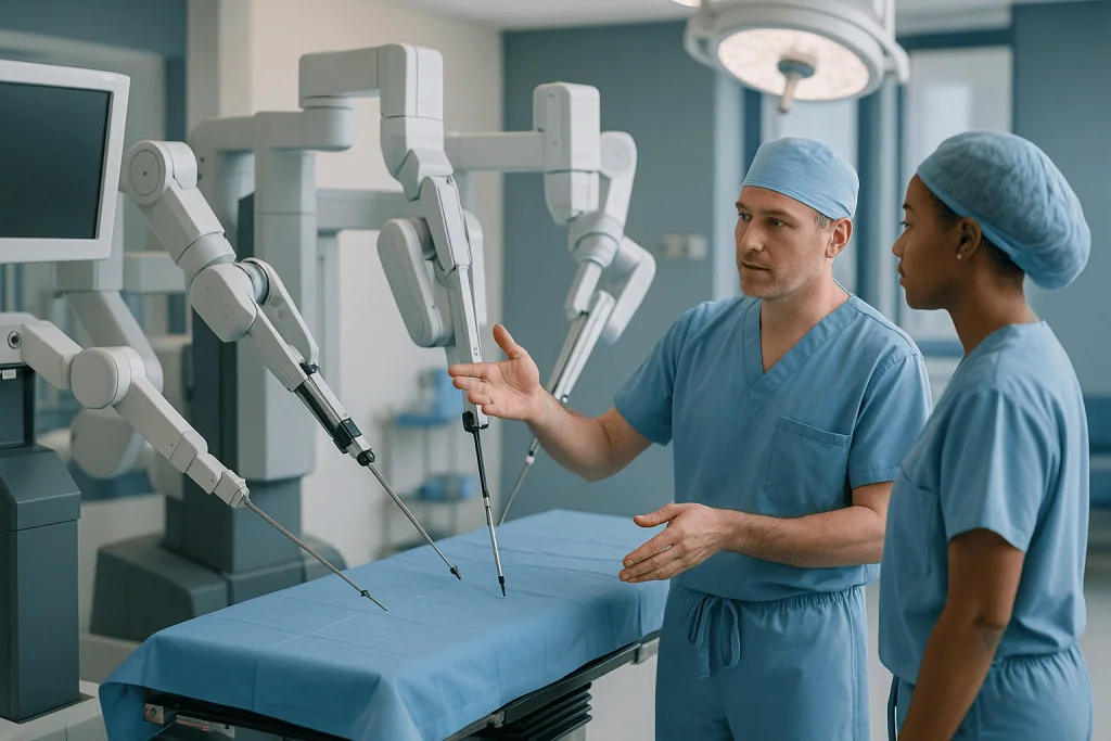
With a robotic approach, the room looks different. After the ports are placed, a cart with several arms is docked to the patient. The surgeon moves to a console, looks into a binocular viewer and sees a magnified, three-dimensional image. The instrument tips have wrists that bend further than a human hand, which matters when a blood vessel curls behind a bronchus or a lymph node sits in a tight corner near the heart. A second surgeon or trained assistant stays at the bedside to swap instruments, manage suction and respond immediately if anything changes.
In both versions the core steps are identical. The lung on the operating side is deflated so there is room to work. The surgeon identifies the artery, vein and airway supplying the piece of lung to be removed, seals and divides each one, usually with a stapling device, then frees the tissue and places it in a bag. The bag is drawn out through one of the small incisions, which may be widened slightly for a large lobe. Lymph nodes are sampled or cleared as part of a cancer operation. A chest tube is left in place to drain air and fluid while the remaining lung re-expands.
Both are keyhole operations. The debate between them is about vision, dexterity, cost of equipment and surgeon preference, not about a fundamentally different operation.
What happens during an open thoracotomy, and why surgeons still use it
An open thoracotomy is not a failure or a relic. It is a deliberate choice for situations where the surgeon needs the widest possible view and the ability to feel the tissue directly.
The incision typically curves below the shoulder blade and along the side of the chest. Muscles of the chest wall are divided or moved aside, and a retractor holds two ribs apart. Occasionally a small piece of rib is removed to create space. The surgeon then works with both hands inside the chest, which allows palpation: pressing on the lung to find a nodule that cannot be seen, or assessing whether a tumor has grown into the chest wall, a major vessel or the lining around the heart.
Why would a team recommend this in an era of keyhole surgery? Several reasons recur in guideline-level descriptions and in the patient resources published by Cleveland Clinic and Mayo Clinic:
- Large or centrally placed tumors that sit close to the main airway or the great vessels, where reconstruction of the airway or vessel may be needed.
- Tumors that invade neighboring structures, where a wider margin of chest wall or diaphragm must come out with the lung.
- Dense scarring from previous chest surgery, serious infection or radiation, which can glue the lung to the chest wall and make camera work hazardous.
- Bleeding or other unexpected findings during a minimally invasive operation that are safest handled through a larger incision.
The trade-off is the chest wall itself. Spreading ribs stretches the intercostal nerves that run beneath each rib, and that stretch is the main reason open surgery tends to hurt more and for longer. MedlinePlus notes that recovery from open lung surgery generally takes longer than after a minimally invasive approach. For the right patient, though, the thoroughness an open approach allows is exactly the point.
Robotic vs VATS lung surgery: what the evidence actually shows
Here is where honesty matters most, because marketing has run ahead of data. Comparative studies and systematic reviews of robotic versus VATS lobectomy consistently find the two approaches to be broadly similar on the outcomes patients care about most: complication rates, length of hospital stay, and the completeness of the cancer operation. Some studies report a modest advantage for robotic surgery in the number of lymph nodes sampled, or a lower rate of conversion to open surgery; others find no difference. Robotic operations tend to take longer in the operating room, especially early in a team’s experience, and they use more expensive equipment.

Neither approach has been shown to be clearly superior for long-term cancer control. Where differences appear, they are small and often depend on the surgeon’s experience with each tool more than on the tool itself.
| Feature | Open thoracotomy | VATS | Robotic (RATS) |
|---|---|---|---|
| Incision | One long incision, ribs spread | One to four small incisions, no rib spreading | Three to five small ports, no rib spreading |
| Surgeon’s view | Direct vision, can feel tissue | Two-dimensional screen | Magnified three-dimensional view |
| Instruments | Handheld, full range of motion | Long, straight, rigid | Wristed, tremor filtered |
| Typical pain and recovery | Longest | Shorter | Shorter, similar to VATS |
| Operating time | Variable | Usually shorter than robotic | Often longer, especially early in experience |
| Best suited to | Complex, invasive or heavily scarred cases | Most early-stage tumors | Most early-stage tumors; tight anatomic spaces |
Read the table as a description of tendencies, not promises. Mayo Clinic frames robotic surgery as one minimally invasive option among others rather than an upgrade, and that framing fits the evidence. What matters is a well-trained team using an approach it knows deeply, on a patient whose anatomy suits it.
VATS vs open thoracotomy: where the real difference lies
If the robotic-versus-VATS gap is narrow, the minimally-invasive-versus-open gap is wide, and this is the comparison that has reshaped thoracic surgery.
The anatomy explains it. Beneath every rib runs a bundle of nerve, artery and vein. A thoracotomy retractor stretches that bundle for hours, and a stretched nerve can keep firing for weeks or months. Small VATS incisions slip between the ribs without spreading them, so the nerve is handled far more gently. The result, according to Johns Hopkins and Mayo Clinic patient resources, is generally less post-operative pain, fewer breathing complications, a shorter hospital stay and a quicker return to ordinary activity for people who are suitable for a keyhole approach.
Less pain is not only a comfort issue. After lung surgery, the single most important recovery task is to breathe deeply and cough so that the remaining lung stays open and clear of secretions. People in significant pain take shallow breaths. Shallow breaths let small airways collapse and mucus pool, and that is the pathway to pneumonia. A less painful incision therefore protects the lung itself.
Guidelines from major cancer and thoracic societies now recommend a minimally invasive approach for early-stage lung cancer whenever it can be done safely and without compromising the completeness of the operation. The caveat in that sentence carries weight. Cancer outcomes between VATS and open lobectomy appear comparable when the same quality of resection and lymph node assessment is achieved, which is why surgeons are trained to switch to open rather than accept a lesser operation through a smaller cut.
Open surgery has not been pushed out. It has been reserved. The shift is from “open by default” to “open when the anatomy or the situation demands it,” and that reservation is a sign of a mature specialty, not an outdated one.
Who is usually offered minimally invasive surgery, and who is asked to wait?
Selection happens long before the operating room, and it starts with imaging. A CT scan shows where the nodule sits, how large it is and how close it lies to the central airways. A PET scan may be used to look for spread. Lung function tests measure how much breathing reserve you have, because removing a lobe permanently reduces capacity and the team needs to know you can afford the loss. Many people also have a heart assessment, since general anesthesia with one lung deflated is demanding on the cardiovascular system.
The typical candidate for a VATS or robotic lobectomy has an early-stage tumor located toward the outer part of the lung, no evidence of invasion into surrounding structures, adequate lung function and no major prior surgery on that side of the chest. These are the cases where the minimally invasive advantages are clearest and the risk of needing to convert is lowest.
Who is asked to wait, and for what? Several groups come up repeatedly:
- People who still smoke are often asked to stop for a period before surgery, because active smoking raises the risk of breathing complications and slows wound healing. The CDC and NHS both emphasize that quitting before any surgery improves recovery.
- People with poorly controlled heart disease, recent heart attack or unstable breathing problems may need those conditions optimized first.
- People with active infection in the lung may be treated for the infection before an elective operation.
- People whose nodule is very small and indeterminate may be offered surveillance scans rather than immediate surgery, since many such nodules turn out to be benign.
Waiting here is not delay for its own sake. It is the team lowering the risk of the operation before taking it on. The decision about timing, as with the approach itself, rests with the treating surgeon and the wider tumor board.
Who should not have robotic surgery?
This is one of the most searched questions on the topic, and the answer is less about the robot than about the chest it is entering.
Robotic and VATS approaches share nearly all the same limits, because both depend on creating a working space inside a chest with one lung deflated. Anything that removes that space, or makes it dangerous to work through small ports, points toward open surgery. Dense adhesions from previous surgery, tuberculosis, empyema or chest radiation can fix the lung to the chest wall so firmly that the camera cannot be safely introduced. A tumor that has grown into the chest wall, the spine, the heart’s lining or a major vessel usually needs the wider exposure and hand control of a thoracotomy.
Patient-level factors matter too. Someone who cannot tolerate having one lung deflated for the length of a longer operation, because of very limited lung reserve, may do better with a shorter procedure or a different plan altogether. Robotic cases frequently take longer than VATS, so that time cost is weighed against the dexterity benefit. People with very high body weight, severe spinal curvature or a very small chest cavity can present positioning and access challenges that the team will assess case by case.
Two practical considerations are easy to overlook. First, robotic surgery requires that the robotic arms be docked to the patient; in a sudden emergency, undocking adds steps compared with a surgeon already standing at the bedside with handheld instruments. Teams rehearse this, but it is one reason some surgeons prefer VATS for anatomy they expect to be difficult. Second, the surgeon’s own experience is a legitimate selection criterion. A surgeon with hundreds of VATS lobectomies and a handful of robotic ones may serve you better through the approach they know, and should say so.
None of this is a list for self-screening. It is the kind of reasoning your surgeon will walk through with you.
Why does a VATS or robotic operation convert to open thoracotomy?
Conversion to thoracotomy during VATS or robotic surgery means the surgeon starts with small incisions and, partway through, makes the larger incision of an open operation. People sometimes hear this as “something went wrong.” More often it means the surgeon exercised exactly the judgment you want.
Conversions fall into roughly three categories. The first is anatomic: the surgeon finds adhesions, a tumor that is larger or more invasive than the scans suggested, or lymph nodes stuck to a vessel in a way that cannot be safely separated with long instruments. The second is bleeding: a tear in a pulmonary artery branch can lose blood quickly, and direct hand pressure through an open incision is the fastest way to control it. The third is technical or physiologic: the lung will not deflate enough to create working space, or the patient does not tolerate single-lung ventilation.
Comparative studies have examined whether robotic surgery converts less often than VATS, and several report a lower conversion rate for the robotic approach, possibly because the wristed instruments and three-dimensional view make dissection around vessels easier. Other series show no meaningful difference. Published rates vary widely between centers and depend heavily on case selection, so no single number applies to you.
What should you take from this? Three things. Your consent form will almost certainly include the possibility of conversion, and that is standard, not a warning about your particular case. Planned conversion for a safe, complete operation is a success of judgment. And if you wake up with a longer incision than expected, your recovery will follow the open-surgery timeline, which your team will explain. Ask beforehand how your surgeon approaches this decision; the quality of the answer tells you a great deal about the team.
Does the approach change how much lung is removed?
In principle, no. In practice, the question deserves a careful answer, because the extent of the operation matters more to your long-term breathing and cancer outcome than the size of the skin incision.
The three standard resections are a wedge, which removes a small piece of lung around a nodule; a segmentectomy, which removes one anatomic segment with its own artery, vein and airway; and a lobectomy, which removes a whole lobe. A pneumonectomy, removal of an entire lung, is now uncommon for early disease. All three smaller operations can be done open, by VATS or robotically. The choice between wedge, segment and lobe is driven by the tumor’s size, type and position, and by your lung reserve, not by which tool the surgeon prefers.
Where approach and extent intersect is in lymph nodes. A proper cancer operation includes assessing the lymph nodes in the center of the chest, because finding cancer there changes the stage and the recommendation for further treatment. Early criticisms of VATS suggested that surgeons might sample fewer nodes through small incisions. Later experience and several comparative studies suggest that experienced VATS and robotic surgeons achieve node assessment comparable to open surgery, with some data favoring the robotic approach for node counts. The honest summary is that technique and training determine the thoroughness of the node dissection far more than the platform does.
Segmentectomy deserves a note. For some very small, peripheral early cancers, removing a segment rather than a whole lobe preserves more breathing capacity, and this smaller operation benefits from the fine dissection that wristed robotic instruments allow. Your surgeon may mention that as a reason for favoring one approach. Whether you are a candidate for a segment rather than a lobe is a tumor board decision based on the specifics of your scan and biopsy, and it should never be driven by a wish to use a particular machine.
How long does it take to recover from a robotic lobectomy?
Recovery timelines are where expectations most often drift from reality, in both directions. Some people expect to be sidelined for months after keyhole surgery; others expect to be back at the gym in a week. Neither matches the typical course.
Hospital stay comes first. After a minimally invasive lobectomy, whether robotic or VATS, patient resources from Cleveland Clinic and Mayo Clinic describe a stay that is usually measured in a few days, and often shorter than after an open operation. The main determinant is the chest tube. It stays until the lung has sealed any small air leaks from the stapled surface and fluid drainage has fallen to a modest level. Some people have the tube out the day after surgery; others need it for several more days if a leak persists. You go home when the tube is out, your pain is controlled with medicines taken by mouth, and you are walking and eating.
At home, the first two weeks are about walking, breathing exercises and rest. Most people notice steady improvement in stamina through weeks two to four. MedlinePlus describes a return to normal activity after lung surgery that generally takes several weeks for minimally invasive approaches and longer, often up to two or three months, after thoracotomy. Robotic and VATS recovery timelines are broadly similar in published comparisons; there is no strong evidence that one gets you back to work faster than the other.
Lifting limits are typical for several weeks to protect the healing incisions and chest wall. Driving usually waits until you can turn and brake without pain and are no longer taking sedating pain medicine; your team will give you a specific guide. Breathlessness on exertion after losing a lobe can persist for weeks and gradually improves as the remaining lung adapts.
These are ranges, not promises. Age, lung function before surgery, complications and whether you need further cancer treatment all shift the curve.
What the first days and weeks after lung surgery usually look like
Day one is louder than people expect. You wake with a chest tube on the operated side connected to a drainage canister, a line in your arm, oxygen by nasal prongs and often a small device at the bedside that measures your breathing. The nursing team will have you sitting up within hours and walking the corridor the same day or the next morning. This feels early. It is also the single most effective thing you can do to prevent pneumonia and blood clots.
Breathing exercises become a rhythm. An incentive spirometer, a plastic device you inhale through to lift a marker, is used many times a day to keep the remaining lung fully inflated. Coughing hurts, so staff teach you to hug a pillow against the incisions for support. Pain is managed with a combination of approaches chosen by the anesthesia and surgical team; many centers use nerve blocks placed during surgery alongside medicines by mouth. Decisions about which medicines and for how long sit with your prescribers.
The chest tube comes out on the ward once the lung is sealed. Its removal is brief and strange rather than agonizing, and most people feel markedly better once it is gone.
The first fortnight at home typically includes:
- Daily walks that gradually lengthen, with rest between.
- Continued breathing exercises, because the lung can still partially collapse in the early weeks.
- Shoulder stretches on the operated side to prevent stiffness.
- Small, frequent meals if appetite is poor.
- A follow-up visit, usually with a chest X-ray, to check the lung and review the pathology report from the removed tissue.
Fatigue is the symptom people underestimate. It is normal to need an afternoon rest for several weeks even after keyhole surgery. Numbness or tingling around the incisions, and sometimes along the chest wall, is also common and usually settles over months as small nerves recover.
What people often get wrong about robotic, VATS and open lung surgery
Myths cluster around new technology, and lung surgery has collected its share. Setting them straight helps you walk into the consultation with the right questions.
“The robot does the operation.” It does not. Every movement inside your chest is made by the surgeon at the console; the system has no independent decision-making. If the surgeon’s hands stop, the instruments stop.
“Robotic surgery is always better than VATS.” The evidence does not support this. Comparative studies show similar complication rates, hospital stays and cancer outcomes. Robotic surgery offers better visualization and dexterity, which some surgeons value for certain anatomy, but those tools have not translated into consistently better results for patients across studies.
“Open surgery means the cancer is worse.” Sometimes it reflects a more advanced or awkwardly placed tumor, but just as often it reflects scar tissue, previous surgery or a surgeon’s considered judgment about safety. The approach is not a prognosis.
“Smaller incisions mean a smaller operation.” A lobectomy removes the same lobe whether the incision is one long cut or four small ones. Keyhole surgery changes how you reach the lung, not how much comes out.
“Keyhole surgery is painless.” It hurts less and for a shorter time than open surgery, but chest tubes, deep breathing and coughing are uncomfortable for everyone. Expect real discomfort for a week or two.
“Converting to open means a mistake was made.” Conversion is usually a safety decision made in the moment. It is discussed in advance precisely because it is an expected possibility.
“You should pick the hospital with the newest robot.” Equipment matters less than the experience of the team using it and the volume of lung operations they perform. A thoughtful surgeon who explains why they favor a particular approach for your anatomy is worth more than any machine.
Questions to ask your care team
A good surgical consultation is a conversation, and the quality of your questions shapes the quality of the answers. Bring a notebook or a companion; most people retain only a fraction of what is said in these appointments.
On the operation itself:
- Which approach are you recommending for me, and what about my scans or history led you there?
- How much lung do you plan to remove: a wedge, a segment or a lobe? What would change that plan during surgery?
- How will lymph nodes be assessed, and how does that influence treatment afterward?
- If you begin with a minimally invasive approach, under what circumstances would you convert to open, and how would that change my recovery?
On the team:
- How many of these operations, by this approach, do you and your team perform each year?
- Who else will be in the operating room, and what are their roles?
- Will my case be reviewed by a multidisciplinary tumor board?
On preparation and recovery:
- What can I do in the weeks before surgery to lower my risk, such as stopping smoking, exercise or breathing training?
- How will pain be managed, and who do I contact if it is not controlled at home?
- What is a realistic timeline for walking, driving, lifting and returning to work for someone with my lung function?
- Which symptoms after discharge should prompt a phone call, and which mean I should go to the emergency department?
On alternatives:
- Are there non-surgical options for my situation, such as targeted radiation, and how do they compare for someone like me?
- What happens if I choose to wait or to decline surgery?
Notice what is missing from this list: questions about which approach is “best” in the abstract. The useful question is always which approach is best for this tumor, in this chest, in these surgeons’ hands.
When to call your doctor
Complications after lung surgery are uncommon but time-sensitive, and the people who do best are the ones who call early rather than waiting to see whether a symptom settles. Your discharge paperwork will include a number to ring at any hour; keep it visible on the refrigerator.
Call your surgical team promptly if you notice any of the following in the days or weeks after discharge:
- Fever, chills or sweats, which can signal pneumonia or a wound infection.
- Increasing redness, warmth, swelling or drainage from any incision, especially pus or a foul smell.
- Breathlessness that is worsening rather than steadily improving, or breathlessness at rest.
- A cough producing thick yellow or green mucus, or any coughing up of blood beyond small streaks in the first days.
- Pain that is getting worse instead of better, or pain not controlled by the plan you were given.
- Swelling, warmth or pain in one calf, which can indicate a blood clot in the leg.
- A crackling feeling under the skin of the chest or neck that is spreading, which can mean air is leaking from the lung.
- Irregular or very fast heartbeat, which is a known issue after lung surgery and usually treatable but needs assessment.
Call emergency services or go to the nearest emergency department without delay for sudden severe breathlessness, chest pain that is new and crushing or spreading to the arm or jaw, coughing up a significant amount of blood, fainting, confusion, or blue or gray lips or fingertips. These are red-flag signs of a collapsed lung, a clot traveling to the lung, serious bleeding or a heart problem, and minutes matter.
Beyond emergencies, keep every scheduled follow-up. The pathology report from your operation, usually reviewed at the first visit, determines whether any further treatment is recommended, and that conversation belongs with your treating team.
Frequently asked questions
What is the difference between RATS vs VATS lobectomy?
Both remove a lobe of lung through small incisions without spreading the ribs. In VATS the surgeon stands at the bedside using straight instruments and a two-dimensional screen. In RATS, robotic-assisted thoracic surgery, the surgeon controls wristed instruments from a console with a magnified three-dimensional view. Studies show similar complications, hospital stays and cancer outcomes; robotic operations often take longer and use costlier equipment.
Who should not have robotic surgery for the lung?
Robotic surgery shares the limits of all keyhole approaches. People with dense adhesions from prior surgery, infection or radiation, tumors invading the chest wall or major vessels, or very limited tolerance for one-lung ventilation during a longer operation are usually steered toward open thoracotomy. Surgeon experience with the platform also matters. The assessment is individual and rests with the treating surgeon.
How long is robotic lobectomy recovery time?
Hospital stays after a robotic or VATS lobectomy are typically measured in a few days, driven mainly by when the chest tube can be removed. Patient resources from MedlinePlus and Mayo Clinic describe a return to ordinary activity over several weeks, with lifting restrictions for a similar period. Open thoracotomy recovery is longer, often two to three months. Individual timelines vary with age, lung function and complications.
Is VATS vs open thoracotomy really that different for the patient?
Yes, and this is the comparison with the clearest evidence. VATS avoids spreading the ribs, so the nerves beneath them are handled gently, which generally means less pain, fewer breathing complications and a shorter hospital stay. Cancer outcomes appear comparable when the same quality of resection is achieved. Open surgery remains the safer choice for complex, invasive or heavily scarred cases.
How often does conversion to thoracotomy during VATS happen?
Published conversion rates vary widely between centers and depend heavily on how cases are selected, so no single figure applies to an individual. Common reasons are unexpected adhesions, bleeding from a lung vessel, or a tumor found to be more invasive than scans suggested. Some studies report fewer conversions with robotic surgery, others no difference. Conversion is a planned safety option discussed during consent.
Does VATS cost less than robotic lung surgery?
This article does not quote prices, because figures vary enormously by country, insurer and hospital and quickly become outdated. In general terms, published comparisons note that robotic systems involve higher equipment and instrument costs and longer operating times than VATS, while hospital stays are similar. Your care team and insurer are the right sources for what any approach would mean for you financially.
Does robotic surgery remove more lymph nodes than VATS?
Some comparative studies report a modestly higher lymph node count with robotic lobectomy, possibly because wristed instruments make dissection in tight spaces easier. Other studies show no meaningful difference. Experienced VATS surgeons achieve node assessment comparable to open surgery. The thoroughness of the lymph node dissection depends far more on the surgeon’s training and technique than on the platform used.
Is the robot doing the operation, or the surgeon?
The surgeon performs every movement. The robotic system translates the surgeon’s hand motions at a console into instrument movements inside the chest, filtering out tremor and allowing a wrist-like range of motion. It has no autonomous function and makes no decisions. A second surgeon or trained assistant remains at the bedside throughout to exchange instruments and respond immediately if anything changes.
Will I lose a lot of breathing capacity after a lobectomy?
Removing one lobe takes out roughly a fifth to a third of total lung tissue, depending on which lobe is removed. Before surgery, lung function tests confirm that you have enough reserve to tolerate the loss. Most people notice breathlessness on exertion for weeks that gradually improves as the remaining lung adapts. The approach, open or keyhole, does not change how much lung is removed.
Can I choose the approach myself?
You can and should express preferences, but the final recommendation sits with your surgeon and the multidisciplinary team, because it depends on tumor position, scarring, lung function and the team’s experience with each approach. A good surgeon will explain why one approach suits your anatomy and what would prompt a change of plan. Seeking a second surgical opinion is reasonable if you remain unsure.
References
This article is for general information only and is not a substitute for professional medical advice. Please consult a qualified doctor about your individual situation.

More from the Blog

How Long Does an Acute Bronchitis Cough Last? The Typical Timeline Week by Week
Acute bronchitis usually lasts about one to three weeks. Fever, aches, and chest heaviness typically ease within the first seven to ten days, while…

How Doctors Diagnose a Lung Infection: Chest Imaging, Blood Tests and Sputum Cultures
Doctors diagnose a lung infection by combining your history and a chest examination with targeted tests: pulse oximetry to check blood oxygen, a chest…

How Pulmonary Edema Is Treated in the Emergency Room: Oxygen, Diuretics and Monitoring
Pulmonary edema emergency treatment focuses on getting oxygen into the blood and fluid out of the lungs quickly. Emergency teams typically sit the person…

Questions to Ask at Your Asthma Review: Triggers, Inhaler Technique and Follow-Up
At an asthma review, the most useful questions to ask cover three areas: which triggers are driving your symptoms and how to reduce exposure,…

Can You Fly With Emphysema? Altitude, In-Flight Oxygen and Planning Long Trips
Many people with emphysema can fly, but it depends on how much oxygen your blood carries at rest and how stable your lungs have…

Living With Bronchiectasis: Exercise, Travel and Fitting Airway Clearance Into Your Day
Living with bronchiectasis usually centers on a daily airway clearance routine, regular physical activity, and prompt attention to flare-ups. Most people can exercise and…