

What a Melanoma Tumor Board Decides and Why Several Specialists Review Your Case
Key Takeaways
- A melanoma tumor board produces a written recommendation to your treating doctor, not a binding order, and the final decision remains yours and your team's together.
- Breslow thickness, the depth of the tumor in millimeters, is the single strongest predictor of behavior for skin melanoma and drives most early board decisions, with staging thresholds at roughly 0.8, 1, 2 and 4 mm.
- Scans cannot detect microscopic melanoma cells in a lymph node, which is why boards often recommend a sentinel lymph node biopsy even when imaging looks clear.
- Roughly half of cutaneous melanomas carry a BRAF mutation, and testing for it becomes relevant once systemic treatment is being considered for stage III or IV disease.
- Melanoma causes most skin cancer deaths but is not the leading cause of cancer death overall, and most cases are diagnosed at an early stage when surgery alone is standard.
- Being presented at a tumor board does not signal an advanced case; many hospitals review every invasive melanoma, including thin ones with an excellent outlook.
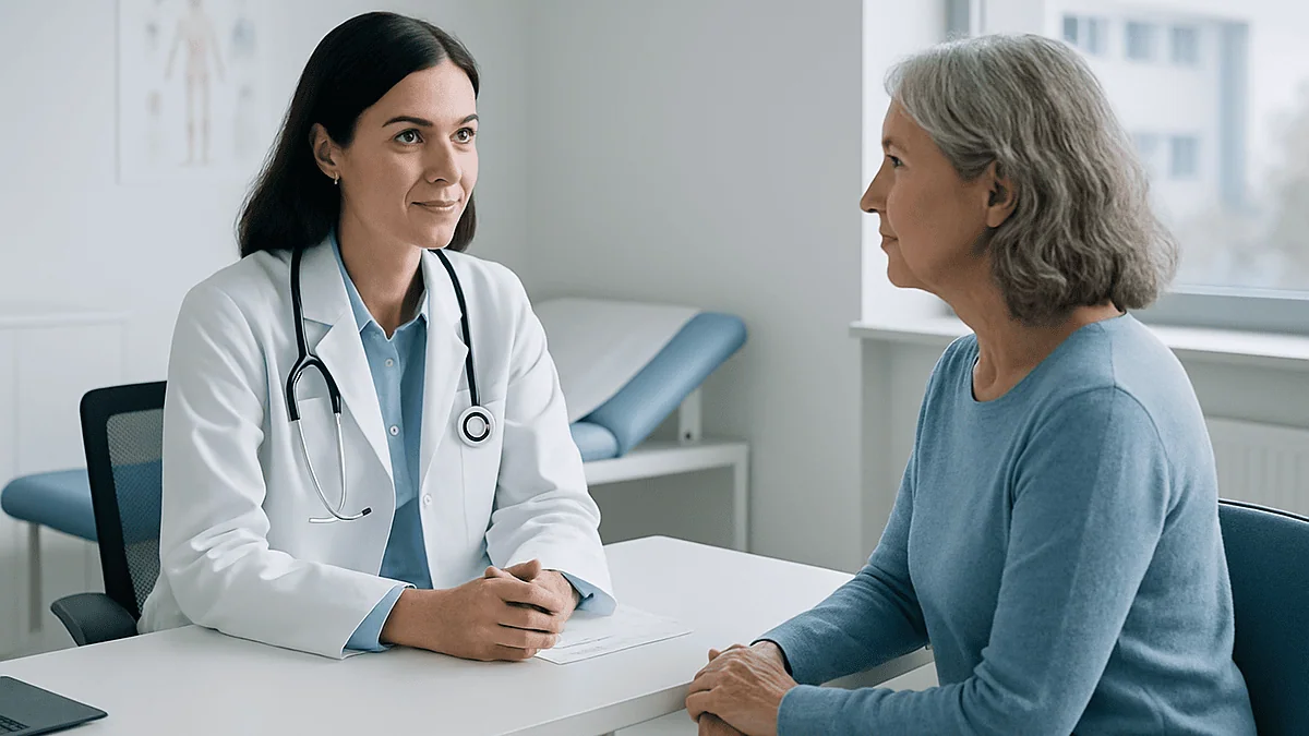
A melanoma tumor board is a scheduled meeting where dermatologists, surgeons, medical and radiation oncologists, pathologists and radiologists review one patient's slides, scans and history together. They confirm the stage, agree on whether more surgery, a sentinel lymph node biopsy, drug therapy, radiation or observation fits best, and send a joint recommendation back to the treating team, who make the final decision with the patient.
The phone call usually comes on an ordinary weekday. A nurse explains that the mole removed two weeks ago was melanoma, and that your case will be “presented at tumor board” on Thursday. Then a pause, and the question most people ask next: does that mean it is bad?
Not necessarily. A melanoma tumor board is closer to a second, third and fourth opinion happening in the same room at the same time than to an alarm bell. The people around that table have seen thousands of melanoma slides and scans between them, and the point of gathering them is to make sure no single specialist’s blind spot shapes your plan.
This article walks through what happens in that room, which details the specialists argue over, what the board can and cannot decide, and how to use the recommendation that comes back to you.
What is a tumor board, and what does it actually produce?
A tumor board is a regular meeting of cancer specialists from different fields who review individual patients’ cases and agree on a recommended plan. The National Cancer Institute defines it as a treatment planning approach in which doctors who are experts in different specialties review and discuss a patient’s medical condition and treatment options. Many hospitals call the same thing a multidisciplinary team meeting, or MDT, and some hold a dedicated melanoma and skin cancer board separate from the boards for breast, lung or blood cancers.
What comes out of the meeting is a written recommendation, not an order. The board might conclude that a melanoma has been fully removed and needs only skin checks, that a wider excision and a sentinel lymph node biopsy should follow, that scans are warranted before any decision, or that drug therapy should be discussed. That note goes into the record and back to the doctor who referred you, usually a dermatologist or surgeon, who then sits down with you to translate it into a plan you agree with.
Two features make the meeting worth the logistics. The first is that the pathologist who examined your tissue and the radiologist who read your scan are physically or virtually present, so a surgeon can ask “how confident are you about that margin?” and get an answer in seconds rather than days. The second is disagreement. Boards are designed so that a medical oncologist who leans toward drug treatment and a surgeon who leans toward operating have to make their case in front of colleagues, with your specific slides on the screen. Consensus reached that way tends to be more carefully reasoned than any one clinic visit.
Patients are almost never in the room. That can feel odd, but it keeps the discussion frank, and you receive the result in a proper consultation where questions are welcome.
Who sits on a multidisciplinary melanoma team?
The cast changes slightly from hospital to hospital, but a melanoma board typically brings together six or seven roles, each of which sees the disease from a different angle.
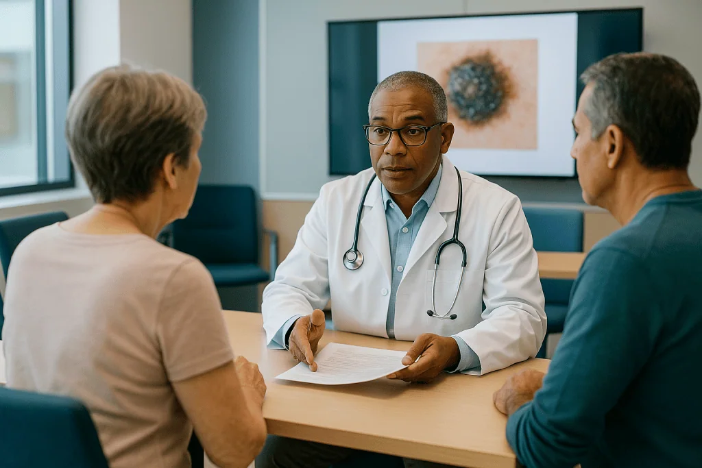
- Dermatologist: a skin specialist, often the person who first suspected the lesion. They speak to the appearance of the original spot, your other moles, sun exposure history and skin type.
- Dermatopathologist or pathologist: the doctor who examined your tissue under the microscope. Their report supplies the measurements that drive most early decisions.
- Surgical oncologist or plastic surgeon: plans the wider excision (removing a rim of normal-looking skin around the scar) and any lymph node procedure, and thinks about function and appearance in tricky areas such as the face, ear or fingertip.
- Medical oncologist: a cancer physician who manages drug treatments, including immunotherapy and targeted therapy, and weighs them for higher-stage disease.
- Radiation oncologist: considers whether radiation has a role, most often for lymph node regions or brain metastases rather than for the primary skin lesion.
- Radiologist: reads CT, PET-CT, MRI or ultrasound and describes what is, and is not, visible.
- Specialist nurse or nurse navigator: keeps track of what you have been told, what you want, and the practical steps that follow.
Larger boards also invite a genetic counselor when there is a strong family history, an ophthalmologist for melanoma of the eye, or a palliative care physician when symptom control matters as much as tumor control. Trainees and students often attend to learn.
The value of the mix is easiest to see in a hard case. A thick melanoma on the scalp of an older adult with heart failure raises a surgical question (how much to remove and how to close it), a medical question (can this person tolerate systemic treatment), and a practical question (who will manage the wound at home). One specialist can answer one of those well. The team can answer all three at once.
How a melanoma tumor board meeting actually works
Picture a conference room, or increasingly a video call, early in the morning before clinics open. A coordinator has circulated a list of cases, sometimes a dozen or more, each with a short summary. Your case is presented by the referring doctor in a few sentences: age, where the lesion was, what the biopsy showed, what has been done so far, and the specific question the team needs to answer.
The pathologist then puts your slides on the screen. This is the part most patients never see and would find surprising: a room full of doctors leaning toward a projected image of stained cells, debating whether a cluster at the deep edge is melanoma or a benign nevus, or whether ulceration is present. If scans exist, the radiologist follows with the images, pointing out enlarged lymph nodes or confirming that nothing suspicious is visible.
Discussion follows. For a thin, completely excised melanoma, it may last under two minutes and end with agreement on the width of the re-excision and a follow-up schedule. For a case involving positive lymph nodes, an uncertain scan finding or a patient with other serious illnesses, the conversation may run much longer and end with a request for more information rather than a decision: a repeat scan, a molecular test on the tumor, or a fitness assessment before any operation.
The coordinator records the outcome. Typical wording is something like “recommend wide local excision with sentinel lymph node biopsy; discuss adjuvant options if node positive; refer to medical oncology.” That sentence becomes the backbone of your next appointment.
Boards also revisit patients. The same person may be discussed after surgery when node results return, again if a scan changes, and again if treatment needs adjusting. Each pass is short, but it means the whole team is kept in the loop rather than one clinic passing notes to another.
What the pathology report tells the board
Almost everything a melanoma board decides in the early stages rests on a single document: the pathology report from the biopsy. Understanding its main lines makes the board’s reasoning far less mysterious.
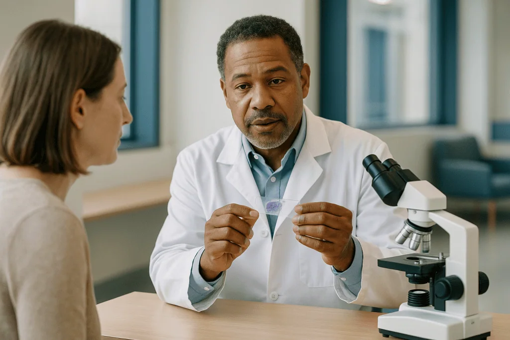
Breslow thickness is the depth of the tumor measured in millimeters from the top layer of skin to its deepest point. It is the strongest single predictor of how a cutaneous melanoma behaves, and the staging system used by the NCI divides the earliest tumors at thresholds of 0.8, 1, 2 and 4 millimeters. A melanoma under 0.8 mm without ulceration is treated very differently from one over 4 mm.
Ulceration means the surface of the tumor has broken down, visible under the microscope. Its presence moves a melanoma into a higher risk category at the same thickness.
Mitotic rate is a count of dividing cells per square millimeter, a rough measure of how quickly the tumor is growing.
Margins describe whether the edges of the removed tissue are free of melanoma cells. A “positive” or “involved” margin means cells reach the cut edge and more tissue needs to come out. Even with clear margins, guidelines recommend a wider excision because microscopic cells can sit beyond the visible tumor.
Other features include the subtype (superficial spreading, nodular, lentigo maligna, acral or desmoplastic), whether tumor cells are seen inside blood or lymph vessels, and any regression.
The pathologist at the board does more than read these lines aloud. They can say how confident they are, whether the biopsy sampled the whole lesion or only part of it (a shave biopsy may underestimate depth), and whether a second opinion on the slides would help. When the room disagrees about a feature, the honest answer is often “we cannot be sure,” and the plan is adjusted to allow for that uncertainty rather than pretending it away.
Staging: the question underneath every other decision
Stage is a shorthand for how far a cancer has spread, and it is the first thing a melanoma board tries to pin down, because nearly every downstream choice depends on it. The system is called TNM: T for the primary tumor’s thickness and ulceration, N for lymph node involvement, M for metastasis to distant organs.
In plain terms, according to the NCI’s patient staging summary:
- Stage 0 (melanoma in situ): cells are confined to the top layer of skin.
- Stages I and II: the melanoma is only in the skin, graded by thickness and ulceration; stage II tumors are thicker or ulcerated.
- Stage III: melanoma has reached nearby lymph nodes, or has formed small deposits in skin or lymph channels near the original site.
- Stage IV: melanoma has spread to distant skin, lymph nodes or organs such as the lungs, liver or brain.
The board’s difficulty is that the initial pathology gives only the T. The N and M often remain unknown until further steps. This is why the recommendation for a stage I or II melanoma frequently includes a sentinel lymph node biopsy, a procedure in which a tracer is injected near the scar to find the first node or nodes the area drains to, so they can be removed and examined. The result can move a person from stage II to stage III, which changes the conversation about drug treatment entirely.
Imaging enters the picture selectively. For thin melanomas with no symptoms, scans are generally not recommended, because they rarely find anything and often flag harmless spots that lead to needless worry and procedures. For thicker tumors, positive nodes or symptoms, CT, PET-CT or MRI become part of the staging package. Deciding who needs imaging, and which kind, is one of the board’s genuinely useful judgment calls.
A staging conclusion is not a prognosis handed down as fate. It is the map the team uses to choose among options, and it can be revised as new information arrives.
What each melanoma tumor board decision usually hinges on
The table below summarizes the questions a board most often works through and which findings tend to tip the balance. It describes typical guideline-level reasoning rather than a rule for any individual; the team weighs each factor against your overall health and preferences.
| Decision point | Findings that push toward “yes” | Findings that push toward “wait” or “no” | Specialist who leads |
|---|---|---|---|
| Wider excision | Any confirmed melanoma; margin width scaled to thickness | Rarely omitted; cosmetic or functional limits shape the approach | Surgeon |
| Sentinel lymph node biopsy | Thickness above roughly 0.8 mm, ulceration, high mitotic rate | Very thin, non-ulcerated tumors; frailty making anesthesia risky | Surgeon and pathologist |
| Staging scans | Thick tumor, positive nodes, symptoms, palpable lump | Thin melanoma with no symptoms | Radiologist |
| Molecular testing (BRAF and others) | Stage III or IV disease where targeted therapy may be considered | Early-stage disease with no plan for systemic treatment | Pathologist and medical oncologist |
| Adjuvant (after-surgery) drug therapy | Node-positive disease or high-risk stage II | Low-risk disease; serious autoimmune illness; patient preference | Medical oncologist |
| Radiation | Bulky node disease with concern for regrowth; brain metastases; inoperable sites | Most primary skin melanomas | Radiation oncologist |
| Clinical trial referral | Eligible stage and a trial open locally or regionally | No suitable trial; patient prefers standard care | Medical oncologist |
Notice how often the pathologist appears in the middle column. Numbers on a report, not opinions, drive the first several rows. Notice too that “patient preference” sits in the wait column for systemic therapy. A board can recommend, but a person with a good understanding of the trade-offs can decline, and a good team records and respects that.
The thresholds in the table, including the approximate 0.8 mm figure for considering sentinel node biopsy, reflect the staging categories described by the NCI and the treatment overviews published by Mayo Clinic and the NHS. Your team may apply them with some flexibility based on features not captured in a table.
Is a tumor board a good thing before surgery?
The short answer is yes, and the reasons are concrete rather than reassuring platitudes.
Melanoma surgery is not a single operation with a fixed recipe. The width of skin removed around the scar depends on tumor thickness; the NHS and Mayo Clinic both describe wider margins for thicker tumors. Whether to add a sentinel lymph node biopsy depends on the pathology features discussed earlier. Whether to remove additional nodes if the sentinel is positive has changed over the past decade as evidence showed that close ultrasound surveillance is a reasonable alternative for many people. Getting these choices right the first time avoids a second trip to the operating room.
A board before surgery also catches the errors that are easiest to make alone. A pathology report may have been based on a partial biopsy that understated depth. A lesion on the ear or eyelid may need a plastic surgeon rather than a general approach. An older adult on blood thinners for a heart valve needs cardiology input before a node procedure. A person with a second suspicious mole may benefit from having both addressed in one session. Each of these is obvious to someone in the room and easy to miss in a busy clinic.
Timing matters too. For most early melanomas, guidelines do not treat surgery as an emergency; a wait of a couple of weeks to allow a board review does not change the biology of a tumor that has already been present for months. Your surgeon can tell you whether anything about your case makes waiting unwise.
What a pre-operative board cannot do is guarantee a smooth operation or a particular result. Surgical complications, wound healing problems and the small chance of a false-negative sentinel node exist regardless of how many people planned the procedure. The board reduces avoidable error; it does not remove risk.
Which treatment classes the board weighs for higher-stage disease
When lymph nodes are involved or the melanoma has spread further, the conversation moves from the surgeon to the medical oncologist, and the options fall into a handful of classes. Understanding the categories, without needing product names, helps you follow your own consultation.
Immunotherapy with checkpoint inhibitors. Melanoma is one of the cancers most visible to the immune system, and these drugs block proteins (such as PD-1 or CTLA-4) that tumors use to switch off attacking immune cells. They are given by infusion over months and can be used after surgery to lower the risk of recurrence in stage III and some stage II disease, or as the main treatment for stage IV. Side effects come from the immune system overreacting against normal tissue, which is why your team screens for autoimmune conditions and monitors thyroid, liver and bowel function.
Targeted therapy. Roughly half of cutaneous melanomas carry a mutation in a gene called BRAF, according to the NCI, which drives cell growth. Pills that block BRAF and a partner protein, MEK, can shrink these tumors. Testing the tumor for the mutation is therefore a routine board recommendation once systemic treatment is on the table.
Neoadjuvant therapy. This means giving drug treatment before surgery rather than after. It is an area of active research and increasing use for bulky stage III disease, and a board is exactly where the sequencing question, drugs first or surgery first, gets argued out.
Radiation. Used selectively for node regions and brain metastases; stereotactic radiosurgery, a highly focused form, is common for small brain lesions.
Clinical trials. Boards keep a running list of open studies and flag patients who may be eligible.
None of this article is a recommendation for or against any therapy. Whether a class fits you, in what order, and for how long, rests with your prescribing oncologist after a discussion of benefits, side effects and your own priorities.
Who is usually reviewed, and who is usually asked to wait
Not every melanoma passes through a formal board, and being reviewed or not reviewed says less about your outlook than people assume.
Cases that are almost always presented include melanomas over about 1 mm thick, any tumor with ulceration or a high mitotic rate, any case with a positive or uncertain margin, melanomas in difficult anatomical locations, cases with a positive sentinel node or suspicious scan, all stage IV disease, and any situation where the referring doctor is simply unsure. Rare subtypes, such as melanoma of the eye, the mucous membranes or the nail bed, are presented because so few doctors see them regularly.
Cases that may be handled in clinic include melanoma in situ and very thin invasive melanomas with clear margins in a straightforward location, where guidelines are unambiguous: a wider excision and skin surveillance. Some hospitals present these anyway for completeness; others reserve the meeting for cases with a real question to answer. Either approach is standard.
“Asked to wait” carries two different meanings here. The first is a short delay, typically one to two weeks, so that a board can meet or a molecular result can return before surgery is scheduled. Both the NHS and Mayo Clinic describe pathology and staging as steps that take days to weeks, and for most early melanomas this interval is considered safe. The second meaning is more substantial: a board may recommend active surveillance rather than immediate intervention, for example ultrasound checks of a node basin instead of removing more nodes, or observation instead of adjuvant drug therapy for lower-risk stage II disease. Waiting in this sense is a deliberate, evidence-based choice, not neglect.
People sometimes ask whether they can request a board review. You can. A referring doctor will usually agree, especially when you have questions the clinic cannot fully answer, and many hospitals will also review outside cases as a formal second opinion.
Rare and complex melanoma cases that need the whole room
A general melanoma board earns its keep most clearly on cases that fall outside the common pattern of a sun-exposed skin lesion in a fair-skinned adult.
Uveal (eye) melanoma arises in the pigmented layers inside the eye. It behaves differently from skin melanoma, rarely carries BRAF mutations, and when it spreads it tends to go to the liver. An ophthalmologist who specializes in eye tumors joins the discussion, and the treatment question centers on preserving vision while controlling the tumor, using approaches such as targeted radiation to the eye or surgery. Systemic options for spread uveal melanoma differ from those for skin melanoma, which is precisely why the distinction matters at board.
Mucosal melanoma, on membranes such as the nasal passages, mouth or genital tract, is often found later because it is hidden, and its molecular profile leans toward mutations in a gene called KIT rather than BRAF. Head and neck surgeons and gynecologic oncologists may be pulled in.
Acral melanoma, on palms, soles or under nails, is proportionally more common in people with darker skin and is frequently mistaken for a bruise, wart or fungal infection at first. Surgery near a nail or on a weight-bearing sole raises real functional questions.
Melanoma of unknown primary presents as a lymph node or organ deposit with no skin lesion found. The board weighs how hard to search for the original site and treats the case by its stage.
Molecular findings beyond BRAF also come up. A patient’s report may mention CDKN2A, a tumor suppressor gene whose inherited variants raise melanoma risk within families and can prompt a genetics referral, or PD-L1, a protein whose expression on tumor cells is sometimes measured as an immune marker. The NCI notes that PD-L1 status does not by itself determine whether immunotherapy is offered in melanoma, and a board is where such results are placed in context rather than over-interpreted.
Brain metastases bring neurosurgery and radiation oncology into the discussion, and the team must sequence local treatment with systemic therapy carefully.
Melanoma treatment planning: what the next days and weeks look like
The board meets, the note is written, and then the process shifts back to you. The following sequence is typical, though your team’s version may differ.
Within a few days, you have an appointment or a call from the referring doctor or nurse navigator explaining the recommendation. Ask for the wording of the board note itself; most teams will share it or paraphrase it closely. This is the moment to raise anything the board may not have known: a new symptom, a medication you take, a strong preference against a particular treatment.
Within roughly one to three weeks, surgery is scheduled if that is the plan. Mayo Clinic and the NHS describe wide excision as a procedure usually done under local or general anesthesia, often as a day case. If a sentinel node biopsy is included, expect a tracer injection and imaging beforehand, a second small incision, and a week or two before the node results return.
After results return, the board frequently meets on your case again. A negative node keeps you in stage I or II and shifts the plan toward surveillance. A positive node raises the question of adjuvant drug therapy, and a medical oncology consultation follows, often with a molecular test on the tumor if it has not already been done.
If systemic therapy begins, infusions of checkpoint inhibitors are usually spaced weeks apart over a period of months, with blood tests at each visit; targeted pills are taken daily with regular clinic reviews. Exact schedules are set by your oncologist.
Surveillance then stretches over years: skin examinations at intervals set by stage, lymph node checks, and for higher stages, periodic imaging. Both Mayo Clinic and the NHS emphasize that most recurrences are found by patients or clinicians noticing a change, which makes learning your own skin a genuine part of treatment.
Throughout, one person, often the nurse navigator, should be your single point of contact. If you do not know who that is, ask at the first post-board appointment.
What people often get wrong about melanoma and tumor boards
“If my case is going to tumor board, it must be advanced.” Many boards review every invasive melanoma, including thin ones with an excellent outlook. Presentation reflects hospital policy and the presence of a question, not severity.
“The board decides and I have to comply.” The recommendation is advice to your treating doctor, who discusses it with you. You can accept, decline, ask for alternatives or seek another opinion. The decision is yours and your team’s together.
“Melanoma is the deadliest cancer.” Melanoma is the most dangerous common form of skin cancer and accounts for the large majority of skin cancer deaths, according to the NCI and MedlinePlus. It is not the leading cause of cancer death overall; lung cancer holds that position in CDC statistics. Most melanomas are found at an early stage, when surgery alone is standard treatment. Fear is understandable; catastrophizing is not warranted.
“A clear scan means I am fine, so why did the board recommend a node biopsy?” Scans cannot see microscopic clusters of cells in a lymph node. Only removing and examining the node can.
“There is a newest breakthrough my board is not offering me.” Headlines about melanoma treatment have been frequent, and the field has advanced: checkpoint immunotherapy, targeted therapy for BRAF-mutated tumors, cellular therapies using a patient’s own immune cells, and treatment given before surgery are all part of current practice or active study. A board is the group most likely to know what applies to your stage and what is still experimental. If you read about something, bring it to your appointment; a good team will tell you honestly whether it fits.
“I had melanoma removed, so I am not a cancer survivor.” The NCI defines a survivor as anyone with a cancer diagnosis, from that day onward, regardless of stage or treatment. If the word helps you, it is yours to use.
“Once treated, I am done with skin checks.” People who have had one melanoma carry a higher risk of another. Surveillance is part of the plan, not an optional extra.
Questions to ask your care team after the board meets
A board note is dense and brief. These questions turn it into something you can act on. Bring them written down, and bring someone to take notes if you can.
- What stage does the board believe I have now, and what information is still missing to be sure?
- Which findings on my pathology report drove the recommendation, and were there any features the pathologist was uncertain about?
- If surgery is recommended, how wide an excision is planned, will it include a sentinel lymph node biopsy, and how will the wound be closed?
- If a sentinel node biopsy is not recommended, why not in my case?
- Were scans discussed, and if none are planned, what was the reasoning?
- Has my tumor been tested for BRAF or other mutations, and would the result change anything at my stage?
- If drug therapy is on the table, what are we hoping it will achieve, what side effects should I watch for, and what happens if I decide against it?
- Did the board consider active surveillance or observation as an alternative, and what would that involve?
- Are there clinical trials I might be eligible for, and what would joining one mean day to day?
- Was there disagreement in the room, and if so, about what?
- How soon do things need to happen, and what would be the consequence of waiting a few weeks to think or seek another opinion?
- Who is my main point of contact, and how do I reach the team between appointments?
- How often will my case return to the board, and will I be told each time?
The question about disagreement is the one patients skip and doctors most respect. If two specialists saw your case differently, you deserve to know the shape of that difference, because it often marks exactly the point where your own values should tip the balance.
When to call your doctor
A tumor board works on the information available on the day it meets. Anything that changes afterward needs to reach your team, and some changes should not wait for the next scheduled visit.
Contact your team promptly, the same day or next working day, if you notice:
- A new lump under the skin near the surgical scar, or in the groin, armpit or neck on the same side, especially one that is firm and growing.
- A new dark spot, or a change in size, shape, color or texture of an existing mole, or a spot that itches, bleeds or will not heal.
- Signs of wound infection after surgery: spreading redness, warmth, increasing pain, pus, or a fever.
- Unexplained weight loss, persistent fatigue out of proportion to your activity, or a cough or shortness of breath that lingers.
- Persistent abdominal pain, yellowing of the skin or eyes, or a change in bowel habit that does not settle, which can relate to the liver or bowel in advanced disease or to immunotherapy side effects.
Seek urgent or emergency care if you experience:
- A sudden severe headache, new confusion, weakness or numbness on one side, difficulty speaking, a seizure, or new visual disturbance. These can signal a problem in the brain and need same-day assessment.
- Severe or bloody diarrhea, or severe abdominal pain, while receiving immunotherapy; the immune system can inflame the bowel and this requires rapid treatment.
- Chest pain, sudden breathlessness, or a swollen, painful calf, which can indicate a blood clot; people with cancer and recent surgery carry a higher clot risk.
- A high fever during any drug treatment.
If you are on any systemic therapy, your oncology team will have given you an emergency contact number and a card describing your treatment. Carry it, and show it to any emergency clinician you see. None of these signs means the melanoma has returned; most have ordinary explanations. The point of calling is to let the people who know your case sort that out quickly, and to let the board revisit your plan if something has genuinely changed.
Frequently asked questions
Is a tumor board a good thing before melanoma surgery?
Yes. Reviewing a case before surgery lets the surgeon, pathologist and oncologists agree on the width of excision, whether a sentinel lymph node biopsy is needed, and whether scans or molecular tests should come first. That reduces the chance of a second operation. For most early melanomas, the one-to-two-week delay for a board to meet is considered safe; your surgeon can confirm whether anything about your case makes waiting unwise.
What is a tumor board in cancer care, in plain terms?
A tumor board is a scheduled meeting where specialists from several fields review individual patients’ cases together and agree on a recommended plan. For melanoma, the group usually includes a dermatologist, pathologist, surgeon, medical oncologist, radiation oncologist and radiologist. The National Cancer Institute describes it as a treatment planning approach. Patients are rarely present; the recommendation is delivered afterward at a consultation where you can ask questions and weigh alternatives.
Am I considered a cancer survivor if I had melanoma?
Yes. The National Cancer Institute defines a cancer survivor as anyone who has been diagnosed with cancer, from the moment of diagnosis through the rest of their life, regardless of stage or whether treatment was surgery alone. Whether you choose to use the word is personal. What matters medically is that a history of melanoma raises the risk of a second one, so long-term skin surveillance stays part of your care.
Is melanoma the deadliest type of cancer?
No. Melanoma is the most dangerous common form of skin cancer and accounts for the large majority of skin cancer deaths, according to the NCI and MedlinePlus, but lung cancer causes the most cancer deaths overall in CDC statistics. Most melanomas are diagnosed at an early stage, when surgery is the standard treatment. Outlook depends heavily on stage at diagnosis, which is exactly what a tumor board works to establish.
What is the newest breakthrough in melanoma treatment?
The field has moved quickly, and current practice or active research includes checkpoint immunotherapy, targeted pills for BRAF-mutated tumors, cellular therapies using a patient’s own immune cells, and giving drug treatment before surgery for bulky stage III disease. Which of these applies depends entirely on stage and tumor features. A tumor board is the group best placed to say what is standard for your situation and what remains experimental or available only in a trial.
How does a multidisciplinary melanoma team reach a decision when specialists disagree?
Disagreement is expected and useful. Each specialist argues from their own evidence, with the patient’s slides and scans on screen, and the group either reaches consensus or records the options with the reasoning for each. The referring doctor then presents that range to the patient. Asking your team whether there was disagreement, and about what, often reveals the point where your own preferences should tip the balance.
Why does the board recommend a sentinel lymph node biopsy if my scan is clear?
Because scans cannot see microscopic clusters of melanoma cells inside a normal-sized lymph node. A sentinel node biopsy removes the first node or nodes draining the tumor site so a pathologist can examine them directly. Guidelines generally consider it for melanomas thicker than about 0.8 mm or with ulceration. The result can change stage from II to III, which changes whether drug therapy is discussed.
How long does melanoma treatment planning take after the tumor board meets?
Typically you hear the recommendation within a few days, and surgery, if planned, is scheduled within roughly one to three weeks. Sentinel node results take a week or two after the operation, and the board often reviews the case again at that point. Mayo Clinic and the NHS describe pathology and staging as steps measured in days to weeks. Your team sets the exact timeline based on urgency and your circumstances.
Can I ask for my case to be discussed at a tumor board before surgery?
Yes. Most referring doctors will agree to present a case on request, especially when you have questions the clinic cannot fully answer or when the pathology is borderline. Many hospitals also review cases from other institutions as a formal second opinion. Requesting a board review does not commit you to any treatment; it simply brings more expert eyes to your slides, scans and options before decisions are made.
What happens if I decline the melanoma tumor board's recommendation?
The recommendation is advice, and declining it is your right. A good team will ask about your reasons, explain the trade-offs of the alternative you prefer, and record the decision. Common examples include choosing ultrasound surveillance of lymph nodes over further node surgery, or observation instead of adjuvant drug therapy. Your case can return to the board with your preference noted so the plan is adjusted around it.
References
This article is for general information only and is not a substitute for professional medical advice. Please consult a qualified doctor about your individual situation.

More from the Blog
How Doctors Check Whether CAR-T Is Working: Scans, Blood Tests and the Waiting Period
Doctors judge whether CAR-T has worked mainly with imaging and laboratory tests done on a schedule, not in the first days after infusion. For…

Who Is a Candidate for CyberKnife? Tumor Size, Location and Prior Treatment Explained
A candidate for CyberKnife is usually someone with a small, well-defined tumor, benign or cancerous, that imaging can outline clearly, often sitting near sensitive…
Brachytherapy for Cervical Cancer: How Internal Radiation Sessions Are Delivered
Brachytherapy for cervical cancer delivers radiation from a sealed source placed inside the uterus and vagina through a hollow applicator. Each session usually involves…
Selective, Modified or Radical Neck Dissection: How Each Approach Is Performed
Neck dissection removes lymph nodes when cancer has spread to the neck or is likely to. A radical neck dissection takes all node groups…

What Happens on Bone Marrow Transplant Day: Why the Infusion Looks Like a Transfusion
On bone marrow transplant day, often called Day Zero, donated or previously collected blood-forming stem cells are given through a central line, much like…

Acute vs Chronic Graft-Versus-Host Disease: How Timing and Organs Involved Shape Treatment
Acute and chronic graft-versus-host disease are distinguished mainly by their clinical features, not just by timing. Acute GVHD usually appears within the first weeks…