

Which Eye Surgery Treats Which Problem? Cataract, Laser, Glaucoma, Retina and Cornea Explained
Key Takeaways
- Cataract surgery uses ultrasound, not a laser, to remove the clouded lens, and the NHS puts the operation at 30 to 45 minutes with a typical full recovery of 2 to 6 weeks.
- Laser vision correction reshapes only the cornea, so it does not prevent cataract or glaucoma later and cannot stop the loss of near focus that arrives in the forties.
- Every glaucoma treatment, from drops to trabeculectomy, works by lowering eye pressure to protect remaining nerve fibers; none restores sight already lost.
- A sudden shower of floaters, flashes or a spreading shadow can mean a retinal tear or detachment, and the NHS advises same-day emergency eye assessment because repair works best within days.
- Anyone with a gas bubble in the eye after retinal surgery must not fly or go to high altitude until the surgeon confirms it has absorbed, because expanding gas can push pressure dangerously high.
- Mayo Clinic estimates roughly 1 in 10 corneal transplants face rejection, which is why redness, light sensitivity, pain or blurring after a graft is treated as urgent.
Different eye surgeries treat different parts of the eye. Cataract surgery replaces a clouded natural lens; laser vision correction reshapes the cornea to reduce glasses dependence; glaucoma surgery and laser lower eye pressure; retinal surgery repairs tears, detachments and bleeding at the back of the eye; and corneal procedures replace or strengthen the eye's clear front window. The right one depends on the diagnosis, and only an eye specialist can match them.
Two people sit three chairs apart in the same waiting room. One is 71 and has just been told the word “cataract.” The other is 28, has worn glasses since fourth grade, and has come to ask about laser. Both will hear the phrase “eye surgery” before they leave. Neither will mean the same thing.
That is the quiet confusion at the heart of this topic. The eye is small, but it is built in layers, and each layer has its own way of failing and its own way of being repaired. When someone searches for types of eye surgery, they are usually not asking for a catalog. They are asking a more personal question: which of these is the one for me, or for my mother, or for my child?
This guide walks through the main procedures by the part of the eye they fix, what actually happens in the room, what recovery tends to look like, and where the myths creep in. The choosing, always, stays with the specialist who has examined the eye.
What are the main types of eye surgery, and which part of the eye does each fix?
Think of the eye as a camera. At the front is the cornea, the clear dome that does most of the focusing. Behind it sits the iris and, in the center, the natural lens, which fine-tunes focus. Fluid inside the eye keeps the whole structure inflated at a steady pressure. At the back is the retina, the light-sensitive film, with the macula, the small central patch responsible for reading and faces, at its heart. Nearly every type of eye surgery targets one of those layers.
That is why a single ranked list of “the best eye surgeries” makes no sense. A cataract operation does nothing for glaucoma. A laser that reshapes the cornea does nothing for a detached retina. The table below is the map most people wish they had before their first appointment.
| Part of the eye | Common problem | Procedure family | What it aims to do |
|---|---|---|---|
| Natural lens | Cataract (clouding) | Cataract surgery with artificial lens | Restore clarity by replacing the lens |
| Cornea (shape) | Short sight, long sight, astigmatism | Laser vision correction | Reduce dependence on glasses or contacts |
| Fluid drainage | Glaucoma (high pressure damaging the nerve) | Laser or drainage surgery | Lower pressure to protect remaining vision |
| Retina and vitreous | Detachment, tear, macular hole, diabetic bleeding | Vitrectomy, laser, gas or oil | Reattach or repair the light-sensing layer |
| Cornea (tissue) | Scarring, swelling, keratoconus | Transplant or cross-linking | Replace or stiffen the clear window |
One more distinction matters. Some operations are about preserving sight that would otherwise be lost, such as glaucoma and retinal surgery. Others are about improving sight that is already fixable another way, such as laser correction for someone who could simply keep wearing glasses. The risk-benefit conversation is very different for each, and a good specialist will say so plainly.
Cataract surgery: what actually happens in the room
A cataract is the natural lens turning cloudy, usually with age, so that light scatters instead of focusing. The World Health Organization lists it among the leading causes of blindness worldwide, and it is also one of the most reliably treatable, because the fix is mechanical rather than biological: take out the clouded lens, put in a clear one.
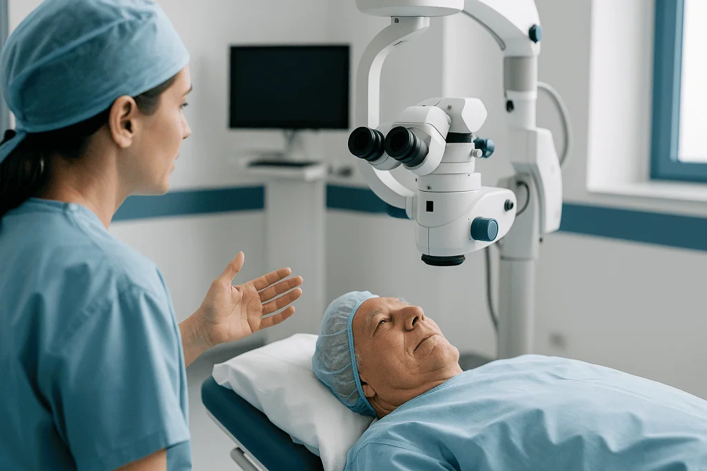
The operation is almost always done awake, with numbing drops or a small injection around the eye, and sometimes a light sedative. The surgeon makes a cut at the edge of the cornea so small it usually seals itself without stitches. Through it, a fine probe uses ultrasound to break the cloudy lens into fragments and vacuum them out, a technique called phacoemulsification. A folded artificial lens, the intraocular lens or IOL, is slid through the same opening and unfolds into the thin capsule that held the original. According to the NHS, the procedure typically takes 30 to 45 minutes, and most people go home the same day.
Two questions come up in almost every consultation. First, which artificial lens? A standard monofocal lens is set for one distance, usually far, with reading glasses afterwards. Toric lenses correct astigmatism. Multifocal or extended-depth lenses aim to reduce glasses use at several distances but can add halos at night; whether that trade suits a particular person is a judgment the surgeon makes with them, not a ranking. Second, both eyes at once? Surgeons often operate on one eye and then the other some weeks later, so the first eye can be checked and the lens choice for the second refined.
Some centers use a femtosecond laser to make the initial cuts and soften the lens before the ultrasound step. It is a variation in tooling rather than a different operation, and mainstream evidence has not shown it to be clearly better than the standard manual technique for most patients.
Laser eye surgery types: LASIK, surface laser and keyhole lenticule extraction
Laser vision correction does not touch the inside of the eye at all. It reshapes the cornea by a few hundredths of a millimeter so that light lands on the retina without help from glasses. The three main laser eye surgery types differ mainly in how the surgeon reaches the tissue to be reshaped.
In LASIK, a thin flap is lifted from the surface of the cornea, an excimer laser sculpts the layer beneath, and the flap is laid back down like a hinged lid. Mayo Clinic describes the procedure as taking about 30 minutes or less for both eyes, with vision often usable the next day. The flap is the strength and the weakness: fast comfort, but a structure that can, rarely, shift after a blow to the eye years later.
Surface laser, often called PRK or LASEK, skips the flap. The outer skin of the cornea is removed or moved aside and the laser works directly on the surface. Healing is slower and more uncomfortable for the first few days because that skin must grow back, but there is no flap to worry about, which is why it is sometimes preferred for people with thinner corneas or physically demanding jobs.
The newer keyhole approach, small-incision lenticule extraction, uses a femtosecond laser to cut a thin disc of tissue inside the cornea, which is drawn out through a tiny side opening. No flap, no surface removal. It is currently used mainly for short sight with or without astigmatism.
None of these treats the age-related loss of near focus that arrives in the forties, and none stops cataract or glaucoma from developing later. The NHS notes that laser correction is generally not offered to people under 21, whose prescriptions may still be changing, or to anyone whose prescription has shifted recently. Dry eye, unstable cornea shape and certain autoimmune conditions can also rule it out.
Lens-based vision correction when laser is not the right tool
Not every prescription can be carved safely into a cornea. Very high short sight, significant long sight, or a cornea that is simply too thin push the numbers beyond what laser reshaping can do without weakening the eye. For those people, surgeons turn to the lens instead.

The first option is an implantable lens placed inside the eye in front of the natural lens, behind the iris. It works like a permanent contact lens that never dries out or falls out, and the natural lens keeps doing its job of focusing near and far. Because nothing is removed, the procedure is in principle reversible. The trade-off is that it involves entering the eye, which carries the small risks of any internal procedure, including cataract forming earlier than it otherwise would and pressure changes that need monitoring.
The second option is refractive lens exchange, which is cataract surgery performed before a cataract exists. The natural lens is removed and an artificial lens of the chosen power takes its place. It is usually reserved for people in middle age or older whose natural lens has already lost its ability to change focus, so there is less to lose by removing it. In a younger eye, taking out a healthy lens means giving up all natural near focus, which is why specialists are cautious.
The NHS groups these under “lens surgery” and is candid that, like laser, they are elective procedures for a problem glasses already solve. That framing matters for how a person weighs risk. Someone facing sight-threatening glaucoma accepts risks a person with a moderate glasses prescription may reasonably decline. A thorough assessment, including measurement of corneal thickness, pupil size, tear quality and the health of the retina, is what tells the surgeon which of these tools, if any, fits.
Glaucoma surgery options: lowering pressure to protect the nerve
Glaucoma is damage to the optic nerve, the cable carrying images from eye to brain, most often driven by fluid pressure inside the eye that is too high for that particular nerve to tolerate. Every treatment, from drops to laser to surgery, has a single mechanism: lower the pressure. None can restore nerve fibers already lost, which is why glaucoma surgery is about keeping the sight a person still has.
Drops are usually first, and for many people they are enough. The NHS describes several laser treatments as the next rung. Laser trabeculoplasty applies gentle laser energy to the eye’s drainage meshwork to help fluid leave more easily. Laser iridotomy makes a tiny hole in the iris to open a blocked drainage angle, the standard treatment for angle-closure glaucoma. Cyclodiode laser reduces the production of fluid by treating the tissue that makes it. These are quick, done with numbing drops, and often used to reduce reliance on drops rather than replace them entirely.
When pressure remains too high, drainage surgery follows. Trabeculectomy, which the NHS calls the most common glaucoma operation, creates a new channel under the upper eyelid so fluid can seep out into a small reservoir beneath the surface of the eye. Tube shunts do a similar job with a tiny implanted drain. Both require weeks of close follow-up because the new channel can scar shut or, less often, drain too much.
Minimally invasive glaucoma surgery, or MIGS, is a group of newer micro-devices and techniques that widen or bypass the natural drainage system through very small incisions, often at the same time as cataract surgery. They tend to lower pressure less dramatically than trabeculectomy but with a gentler recovery, which makes them a middle option for mild to moderate disease. Which rung a person needs depends on how fast the nerve is changing and how high the pressure sits.
Retina surgery: detachment, tears, macular holes and diabetic bleeding
The retina is the paper-thin layer that turns light into nerve signals. When it tears or lifts away from the wall of the eye, the cells it contains lose their blood supply and begin to die within days. That is why retinal detachment is treated as an emergency, and why the NHS advises anyone with a sudden shower of floaters, flashes of light or a shadow spreading across vision to seek urgent eye care the same day.
Small tears caught early can often be sealed in the clinic. Laser or a freezing probe, cryotherapy, creates a scar around the tear that welds the retina down before fluid can slip beneath it. Once the retina has actually detached, three operations are used, alone or together.
Vitrectomy removes the vitreous, the clear gel filling the eye, so the surgeon can reach the retina from inside, flatten it and seal the break. The eye is then filled with a gas bubble or silicone oil to hold the retina in place while it heals. Gas absorbs on its own over weeks; oil must be removed in a later procedure. A scleral buckle is a silicone band stitched around the outside of the eye to push the wall inward against the retina. Pneumatic retinopexy injects a gas bubble alone, with the patient positioning their head so the bubble presses on the tear.
Vitrectomy is also the workhorse for other back-of-eye problems: peeling the membrane that causes a macular hole, clearing blood from advanced diabetic eye disease, and removing scar tissue. Anyone with a gas bubble must not fly or travel to high altitude until the bubble has gone, because the gas expands as air pressure falls and can push eye pressure dangerously high; the NHS notes this wait is typically several weeks and the surgeon confirms when it is over.
Cornea surgery: transplant, partial transplant and cross-linking
The cornea is living tissue, and like any tissue it can scar, swell or warp. When glasses and contact lenses can no longer see through the damage, surgery aims either to replace the tissue or to stop it deforming further.
A full-thickness transplant, penetrating keratoplasty, removes a central disc of the patient’s cornea and stitches in a matching disc from a donor. It has been performed for over a century and remains the choice when all layers are damaged. Recovery is long: Mayo Clinic notes vision can take up to a year to settle as stitches are adjusted and removed, and the graft can be rejected by the immune system. Mayo puts the risk of rejection at roughly 1 in 10 transplants, which is why sudden redness, pain, light sensitivity or a drop in vision after a graft is treated urgently.
Partial-thickness transplants replace only the diseased layer. Endothelial keratoplasty, in versions abbreviated DSAEK and DMEK, swaps the thin inner pump layer whose failure causes swelling; the new tissue is floated into place with an air bubble rather than stitched, and vision often returns faster than after a full graft. Deep anterior lamellar keratoplasty replaces the front layers while keeping the patient’s own inner layer, lowering rejection risk for conditions such as keratoconus.
Keratoconus, a progressive thinning that pushes the cornea into a cone, has its own preventive procedure. Corneal cross-linking soaks the cornea in riboflavin, a form of vitamin B2, and exposes it to ultraviolet light to form new bonds between collagen fibers, stiffening the tissue. It does not improve vision on its own; its purpose is to halt progression so a transplant may never be needed. It is generally offered when scans show the cone is still changing, which is often in teenagers and young adults.
What are the most common eye surgeries, and which is the most difficult?
By sheer volume, cataract surgery leads everywhere it is measured. MedlinePlus describes it as one of the most frequently performed operations in the United States, and the NHS lists it as the most common operation it carries out. Laser vision correction is the most common elective eye procedure among younger adults. Behind those two sit a long tail: glaucoma laser, retinal laser, injections into the eye for macular degeneration (a treatment, though not strictly surgery), eyelid procedures, and squint correction in children.
“Most difficult” is a question surgeons answer differently depending on what they mean by difficulty. Cataract surgery is technically demanding, done under a microscope with movements measured in fractions of a millimeter, but it is so standardized that complications are uncommon in routine cases. Retinal surgery is generally regarded as among the most complex, because the surgeon is manipulating tissue thinner than a sheet of paper at the back of a fluid-filled globe, often with the outcome depending on how much of the retina was still viable when the patient arrived. Full-thickness corneal transplants are difficult less for the operation itself than for the years of stitch management and rejection surveillance that follow.
There is a second kind of difficulty: predictability. Cataract and laser correction have reasonably predictable outcomes because the problem is optical and the fix is optical. Glaucoma and retinal surgery are treating a disease that may keep progressing, so the honest goal is stabilization, and the honest answer to “will my vision come back?” is often “some of it, and we will know more over the coming months.” Understanding which kind of surgery you are facing, the optical kind or the disease kind, sets expectations more accurately than any statistic.
What is the newest eye surgery procedure, and does newer mean better?
Eye surgery attracts new technology quickly because the target is visible, measurable and small. The current wave includes femtosecond-laser assistance in cataract surgery, keyhole lenticule extraction in laser correction, micro-stents and micro-shunts in glaucoma, and thinner and thinner grafts in corneal transplantation. Each arrived with the hope of doing the same job through a smaller opening, with less trauma or a faster recovery.
The evidence behind each is uneven, and that matters more than the launch date. Femtosecond cataract surgery is a good example: it is precise and appealing, yet large comparisons have not shown a clear advantage in vision or safety over the standard ultrasound technique for most eyes, which is why many surgeons reserve it for particular situations rather than every case. Endothelial keratoplasty, by contrast, has genuinely changed practice for corneal swelling because it removes the years of stitch problems that came with full grafts.
Newer also means less long-term data. A device implanted into a 55-year-old’s drainage angle will ideally sit there for three decades; a technique in use for six years cannot yet tell us what year twenty looks like. That is not an argument against innovation. It is an argument for asking a specific question: what does the evidence show this newer technique does better, for someone like me, compared with the established one you also offer?
A reliable signal of an honest practice is a surgeon who performs both the older and newer versions and can explain, without marketing language, when they choose each. A specialist who offers only the newest method for every patient is offering a tool, not a judgment. The right procedure is the one matched to the eye in the chair, and sometimes that is the one that has been working quietly for forty years.
Who is usually offered eye surgery, and who is asked to wait?
For cataract, the threshold is not how cloudy the lens looks to the surgeon but how much it interferes with the person’s life. Mayo Clinic frames the decision around whether reduced vision is affecting driving, reading, work or safety, and notes that waiting rarely harms the eye. Someone who reads fine print comfortably with an early cataract is often asked to return in a year. Someone struggling to see road signs at dusk is usually offered a date. A cataract may also be removed earlier when it is blocking the view of the retina in a person with diabetes or macular disease that needs monitoring.
Laser and lens correction have the opposite logic: the eye must be healthy and stable. Candidates are typically over 21 with a prescription unchanged for at least a year, adequate corneal thickness, a regular corneal shape, and no significant dry eye or autoimmune disease. Pregnancy and breastfeeding, which shift prescriptions temporarily, are common reasons to defer. Keratoconus rules out laser reshaping entirely, because thinning a cornea already weakening is the wrong direction.
Glaucoma surgery is offered when pressure stays too high despite drops and laser, when the nerve is visibly worsening on scans, or when someone cannot tolerate or manage drops. Early or stable glaucoma is watched rather than operated on. Retinal detachment is the one area where nobody is asked to wait; the question is only which technique and how soon.
Corneal transplant is reserved for eyes in which contact lenses and other measures no longer give usable vision, because the years of follow-up and the rejection risk are real costs to the patient’s time and attention. Cross-linking, on the other hand, is offered early, while scans show a cone still moving, because stiffening a cornea works best before it has warped.
Cataract surgery recovery time and what the days after other eye surgeries look like
Most eye operations send people home the same day with an eye shield, a schedule of drops and a follow-up appointment. What differs is how quickly vision clears and how long the eye stays fragile.
After cataract surgery, the NHS says vision is often noticeably better within a few days, though it may be blurry and the eye gritty or watery at first, and full recovery typically takes 2 to 6 weeks. Mayo Clinic notes the eye is usually fully healed by about eight weeks, which is when a final glasses prescription is written. Drops, usually an antibiotic and an anti-inflammatory, run for a few weeks; the eye should not be rubbed, and swimming, dusty environments and heavy lifting are avoided in the early period on the surgeon’s advice.
Laser correction is quicker to see through and slower to settle than people expect. Mayo Clinic describes vision as usable within a day or two of LASIK, with dryness and night glare common in the early months and final vision taking 2 to 3 months to stabilize. Surface laser adds several days of discomfort while the outer skin regrows.
Glaucoma drainage surgery involves the most frequent early visits, often weekly at first, because the surgeon may adjust stitches or massage the eye to keep the new channel open. Vision can be worse than before for weeks while the eye recovers, which alarms people who were not warned.
Retinal surgery with a gas bubble comes with positioning instructions, sometimes face-down for much of the day for a week or more, and vision through the bubble is like looking through water until it absorbs. Corneal grafts are the marathon: up to a year for vision to settle and, after a full-thickness graft, stitches managed over many months. Every one of these is a typical range from published guidance, not a promise for any particular eye.
What people often get wrong about types of eye surgery
“Laser surgery removes cataracts.” It does not. Cataract surgery uses ultrasound, and the artificial lens is placed by hand. A laser is sometimes used for the opening cuts, and a different laser treats the film that can cloud the lens capsule months or years later, but the cataract itself is not lasered away.
“A cataract can grow back.” Once the natural lens is out, it cannot cloud again. What can happen is posterior capsule opacification, a haze on the membrane holding the new lens. Mayo Clinic describes clearing it with a brief in-clinic laser treatment taking a few minutes.
“Laser correction is permanent, so I will never need glasses.” The reshaping is permanent, but the eye keeps aging. Nearly everyone loses near focus in their forties and needs reading glasses, laser or not, and cataract can still develop later.
“Glaucoma surgery will bring my sight back.” It aims to stop further loss by lowering pressure. Nerve fibers already gone do not regrow, which is exactly why glaucoma is screened for before symptoms appear.
“Eye surgery means being put to sleep.” The large majority of adult eye operations are done awake with numbing drops or a local injection, sometimes with mild sedation. General anesthesia is mostly reserved for children, for people who cannot lie still, and for some longer retinal or corneal procedures.
“Floaters and flashes are normal aging; they can wait.” Often they are harmless, but a sudden increase, especially with a shadow or curtain in vision, can signal a retinal tear or detachment, and the window for the best repair is measured in days.
“The newest technique is always the safest.” Newer techniques have less long-term data by definition. Some are clear advances; others are alternatives with different trade-offs. The evidence, not the launch date, tells the difference.
Questions to ask your care team before any eye surgery
The most useful consultation questions are the ones that pull the conversation from the general to the particular. A surgeon can recite average figures for a procedure; what a patient needs is how those figures shift for their eye, their other conditions and their life.
- What exactly is the diagnosis, and which part of the eye does this operation treat? If there is more than one problem, which one is this surgery for and what happens to the others?
- What happens if I do nothing for now? Is this preserving sight I would otherwise lose, or improving sight glasses already correct?
- Which technique are you recommending, which alternatives exist, and why this one for me?
- What are the specific risks in my case, given my prescription, corneal thickness, diabetes, previous surgery or medications, and how would each be handled?
- For cataract or lens surgery: which type of artificial lens, set for which distance, and what will I still need glasses for?
- What will my vision be like in the first week, and when should I expect it to settle? What will limit my activities, driving, work and travel, and for how long?
- Will I have one eye done or both, and what is the interval between them?
- Do I need to stop or continue any of my current medicines, including blood thinners and prostate medicines that affect the iris, and who makes that call?
- How many follow-up visits should I plan, and what happens at each?
- Who do I contact, day or night, if something feels wrong, and what specifically should prompt that call?
Writing the answers down during the appointment, or bringing someone to do it, is not excessive. Eye consultations move quickly and dilating drops make reading afterwards impossible for a few hours. A second appointment to decide is a reasonable request, and a surgeon confident in their recommendation will not mind it.
When to call your doctor: red-flag signs after eye surgery or before it
Most eyes after surgery are gritty, watery and a little red for a few days, and vision wobbles before it settles. The signs below are different. They point to infection, a pressure spike, bleeding, a shifted lens or a retinal problem, all of which are treatable when caught early and far harder when not. If any of them appears, contact the surgical team or seek emergency eye care the same day rather than waiting for the next scheduled visit.
- Pain that is worsening rather than easing, or pain not relieved by the pain relief you were advised to use
- Vision that was improving and then drops suddenly, or vision that becomes markedly worse than it was before surgery
- Increasing redness, a sticky or pus-like discharge, or a swollen, hot eyelid
- New floaters, flashes of light, or a dark curtain or shadow moving across part of your vision
- Severe headache with nausea or vomiting and a hard, aching eye, which can signal dangerously high pressure
- After a corneal graft: any combination of redness, light sensitivity, pain and reduced vision, the classic warning of rejection
- After retinal surgery with a gas bubble: any of the above, or if you are unsure whether the bubble has fully gone before a flight
The same urgency applies before any surgery has happened. Sudden painless loss of vision in one eye, a curtain across the visual field, sudden onset of many floaters with flashes, or a red painful eye with blurred vision and halos around lights are emergencies in their own right. The NHS advises same-day assessment for these because the treatments that work best, particularly for retinal detachment and acute angle-closure glaucoma, depend on speed.
None of this replaces the individual instructions you are given at discharge. If your team’s written advice and this list ever differ, follow the team; they have seen your eye.
Frequently asked questions
What are the most common eye surgeries?
Cataract surgery is the most frequently performed eye operation, described by MedlinePlus as one of the most common operations in the United States. Laser vision correction is the most common elective procedure among younger adults. Glaucoma laser treatments, retinal laser, eyelid procedures and squint surgery in children follow. Volume reflects how common each condition is, not which operation is right for a given person.
What is the best type of eye surgery?
There is no single best type, because each operation treats a different part of the eye. Cataract surgery is right for a clouded lens, laser correction for a healthy eye with a stable glasses prescription, drainage surgery for uncontrolled glaucoma, and vitrectomy for a detached retina. The best procedure is the one matched to your diagnosis and eye measurements by a specialist who has examined you.
What is the newest eye surgery procedure?
Recent developments include keyhole lenticule extraction in laser correction, micro-stents for glaucoma, femtosecond-laser assistance in cataract surgery and ultra-thin corneal grafts. Some, such as thin endothelial grafts, have clearly changed practice; others have not shown consistent advantages over established methods in mainstream evidence. Newer also means less long-term data, so ask what a technique does better for an eye like yours.
What is the most difficult eye surgery?
Retinal surgery is generally regarded as among the most complex, because the surgeon works on tissue thinner than paper at the back of a fluid-filled globe, often against the clock. Full-thickness corneal transplants are demanding for a different reason: years of stitch adjustment and rejection surveillance. Cataract surgery is technically fine work but so standardized that routine cases carry relatively predictable outcomes.
How long is cataract surgery recovery time?
The NHS says vision often improves noticeably within a few days and full recovery typically takes 2 to 6 weeks, while Mayo Clinic describes the eye as usually fully healed by around eight weeks, when a final glasses prescription is written. Drops continue for several weeks and rubbing, swimming and dusty environments are avoided early on. Your surgeon’s instructions take precedence over any general range.
What are the different laser eye surgery types?
The three main types are LASIK, which lifts a thin corneal flap before reshaping the tissue beneath; surface laser such as PRK or LASEK, which reshapes the cornea directly after moving its outer skin; and keyhole lenticule extraction, which removes a thin disc of tissue through a tiny side opening. All reshape the cornea; they differ in access, early comfort and suitability for thinner corneas.
What glaucoma surgery options exist if drops are not enough?
Laser treatments come first: trabeculoplasty to improve drainage, iridotomy to open a blocked angle, or cyclodiode to reduce fluid production. If pressure stays high, trabeculectomy, which the NHS calls the most common glaucoma operation, creates a new drainage channel, or a tube shunt is implanted. Minimally invasive micro-devices offer a middle option for milder disease. The choice depends on how fast the nerve is changing.
Can a cataract come back after surgery?
No. Once the natural lens is removed it cannot cloud again. What can happen is posterior capsule opacification, a haze on the thin membrane holding the artificial lens, which develops in some people months or years later. Mayo Clinic describes clearing it with a brief in-clinic laser treatment lasting a few minutes, after which vision usually clears quickly.
Is eye surgery done under general anesthesia?
Usually not for adults. Cataract, laser correction, glaucoma laser and many retinal and corneal procedures are performed awake with numbing drops or a local injection, sometimes with mild sedation. General anesthesia is mainly used for children, for people who cannot lie still, and for some longer or more complex operations. Your anesthesia plan is decided with the surgical team based on your health and the procedure.
Can I fly after eye surgery?
It depends on the operation. After cataract or laser correction, flying is generally not restricted by the eye itself, though follow-up visits may need to come first. After retinal surgery with a gas bubble, flying and high altitude must be avoided until the bubble has completely absorbed, typically several weeks according to the NHS, because expanding gas can raise eye pressure dangerously. Always confirm timing with your surgeon.
References
This article is for general information only and is not a substitute for professional medical advice. Please consult a qualified doctor about your individual situation.

More from the Blog

What Cornea and External Disease Specialists Treat: Infections, Pterygium and Dystrophies
A cornea and external disease specialist is an ophthalmologist with extra fellowship training in the clear front window of the eye and the surface…
What Chalazion Removal Involves: Numbing, the Incision Inside the Eyelid and the Patch
A chalazion removal procedure is a short outpatient operation for a blocked oil gland in the eyelid. The lid is numbed with a local…

How Corneal Repair Is Done: Sutures, Tissue Glue, Membrane Grafts and Transplant Options
Corneal repair is matched to the depth and cause of the damage. Small scratches are usually left to heal under protection; cuts and perforations…
Ophthalmic Lasers Explained: Retina, Glaucoma and Corneal Lasers and What Each One Does
Eye lasers fall into three broad groups. Retinal lasers seal leaking or abnormal blood vessels and barricade retinal tears. Glaucoma lasers improve fluid drainage…
Ocular Surface Disease Explained: How Tear Film, Eyelids and Cornea Fail Together
Ocular surface disease is an umbrella term for conditions in which the tear film, eyelids and cornea stop working as a unit, most often…

Eye Surgery in Older Adults With Heart Disease or Diabetes: How the Plan Is Adjusted
Eye surgery in older adults with heart disease or diabetes is usually still possible, but the plan changes. Most procedures, especially cataract surgery, are…