

Ocular Surface Disease Explained: How Tear Film, Eyelids and Cornea Fail Together
Key Takeaways
- The tear film has three layers, mucin, water and oil, each made by a different tissue, and a fault in any one destabilizes the whole film.
- Meibomian gland dysfunction, clogged oil glands in the eyelids, is the most common driver of evaporative dry eye, which is why lid care sits at the center of most treatment plans.
- Dry eye feeds a vicious cycle: evaporation concentrates the tears, concentrated tears inflame the surface, inflammation damages tear-producing cells, and the film becomes less stable still.
- Watery eyes are often dry eyes; reflex tears lack oil and mucin, so they spill over the lid without coating the cornea.
- Warm compresses and lid hygiene typically need a few weeks to show an effect, and prescription anti-inflammatory drops work over weeks to months, so early reviews rarely tell the full story.
- Sudden pain, light sensitivity or a vision drop, particularly in a contact lens wearer, are red flags for corneal infection and need same-day care rather than more lubricant drops.
Ocular surface disease is an umbrella term for conditions in which the tear film, eyelids and cornea stop working as a unit, most often dry eye and blepharitis. Unstable or insufficient tears leave the cornea exposed, exposure triggers inflammation, and inflammation further disrupts tear production, creating a cycle. Treatment aims to break that cycle with lubrication, eyelid care and, when needed, prescription anti-inflammatory therapy guided by an eye care professional.
By four in the afternoon the spreadsheet has gone slightly soft at the edges. She blinks hard, twice, and the letters sharpen for a second before the grit returns, that sensation of sand under the lid that no amount of rubbing shifts. The drops in her desk drawer help for about ten minutes. Her optometrist says the words dry eye, then blepharitis, then something about the cornea, and she leaves wondering whether she has one problem or three.
She has one. That is the whole point of the phrase ocular surface disease explained properly: the tear film, the eyelids and the cornea are not neighbors that happen to share an address. They are a single organ system with one job, keeping a living, transparent window wet and smooth roughly every four seconds. When one part falters, the others are pulled down with it.
This article walks through how each part works, how they fail together, and what the treatment path usually looks like, so the next conversation with your eye care team starts on firmer ground.
Ocular surface disease explained: what the term actually covers
The ocular surface is everything the outside world touches when you open your eyes: the cornea (the clear dome at the front), the conjunctiva (the thin membrane over the white of the eye and inside the lids), the eyelid margins with their oil glands, and the tear film that coats all of it. Ocular surface disease is the collective name for disorders of that unit. Dry eye disease is the largest member of the family, but blepharitis, meibomian gland dysfunction, allergic surface inflammation and exposure problems from poorly closing lids all belong under the same roof.
The scale is easy to underestimate. The National Eye Institute puts the number of Americans living with dry eye at nearly 16 million, and both the NHS and the Mayo Clinic describe it as one of the most common reasons people visit an eye care professional. Many of those people cycle through over-the-counter drops for years before anyone examines their eyelids.
Why one label rather than several? Because clinicians noticed that treating the tear film alone often fails when the lids are inflamed, and treating the lids alone often fails when the cornea has already been damaged. The parts are wired together by nerves, hormones and inflammatory signals. A single word for the whole system pushes the examination, and the treatment plan, to look at all three.
One caution before going further. The ocular surface can look remarkably normal in someone who feels miserable, and remarkably damaged in someone who feels little. Symptoms and signs do not always line up, which is one reason a proper examination matters more than any list of complaints, and why this article describes mechanisms rather than offering a self-test.
How does the tear film work, and what are the tear film layers?
Tears are not simply salty water. The tear film is a layered fluid only a few thousandths of a millimeter thick, and each layer has a distinct source and a distinct task.
Closest to the eye sits the mucin layer, produced by goblet cells scattered through the conjunctiva. Mucin is a slippery, gel-like protein that lets watery tears cling to a surface that would otherwise repel them, much as a primer lets paint grip glass. Above it lies the aqueous layer, the watery bulk of the film, secreted by the lacrimal gland tucked under the outer part of the upper lid and by smaller accessory glands. This layer carries oxygen, nutrients, antibodies and enzymes that keep the cornea fed and defended. On top floats the lipid layer, a film of oil released from the meibomian glands in the eyelid margins. Oil slows evaporation the way a lid slows a pot on the stove.
A blink is the machine that assembles all this. Each blink squeezes a little oil from the lid glands, sweeps fresh tears across the cornea, and drains the old film toward small openings at the inner corners of the lids. Between blinks the film thins, then breaks up into dry spots. The time it takes to break up is one of the measurements an eye care professional may make during an examination.
Failure can start in any layer. Too little aqueous production, as in Sjögren’s syndrome, produces what clinicians call aqueous-deficient dry eye. A thin or poor-quality oil layer produces evaporative dry eye, which the Cleveland Clinic and the National Eye Institute identify as the more common pattern. In both, the tears left behind become more concentrated, more salty, and that concentrated fluid is itself an irritant to the cells beneath it.
What do the eyelids do, and what is meibomian gland dysfunction?
Think of the eyelids as both windscreen wiper and oil can. The wiper function is obvious: the upper lid sweeps down, spreads the film, and clears debris. The oil can is less visible. Along the edge of each lid, just behind the lashes, run rows of meibomian glands, several dozen in the upper lid and somewhat fewer in the lower. Their openings sit in a neat line you can sometimes see as tiny dots when you pull the lid gently downward in a mirror.
Healthy meibomian oil flows like a light cooking oil at body temperature. When the glands become inflamed or their openings clog with keratin and thickened secretions, the oil turns pasty and stops reaching the tear film. This is meibomian gland dysfunction, often shortened to MGD, and it is the most frequent driver of evaporative dry eye. Glands that stay blocked for long periods can shrink and eventually drop out, which is why early lid care has more to work with than late lid care.
Blepharitis is the broader term for inflammation of the eyelid margins. The NHS describes it as a common, usually long-term condition in which the lids become red, crusted and irritated, frequently alongside dry eye. Bacteria that normally live harmlessly on the skin can multiply in stagnant oil and release enzymes that break the oil down into irritating fatty acids. Tiny Demodex mites, which live in the lash follicles of many adults, are implicated in some cases. Skin conditions such as rosacea and seborrheic dermatitis raise the odds.
Blinking itself can fail quietly. Studies of screen use cited by the Mayo Clinic and others note that blink rate falls and blinks become incomplete during concentrated near work, so the lower third of the cornea is never fully re-coated and the glands are never fully expressed.
How does the cornea get caught in the crossfire?
The cornea is a marvel of engineering: a living, transparent tissue with no blood vessels, receiving oxygen directly from the air through the tear film and nutrients from the tears and the fluid behind it. It is also among the most densely innervated tissues in the body, which is why a single eyelash on its surface feels like a splinter. Those nerves are not only alarm bells. They form the sensing arm of a reflex loop: irritation on the cornea signals the brainstem, which signals the lacrimal gland to release more tears and the lids to blink.
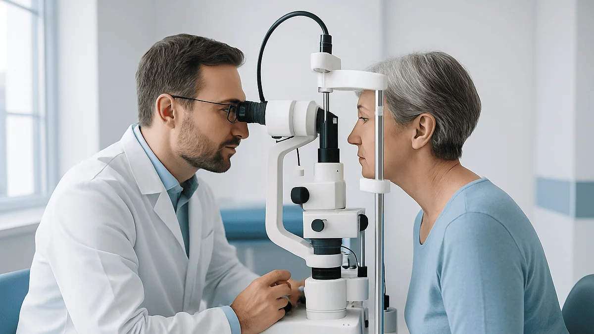
The outermost corneal layer, the epithelium, is a sheet of cells a few layers deep that renews itself constantly. When the tear film breaks up too quickly or contains too little water, patches of these surface cells dry, lose their attachments and lift away. Under a slit lamp (the table-mounted microscope used in eye examinations) an eye care professional highlights these damaged spots with a fluorescein dye that glows under blue light. Small, scattered spots are common in dry eye; larger confluent areas signal a surface in real trouble.
Each damaged patch does two things. It hurts, or at least produces the burning, foreign-body sensation that people describe. It also leaks inflammatory signals into the tears and the tissue beneath, recruiting immune cells that release still more.
Over months and years the corneal nerves themselves can change. Some become less sensitive, so the reflex that should be driving tear production weakens, and the surface dries further without the person feeling it. Others become oversensitive, so the eye aches even when the surface looks reasonably intact, a state sometimes called neuropathic ocular pain. Both patterns help explain why comfort and appearance so often disagree.
Why do tear film, eyelids and cornea fail together?
The central idea in modern thinking about dry eye is a vicious cycle, and it is worth walking around the loop once slowly.
Start anywhere. Suppose the meibomian glands clog and the oil layer thins. Tears evaporate faster, so the fluid left on the eye becomes more concentrated in salts, a state clinicians call tear hyperosmolarity. Concentrated tears stress the surface cells, which respond by releasing inflammatory messengers. Inflammation damages goblet cells in the conjunctiva, so less mucin is produced and the tear film can no longer cling evenly to the cornea. An unstable film breaks up sooner between blinks, exposing more of the cornea to air, which raises evaporation again. The loop has closed and now feeds itself.
Inflammation does not stay put. It spreads along the lid margin and into the meibomian glands, thickening the oil further. It reaches the lacrimal gland, where chronic inflammation is associated with reduced watery tear output. It affects the corneal nerves, blunting the reflex that should be calling for more tears. The three structures fail together not by coincidence but because each one’s failure is the next one’s cause.
This is also why quick fixes disappoint. A lubricating drop replaces water for a few minutes but does nothing about clogged glands or an inflamed lid margin. A warm compress softens oil but cannot, on its own, calm a cornea already peppered with erosions. The cycle has to be interrupted at more than one point, and the points chosen depend on where the examination shows the damage is greatest. An eye care professional, not a symptom, decides that.
One consequence follows: the earlier the loop is interrupted, the fewer structural changes have accumulated, because glands that have dropped out and goblet cells that have been lost do not simply return.
Dry eye vs blepharitis vs ocular surface disease: what is the difference?
People often arrive with one label and leave with another, which is confusing until the relationship is drawn out. Dry eye and blepharitis are both members of the ocular surface disease family; they overlap heavily, and a large share of people have both. The table below sets out where each pattern tends to begin, what the cornea experiences, and the general direction treatment usually takes. It is a map, not a diagnosis.
| Pattern | Where the failure usually starts | What the cornea experiences | Typical first steps discussed with a clinician |
|---|---|---|---|
| Aqueous-deficient dry eye | Lacrimal gland produces too little watery tear (e.g. Sjögren’s syndrome, some medicines, aging) | Thin film, concentrated tears, surface cell damage | Lubricants, tear conservation, assessment for underlying causes |
| Evaporative dry eye with meibomian gland dysfunction | Meibomian glands clog; oil layer thins | Rapid tear breakup, dry spots between blinks | Warm compresses, lid hygiene, blink habits, lubricants |
| Blepharitis (anterior or posterior) | Inflammation of lid margin, often with bacterial overgrowth or Demodex | Irritant by-products in tears; often coexisting MGD | Daily lid cleaning routine; treatment of skin conditions such as rosacea |
| Exposure-related surface disease | Lids do not close fully (facial nerve weakness, eyelid position, nocturnal lagophthalmos) | Localized drying, usually lower cornea | Protection of the exposed area, lid management by a specialist |
Notice how often the right-hand column mentions the lids even when the label says dry eye. That reflects the Cleveland Clinic’s and the National Eye Institute’s shared observation that meibomian gland dysfunction underlies most evaporative disease. In practice, the categories blur: an inflamed lid margin thins the oil layer, thin oil leads to evaporation, evaporation inflames the cornea, and inflammation spreads back to the lids.
The value of the umbrella term is that it stops the question being which box you belong in and starts the more useful one: which parts of your surface are failing, and in what order should they be addressed?
What causes dry eye syndrome and other ocular surface disease?
Causes cluster into things about the person, things about their environment, and things they put in or on their eyes.
Age matters most. The NHS notes that dry eye becomes more common over 50, as tear production and gland function decline. Hormonal shifts play a role; the condition is more common in women, particularly around and after menopause, according to the Mayo Clinic and the National Eye Institute. Autoimmune disease is a heavyweight cause: Sjögren’s syndrome directly attacks tear and saliva glands, and rheumatoid arthritis, lupus and thyroid eye disease are all associated with surface problems. Skin conditions such as rosacea and seborrheic dermatitis drive blepharitis. Diabetes can blunt corneal nerve function, and some people are simply born with fewer or less robust meibomian glands.
Environment does the rest of the damage. Air conditioning, heating, wind, aircraft cabins and low-humidity offices all raise evaporation. Prolonged screen use reduces blink rate and blink completeness. Smoke, both first-hand and second-hand, irritates the surface.
Then there are exposures. Contact lenses sit on the tear film and disrupt it; long daily wear is a well-documented risk factor. Refractive surgery such as LASIK cuts corneal nerves and commonly causes dry eye for months afterward. Several classes of medicine reduce tear production or alter the surface, including antihistamines, decongestants, some antidepressants, some blood pressure medicines, hormone therapies and the acne medicine isotretinoin. Preservatives in eye drops, used many times a day over long periods, can themselves irritate the surface.
That last group deserves a plain warning. If you suspect a medicine you take is drying your eyes, that is a conversation for the clinician who prescribed it. Stopping or changing a prescribed medicine without that conversation can carry risks far greater than dry eyes.
Who is usually treated early, and who is usually asked to watch and wait?
Eye care professionals tend to sort people by two things: how much damage the examination shows, and how much the problem is interfering with life. Those two axes decide the pace.
People moved along quickly usually have visible surface damage, meaning significant corneal staining, filaments of mucus stuck to the cornea, or signs of lid margin disease that is scarring the gland openings. So do those with an underlying condition that makes the surface fragile, such as Sjögren’s syndrome, thyroid eye disease, graft-versus-host disease, or lids that do not close fully after facial nerve weakness or eyelid surgery. Contact lens wearers with surface disease are watched closely because a compromised cornea under a lens is at higher risk of infection. Anyone scheduled for cataract or refractive surgery is often asked to optimize their surface first, because measurements taken on an unstable tear film are less reliable and surgery itself can worsen symptoms for a time.
People asked to watch and wait, or rather to work and wait, typically have mild symptoms with little or no staining. For them the first phase is almost always behavioral and mechanical: humidifying the environment, deliberate blinking during screen work, warm compresses, a daily lid cleaning routine, and preservative-free lubricants if drops are used frequently. Sources such as the NHS suggest these measures need weeks, not days, to show what they can do, so the review is usually set some weeks ahead rather than the following week.
Waiting here does not mean neglect. It means gathering the information that decides whether the next rung, prescription anti-inflammatory treatment or procedures aimed at the glands or tear drainage, is worth its own risks. That judgment belongs to the treating team, who can see the surface, weigh other conditions, and revisit the plan if the surface changes.
How is ocular surface disease diagnosed?
Diagnosis begins with a conversation and a questionnaire. Clinicians commonly use standardized symptom scores, not to label anyone, but to give a baseline that can be compared against later visits. They will ask about medicines, autoimmune conditions, contact lens habits, screen hours and the environments where symptoms peak.
The examination then moves to the slit lamp, the binocular microscope mounted on the table that lets the clinician see the lid margins, gland openings and cornea in fine detail. Several observations are made in sequence. The lid margins are inspected for redness, crusting, telangiectasia (fine dilated blood vessels) and plugged gland openings; gentle pressure on the lid shows whether oil flows and what consistency it has. A drop of fluorescein dye is applied and the clinician watches how long the tear film stays intact after a blink, the tear breakup time, and then looks under blue light for the speckled pattern of damaged surface cells. Another dye, lissamine green, shows damage on the conjunctiva more clearly.
Tear volume can be assessed by observing the strip of tears along the lower lid, or with a Schirmer test, in which a small paper strip is rested inside the lower lid to measure how much it wets over a fixed time. Some practices measure tear osmolarity, the salt concentration of the tears, from a tiny sample, or image the meibomian glands with infrared light (meibography) to see how many glands remain and whether they have shortened. Testing for inflammatory markers in tears is available in some settings.
None of these tests is decisive on its own, and results can vary from day to day. The picture assembled from several of them, interpreted alongside how you feel, is what leads to a working diagnosis. When an underlying systemic cause is suspected, blood tests or referral to a rheumatologist may follow.
How is ocular surface disease treated? Treatment explained step by step
Treatment is layered, and the layers map onto the vicious cycle described earlier: replace what is missing, restore lid function, and calm inflammation.
Replacing what is missing means lubricant drops, gels or ointments. These add water, mimic mucin or add oil depending on their formulation. When drops are used many times a day, clinicians often steer toward preservative-free versions, because preservatives can irritate an already stressed surface. Lubricants soothe; they do not alter the underlying disease.
Restoring lid function is the unglamorous heart of most plans. Warm compresses soften thickened meibomian oil so it can flow again; gentle lid massage afterward helps express it. A daily lid cleaning routine, described in detail by the NHS blepharitis guidance, reduces the crusting and bacterial load at the margin. Deliberate, complete blinks during screen work re-coat the cornea and pump the glands. In-office procedures that heat and express the glands, or that apply intense pulsed light to the skin around the lids, exist; evidence for them is still developing and they are options to discuss, not standard first steps.
Calming inflammation is where prescription medicines enter. Anti-inflammatory eye drops such as cyclosporine and lifitegrast act on the immune cells on the surface and, according to the Mayo Clinic, work gradually over weeks to months rather than instantly. Short courses of corticosteroid drops may be used to settle a flare, with monitoring because of their risks to eye pressure and the lens. Oral antibiotics from the tetracycline class are sometimes used for their anti-inflammatory action on the glands rather than their antibacterial effect. Whether any of these is appropriate, and for how long, is a decision for the prescribing clinician.
Further options for stubborn disease include punctal plugs that slow tear drainage, drops made from the person’s own blood serum, and scleral contact lenses that vault over the cornea holding a reservoir of fluid. Omega-3 supplements are often mentioned; a large NIH-funded trial found no benefit over placebo, so the evidence is at best mixed.
What do the following weeks usually look like after starting treatment?
Progress in ocular surface disease is measured in weeks, and the first honest thing to say is that the early weeks can feel like nothing is happening.
Lid care is the clearest example. The NHS notes that a warm compress and cleaning routine for blepharitis may take a few weeks to make a noticeable difference, and that stopping the routine when things improve usually lets the problem return. The glands need repeated softening and expression before their oil changes character. Many people give up in week two, precisely when the routine is beginning to work.
Prescription anti-inflammatory drops follow a slower arc still. Mayo Clinic guidance describes their effect as building over weeks to months, so a review is typically scheduled a couple of months out rather than a couple of weeks. Some people notice stinging when they first start these drops; that is worth reporting but is not automatically a reason to stop, and the decision to continue or change belongs to the prescriber.
Signs and symptoms tend to improve on different schedules. The corneal staining an eye care professional sees under the slit lamp often begins to clear before comfort improves, because oversensitized nerves take time to settle. The reverse also happens: someone feels much better while the examination still shows damage. Both are reasons to keep the follow-up appointment even when you feel you know how things are going.
Flares are part of the pattern. Air travel, a cold, allergy season, a long stretch of screen work or a dry hotel room can undo a month of gains for a week. A simple diary noting environments, screen hours and symptom intensity turns those setbacks into useful information at the next visit rather than a sense of failure.
The realistic aim is control: a stable surface with fewer, milder flares, and a routine that has become as automatic as brushing teeth.
What people often get wrong about dry eye and blepharitis
Watery eyes cannot be dry eyes. This is the most common misunderstanding, and it runs backward. When the surface is irritated, the cornea’s nerves fire the reflex loop and the lacrimal gland floods the eye with watery tears. Those tears lack the oil and mucin that make them stay put, so they spill over the lid while the cornea underneath remains poorly coated. Streaming eyes in cold wind are a classic example of evaporative dry eye.
Redness-relief drops treat dry eye. They do not. Most of them contain vasoconstrictors, medicines that narrow the blood vessels in the conjunctiva to make the eye look whiter. They do nothing for the tear film, and with repeated use the vessels can dilate again when the drop wears off, a rebound effect. Lubricant drops are a different category.
More drops are always better. Frequent use of drops that contain preservatives can add irritation to an already stressed surface, which is why clinicians ask how many times a day you use them before deciding what to suggest.
It is just getting older, so nothing can be done. Age is a risk factor, but the vicious cycle can be interrupted at any age. Early lid care preserves glands that cannot be replaced later.
Blepharitis is an infection you can clear with one course of treatment. The NHS describes it as a long-term condition that is managed rather than eliminated; bacterial overgrowth is one contributor among several.
Only the eye needs attention. Lids are half the story. A plan that never touches the eyelid margin is, for most people with evaporative disease, addressing the wrong end of the problem.
LASIK fixes dry eye. Refractive surgery cuts corneal nerves and commonly causes or worsens dry eye for months afterward. Surface disease is usually optimized before, not treated by, such surgery.
Questions to ask your care team about ocular surface disease
A good consultation is a two-way exchange, and the right questions steer it toward the parts of the surface that matter most in your case. Bring the ones below that apply, and write down the answers; details fade quickly once you leave the room.
- Which part of my ocular surface is failing most: tear production, the oil layer, the lid margins, or the cornea itself? Is my pattern mainly aqueous-deficient, evaporative, or mixed?
- Did you see staining on my cornea, and if so, how much and where? How will we know at the next visit whether it has improved?
- How many of my meibomian glands look healthy, and is any gland loss already visible?
- Is there any sign that a systemic condition, such as an autoimmune disease or a skin condition, is contributing? Would blood tests or another specialist’s opinion help?
- Could any of my current medicines be drying my eyes, and if so, who should I speak to about that? (Never stop a prescribed medicine without that conversation.)
- What exactly should my daily lid routine involve, how warm and how long, and what mistakes do people commonly make with it?
- If I use lubricant drops frequently, should I be using a preservative-free type?
- What is the plan if the first phase does not help: which options would you consider next, what are their risks, and how long before we judge them?
- Is it safe for me to keep wearing contact lenses while my surface is being treated?
- Are there procedures or supplements you would advise against for me, and why?
- What symptoms should prompt me to contact you before the next scheduled visit?
Notice that several of these questions ask for numbers or descriptions you can compare later: how much staining, how many glands, how long before review. Those anchors turn a vague sense of better or worse into evidence, and they help you and your care team decide together whether the current plan is doing its job.
When to call your doctor
Ocular surface disease is usually a slow, grumbling condition, and most of its ups and downs can wait for a scheduled review. A few situations should not wait, because they may signal something other than dry eye, and because a compromised surface is more vulnerable to infection and injury.
Contact the same day, or use urgent eye care services, for any of the following red-flag signs:
- Sudden or severe eye pain, especially if you wear contact lenses. Pain, redness and light sensitivity in a lens wearer can indicate microbial keratitis, a corneal infection that can threaten sight within days. Remove the lens and seek care.
- A sudden drop in vision, new blurring that does not clear with blinking, or new halos around lights.
- Marked sensitivity to light that is new or worsening.
- Thick yellow or green discharge, or the lids stuck shut on waking.
- A white or gray spot on the cornea that you can see in a mirror.
- Eyelid swelling with fever, or swelling that spreads onto the cheek or forehead.
- Any chemical splash, scratch or foreign body in the eye.
- A red eye that is painful and feels deep or aching rather than gritty, or that comes with nausea or a fixed, mid-sized pupil.
Call within a few days, without urgency but without delay, if your usual symptoms escalate sharply and stay that way, if a new medicine coincides with a clear change in your eyes, if lubricants that used to help stop helping, or if you notice stinging or blurring after starting a prescription drop and are unsure whether to continue. Report these to the prescriber rather than deciding alone.
Between those extremes sits the ordinary work of managing a long-term condition: keeping the follow-up appointment even when things feel stable, bringing your symptom diary, and asking for a review sooner if the pattern changes. Every decision about escalating, changing or stopping treatment sits with your treating team, who can see what you cannot.
Frequently asked questions
What are the three tear film layers and what does each one do?
The tear film has a mucin layer, an aqueous layer and a lipid layer. Mucin, made by goblet cells in the conjunctiva, lets tears cling to the cornea. The aqueous layer, from the lacrimal gland, supplies water, oxygen, nutrients and antibodies. The lipid layer, oil from the meibomian glands in the eyelids, floats on top and slows evaporation. A blink spreads all three across the eye at once.
Can blepharitis and dry eye happen at the same time?
Yes, and they very often do. Blepharitis inflames the eyelid margins, which clogs the meibomian glands and thins the oil layer of the tear film, leading to evaporative dry eye. Dry eye in turn irritates the lids and worsens inflammation. Because the two conditions feed each other, eye care professionals usually treat the eyelids and the tear film together rather than picking one.
What is meibomian gland dysfunction?
Meibomian gland dysfunction is a condition in which the oil glands along the eyelid margins become blocked or inflamed, so their oil turns thick and stops reaching the tear film. Without enough oil, tears evaporate quickly and the cornea dries between blinks. It is the most common cause of evaporative dry eye, and glands that stay blocked for long periods can shrink or drop out.
What causes dry eye syndrome in younger people who spend hours on screens?
Concentrated screen work lowers the blink rate and makes many blinks incomplete, so the lower cornea is never fully re-coated and the meibomian glands are not squeezed regularly. Add air conditioning, contact lenses and, in some people, medicines such as antihistamines or acne treatments, and a young, otherwise healthy eye can develop an unstable tear film. Deliberate full blinks and regular breaks are usually the first things discussed.
Why do my eyes water if they are supposed to be dry?
Reflex tearing is the eye’s alarm response. When the surface is irritated by an unstable tear film, corneal nerves signal the lacrimal gland to release a flood of watery tears. Those tears lack oil and mucin, so they do not spread or stay on the cornea; they spill over the lid instead. Streaming eyes in cold wind or when reading are common signs of this pattern.
How long does it take for dry eye treatment to work?
Longer than most people expect. The NHS notes that a warm compress and lid cleaning routine for blepharitis can take a few weeks to make a difference, and Mayo Clinic guidance describes prescription anti-inflammatory drops as working gradually over weeks to months. Lubricant drops soothe within minutes but do not change the underlying disease. Reviews are usually set weeks or months ahead for this reason.
Does ocular surface disease damage the cornea permanently?
Mild disease usually causes scattered damage to the surface cells of the cornea, which renew themselves continuously once the tear film stabilizes. Severe or prolonged disease can lead to persistent defects, scarring, thinning or infection, particularly in people with autoimmune conditions or lids that do not close fully. Nerve changes in the cornea can also alter sensation over time. Regular examination lets a clinician catch the shift from reversible to structural change.
Are omega-3 supplements effective for ocular surface disease?
The evidence is mixed at best. A large trial funded by the National Institutes of Health found that omega-3 fatty acid supplements did not improve dry eye symptoms or signs more than a placebo. Some smaller studies reported benefit. Because supplements can interact with medicines and conditions, anyone considering them should raise it with their clinician rather than treating them as a proven therapy.
Can I keep wearing contact lenses with dry eye or blepharitis?
Sometimes, but this needs an individual decision from your eye care professional. Lenses sit on and disrupt the tear film, and a cornea already compromised by surface disease is at higher risk of infection under a lens. Some people are asked to pause lens wear while the surface settles, switch lens type or wearing schedule, or be examined more often. Pain, redness or light sensitivity in a lens wearer needs same-day care.
Is ocular surface disease a sign of a more serious illness?
Usually not, but it can be. Most dry eye and blepharitis is related to age, environment, screen habits, contact lenses or skin conditions such as rosacea. In some people it is the first noticeable sign of an autoimmune condition such as Sjögren’s syndrome, thyroid eye disease or rheumatoid arthritis, or a side effect of medicines. Dry mouth, joint pain or unexplained fatigue alongside dry eyes are worth mentioning to your clinician.
References
This article is for general information only and is not a substitute for professional medical advice. Please consult a qualified doctor about your individual situation.

More from the Blog
Ophthalmic Lasers Explained: Retina, Glaucoma and Corneal Lasers and What Each One Does
Eye lasers fall into three broad groups. Retinal lasers seal leaking or abnormal blood vessels and barricade retinal tears. Glaucoma lasers improve fluid drainage…

Eye Surgery in Older Adults With Heart Disease or Diabetes: How the Plan Is Adjusted
Eye surgery in older adults with heart disease or diabetes is usually still possible, but the plan changes. Most procedures, especially cataract surgery, are…

Why You Must Pause Contact Lenses Before a Myopia Laser Assessment (and for How Long)
Contact lenses press on the cornea and temporarily change its shape, so measurements taken too soon after removing them can be inaccurate and could…

Injections, Laser or Surgery for Macular Diseases: How the Retina Team Picks a Path
Macular disease treatment depends on what is happening in the macula. Leaking new blood vessels and swelling are usually treated with repeated anti-VEGF eye…

How Tear Duct Irrigation and Imaging Confirm Where the Blockage Sits
A blocked tear duct test locates where tears stop draining rather than simply confirming that they overflow. An eye specialist usually starts with a…

What Happens in the Operating Room During Cataract Surgery, Minute by Minute
During cataract surgery, a surgeon numbs the eye with drops or a small injection, makes an incision of a few millimeters at the edge…