

Why CyberKnife Planning Starts With MRI, CT or PET-CT: Mapping the Target First

Key Takeaways
- CT is used in every radiosurgery plan because it provides distortion-free geometry and the tissue density values the dose calculation requires, which MRI cannot supply.
- The planning MRI usually differs from a diagnostic MRI: thinner, gap-free slices, a contrast-enhanced 3D sequence and a position matched to the CT so the scans can be fused accurately.
- PET-CT is added selectively to confirm that tissue is metabolically active disease rather than scar, and is rarely useful for benign targets such as acoustic neuromas.
- Margins around a radiosurgery target are often only a millimeter or two, which is why fusion and contouring are checked slice by slice and frequently peer reviewed.
- Treatment commonly starts a few days to a few weeks after the planning scans, and each session typically lasts from under an hour to several hours, with one to five sessions in most courses.
- Response to radiosurgery unfolds over weeks to months, and the planning scans become the baseline against which every follow-up MRI or PET-CT is compared.
CyberKnife planning starts with MRI, CT or PET-CT because the robot can only aim at what has been mapped in three dimensions. CT provides the geometric and tissue-density framework the dose calculation needs; MRI sharpens the edges of soft-tissue targets such as brain, spine and prostate lesions; PET-CT shows metabolically active disease. Fusing these scans lets the team outline the target and nearby organs before any radiation is delivered.
The appointment letter has three dates on it, and none of them is the treatment. First a CT with a custom mask, then an MRI with contrast, then possibly a PET-CT. One patient described it as being asked to sit for a portrait three times before anyone picked up a brush. That is, in fact, a fair description of how CyberKnife treatment planning with MRI, CT and sometimes PET-CT works.
Radiosurgery is unusual among cancer treatments in that almost all of the skill is spent before the machine switches on. The beams are narrow, the dose per session is high, and the margins drawn around a target are often measured in millimeters. Nobody can hit a target that precisely without a map that precise.
This explainer walks through what each scan contributes, why the sequence matters, how long the planning phase typically takes, and what questions are worth asking before you lie down on the table.
What CyberKnife treatment planning with MRI and CT actually involves
Stereotactic radiosurgery is a way of delivering a high dose of focused radiation to a small target, usually in one to five sessions, while sparing the tissue around it. A CyberKnife is one type of system that does this using a compact linear accelerator mounted on a robotic arm; the arm can approach the target from many angles and adjust for small movements during treatment. The name describes a robotic radiosurgery system, not a blade; nothing is cut (MedlinePlus).
Planning is the phase in which a radiation oncologist, a medical physicist and a dosimetrist build a three-dimensional model of the patient, define the target inside it, define every structure that must be protected, and then calculate how dozens or hundreds of individual beams should be arranged so the dose piles up where it is wanted and falls away sharply everywhere else. Every one of those steps depends on imaging.
Think of it as three separate questions, each answered best by a different scan. Where exactly is the body, and how dense is each tissue the beams will pass through? That is the CT question. Where does the abnormal tissue end and normal tissue begin? That is usually the MRI question. Is this tissue biologically active disease, or scar, or something else? That is often the PET-CT question.
The scans are then aligned to one another in planning software, a process called image fusion, so the physician can draw contours while looking at all the information at once. Only after the contours are approved does dose calculation begin. The order is not bureaucratic; it reflects the fact that a plan can only be as accurate as the map underneath it (Mayo Clinic).
Why the CT scan is the backbone of every radiosurgery plan
Every radiosurgery plan is built on a CT, even when the MRI shows the tumor far more clearly. Two properties of CT make it indispensable.

The first is geometry. CT images are acquired in a way that produces almost no spatial distortion, so a point that appears 40 millimeters from the skull edge on the screen really is 40 millimeters away in the patient. MRI, by contrast, can warp slightly at the edges of the field of view or near air-filled sinuses. When margins are being set at a millimeter or two, that difference matters.
The second is tissue density. CT measures how much each voxel of tissue attenuates X-rays, expressed in Hounsfield units, a scale on which water sits at zero, bone is strongly positive and air is strongly negative. The planning software converts those values into electron density and uses them to calculate how each beam will be absorbed as it travels through skin, fat, bone, lung and the target itself. MRI provides no such information, because it maps hydrogen signal rather than X-ray attenuation.
The planning CT is also the scan on which the patient’s position is fixed. Before it is taken, the team makes an immobilization device: a thermoplastic mask for the head and neck, or a vacuum cushion or body mold for the trunk. The patient is scanned in that device, in the exact position that will be reproduced on treatment day (NHS).
Slice thickness is usually thinner than a routine diagnostic CT, commonly around 1 to 1.5 millimeters for cranial targets, because the software reconstructs the three-dimensional model from those slices. For targets that move with breathing, the CT may be acquired in several breathing phases so the team can see the full path the tumor travels.
What is the MRI protocol used in CyberKnife treatment planning?
People searching for the CyberKnife MRI protocol usually want to know whether the planning MRI is different from the one that found the problem. Often it is, in small but important ways.
MRI is the scan of choice for soft tissue. It separates gray matter from white matter, a tumor from the swelling around it, a nerve from the vessel beside it, and the prostate from the rectal wall in a way CT simply cannot. For brain metastases, acoustic neuromas, meningiomas, spinal lesions and prostate cancer, the MRI is usually where the target is actually drawn (Johns Hopkins Medicine).
A typical planning protocol for the brain includes a thin-slice, three-dimensional T1-weighted sequence after gadolinium contrast. Gadolinium is a contrast agent injected into a vein that makes areas with a leaky blood supply, such as many tumors, appear bright. A T2 or FLAIR sequence, which highlights fluid and swelling, is usually added so the team can tell active tumor from surrounding edema. Slices are kept thin and continuous, with no gaps, and the field of view is set so the whole head is included for accurate fusion with the CT.
Other sites call for other sequences. Spine protocols may add fat-suppressed images to show marrow involvement. Prostate protocols rely heavily on T2-weighted images and sometimes diffusion-weighted imaging, which reflects how freely water molecules move within tissue. Some centers acquire the planning MRI in the same immobilization device as the CT to reduce alignment errors.
Timing matters as well. Brain metastases can grow measurably within weeks, so many teams prefer the planning MRI to be recent, often within a couple of weeks of treatment. If your diagnostic MRI is older or was done with thicker slices, you may be asked to repeat it; this is standard practice, not a sign that anything went wrong.
When PET-CT joins the picture, and when it does not
PET-CT combines two scans in one appointment. The PET component tracks a radioactive tracer, most commonly a form of glucose labeled with fluorine-18 (FDG), that collects in tissues using a lot of energy; the CT component provides the anatomical backdrop. Tumors that burn glucose quickly light up, while scar tissue and many benign changes do not (Mayo Clinic).
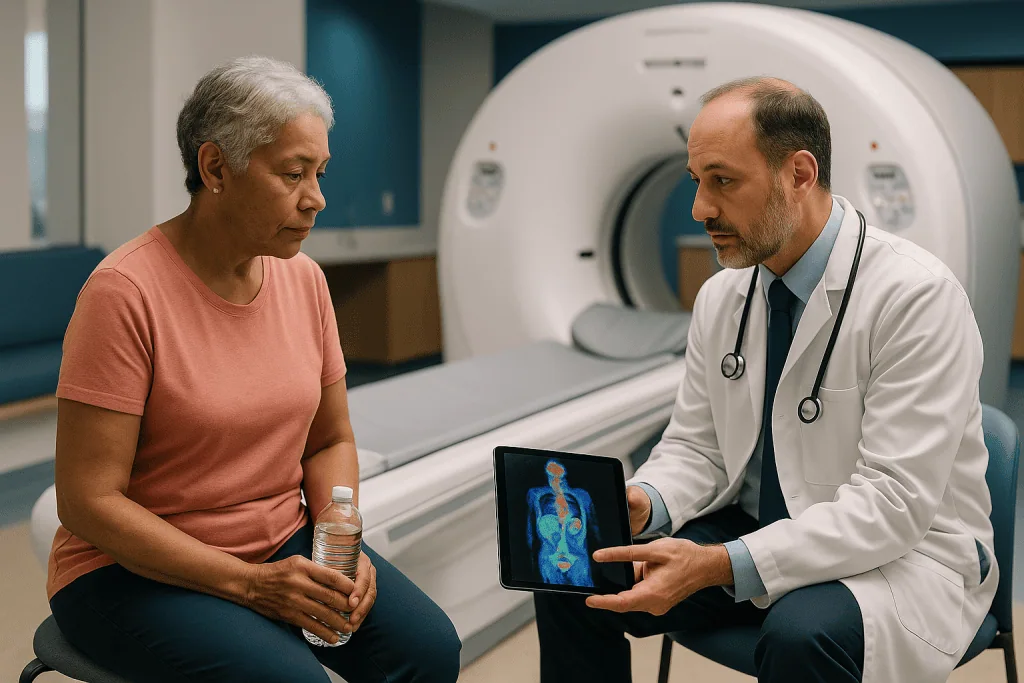
In radiosurgery planning, PET-CT answers a question CT and MRI cannot: is this mass biologically active? That is useful in several situations. In lung cancer, it helps distinguish tumor from collapsed lung or post-infection scarring, which can look identical on CT. After previous radiation, it can help separate recurrent disease from radiation-related change. In prostate cancer, newer tracers that bind to prostate-specific membrane antigen, a protein on the surface of prostate cancer cells, can reveal small deposits in lymph nodes or bone that conventional imaging misses, which affects whether focused treatment makes sense at all.
PET-CT has limits. Its resolution is coarser than MRI or CT, commonly several millimeters, so it is rarely used alone to draw a target edge. Inflammation, infection and healing tissue can take up FDG and mimic cancer. Some slow-growing tumors use little glucose and barely register. For most benign brain conditions treated with radiosurgery, such as acoustic neuroma or trigeminal neuralgia, PET adds nothing and is not ordered.
When it is used, PET-CT is fused to the planning CT in the same way the MRI is, and the physician typically uses it to confirm that the region drawn on anatomical imaging contains the active disease, or to extend the contour to include activity that anatomical imaging underestimated. The decision to include PET-CT rests with the treating team and depends on the tumor type, prior treatment and what questions remain unanswered.
Image fusion: how two or three scans become one map
Image fusion, sometimes called registration, is the step where the planning software overlays the MRI and any PET-CT onto the planning CT so that each point in one scan corresponds to the same point in the body on the others. It sounds like a formality. It is one of the places where accuracy is won or lost.
The software usually starts by matching rigid structures that do not change shape between scans, chiefly bone. The skull is ideal for this, which is one reason cranial radiosurgery can achieve such tight margins. For the spine, individual vertebrae are matched. Soft-tissue targets elsewhere in the body are harder, because the prostate, liver or lung may sit differently depending on bladder filling, bowel contents or breathing, so fusion there is checked more carefully and sometimes limited to a small region around the target.
A physicist or dosimetrist reviews the fusion slice by slice, toggling between scans, looking for any misalignment at known landmarks such as the optic nerves, the brainstem outline or the inner ear. The radiation oncologist signs off before contouring begins. Where distortion in the MRI is suspected, some teams apply distortion-correction algorithms or rely more heavily on the CT for the final geometry.
The practical consequence for patients is that the scans need to be compatible. Thin slices, complete coverage of the region, and the same head position all make fusion more reliable. That is why a diagnostic MRI done at a different time, with thicker slices or a different tilt of the head, may be acceptable for diagnosis yet still need to be repeated for planning.
Once fused, the composite image is what the physician works from. Scrolling through it, the team sees the CT’s bone and density, the MRI’s soft-tissue detail and the PET’s metabolic signal at every level, all in one coordinate system.
Contouring: drawing the target and everything that must be protected
Contouring is the painstaking process of outlining structures on every slice of the fused images. It is done by hand, slice by slice, often with software assistance, and it is where the physician’s judgment is most visible.
Several volumes are defined. The gross tumor volume is the visible disease. A clinical target volume may add a small rim where microscopic spread is suspected, though in radiosurgery this rim is often minimal or absent. The planning target volume adds a margin for setup uncertainty and movement; with robotic tracking this margin is typically small, frequently a millimeter or two for cranial targets, because the system corrects position during treatment rather than relying on a wide buffer (Cleveland Clinic).
Equally important are the organs at risk. These are the structures near the target that have their own dose limits: optic nerves and chiasm, brainstem, cochlea, spinal cord, lenses of the eyes, and in the body the esophagus, bowel, rectum, bladder, heart, major airways and kidneys. Each has a published tolerance derived from decades of clinical experience, and the plan must keep the dose to each below its threshold. The MRI is often the scan that makes these structures visible enough to draw accurately.
Contouring takes time because the stakes of a boundary error are asymmetric. Draw the target too small and part of the disease receives a low dose. Draw it too large and healthy tissue receives a high one. Neither mistake is visible on treatment day; both become apparent months later.
Many centers use peer review, in which a second radiation oncologist checks the contours before planning proceeds. If your team mentions that your case is going to a planning meeting or tumor board, this is usually what they mean, and it is a sign of a careful process rather than uncertainty about your care.
Fiducials, masks and tracking: why the planning scan must match treatment day
A plan is a promise that the beams will meet the target drawn on the scan. That promise only holds if the patient on treatment day is positioned exactly as they were on the planning CT, or if the system can detect and correct any difference. Robotic radiosurgery relies on the second approach, and it starts during planning.
For the head, the skull itself is the reference. Two X-ray cameras in the treatment room take images at intervals, the software compares the bony anatomy with digitally reconstructed radiographs generated from the planning CT, and the robot adjusts its aim if the head has shifted by even a fraction of a millimeter. The thermoplastic mask keeps movement small; it is not screwed into the skull, which distinguishes this approach from older frame-based radiosurgery (MedlinePlus).
For the spine, vertebrae serve the same purpose. For soft-tissue targets that move, such as prostate, liver, pancreas or lung, bone is not a reliable guide because the organ moves independently of the skeleton. Here small gold markers called fiducials are often placed in or near the tumor before the planning CT, usually with a needle under ultrasound or CT guidance. The planning CT then captures the fiducials in their relationship to the target, and the tracking cameras follow them during treatment.
Fiducial placement typically happens about a week before the planning CT so that any swelling settles and the markers stop shifting. Some lung tumors can be tracked without markers by using the tumor’s own density, and a chest marker with infrared sensors can be used to model breathing so the robot follows the target as it rises and falls.
This is the deepest reason the planning scan comes first: every correction made on treatment day is a correction back toward the geometry captured in that CT.
Who is usually offered radiosurgery, and when is CyberKnife not recommended?
Robotic radiosurgery is usually considered for small, well-defined targets that imaging can delineate clearly. Common indications include a limited number of brain metastases, benign brain tumors such as meningiomas and acoustic neuromas, arteriovenous malformations, trigeminal neuralgia, spinal metastases, early-stage lung cancer in people who cannot or prefer not to have surgery, and selected prostate, liver and pancreatic tumors (Mayo Clinic). It is also used when a tumor sits near a previous radiation field or in a location where open surgery carries high risk.
People asking when CyberKnife is not recommended usually encounter a handful of recurring reasons. The target may be too large; as a tumor grows, the volume of normal tissue that would receive a high dose grows faster, and at some size conventional fractionated radiotherapy or surgery becomes safer. The disease may be too widespread, with many deposits or diffuse infiltration that focused beams cannot reasonably cover. The edges may be indistinct on imaging, as with some infiltrating gliomas, so there is no clear boundary to aim at. The target may sit directly against a critical structure already close to its dose limit. Or the person may be unable to lie still for the required time, which is a practical rather than a biological barrier.
Some patients are asked to wait rather than told no. A recent surgery may need to heal so the cavity settles into its final shape before it is imaged. Swelling may need to subside. A fiducial placement may need a week to stabilize. Imaging may need to be repeated because the available scan is too old or too coarse. Systemic treatment timing may need coordination with the medical oncologist.
None of these decisions can be made from a description; they depend on the individual scan and on a multidisciplinary review. The treating team weighs tumor type, location, size, prior treatment and overall health before recommending any approach.
MRI, CT and PET-CT at a glance: what each scan contributes to the plan
The three scans are complementary rather than interchangeable. The table below summarizes the role each one usually plays in radiosurgery planning; individual protocols vary by site and by team.
| Scan | What it measures | Main role in planning | Strengths | Limits |
|---|---|---|---|---|
| CT | X-ray attenuation (tissue density) | Geometric framework; dose calculation; reference for tracking | Minimal distortion; fast; captures bone and density; done in the treatment position | Poor soft-tissue contrast in brain, spine, prostate |
| MRI | Hydrogen signal in tissue | Defining target edges and nearby nerves, vessels and organs | Superb soft-tissue detail; multiple sequences reveal tumor versus swelling | Possible spatial distortion; no density data; longer scan; contraindicated with some implants |
| PET-CT | Tracer uptake plus CT anatomy | Confirming biological activity; finding disease elsewhere; separating tumor from scar | Shows metabolism; whole-body view | Coarse resolution; false positives from inflammation; not useful for most benign targets |
A few practical notes follow from this. The CT is never skipped, because the dose engine cannot run without it. The MRI is skipped only when the target is well seen on CT alone, which is uncommon for brain and spine but more common for some lung lesions. PET-CT is added selectively.
Preparation differs too. The MRI requires a screening questionnaire about metal implants, pacemakers and prior surgery, and a kidney function check if gadolinium is planned. The PET-CT requires fasting for several hours beforehand so that muscle and blood glucose do not compete with the tracer, and avoiding strenuous exercise the day before (MedlinePlus). The CT requires only that you can hold the planned position while the mask or mold is made. Your scheduling team will give site-specific instructions; it is worth reading them the day before rather than the morning of.
How long does radiation treatment planning take, and how long is a CyberKnife session?
Two time questions dominate searches on this topic, and they have different answers.
Planning time runs from the imaging appointments to the first treatment. The scans themselves are short: a planning CT typically takes 15 to 30 minutes including mask or mold construction, an MRI 30 to 60 minutes, a PET-CT about two hours including the tracer uptake period. The longer interval is the invisible work afterward. Fusion, contouring, peer review, dose calculation, plan optimization and physics quality assurance, in which the plan is delivered to a measurement device to confirm the machine produces what the computer predicted, all happen between appointments. In practice treatment usually begins a few days to a few weeks after the planning scans, depending on complexity and how many specialists need to review the case (NHS).
Session length is the second question. Because the dose per session is high and the beam arrangement complex, a single radiosurgery session is longer than a conventional radiotherapy visit. Mayo Clinic describes stereotactic radiosurgery sessions lasting from under an hour to several hours depending on the target and number of beams, and most treatment courses involve between one and five sessions (Mayo Clinic). Cranial targets are often at the shorter end; prostate or lung treatments with breathing tracking tend toward the longer end.
During a session you lie on the couch in your mask or mold, the robot moves around you, pausing at each beam position, and the imaging cameras take periodic X-rays. You will hear motors and occasional clicks. There is no sensation from the radiation itself. Most people find the hardest part is lying still, and teams usually allow brief breaks if needed.
These are typical ranges, not promises. Your team will give you an estimate specific to your plan once it is complete.
What the following days and weeks usually look like
Radiosurgery does not remove a tumor the way surgery does. It damages the DNA of cells in the target so they lose the ability to divide, and the effect unfolds over weeks to months. Understanding that timeline prevents a great deal of unnecessary worry.
In the first few days, most people feel close to normal. Tiredness is the most common complaint. Mild headache or nausea can occur after brain treatment, and some teams prescribe a short course of a corticosteroid, a medicine that reduces swelling, around the treatment; whether and how that is used is a decision for the treating physician. Skin in the beam path may become slightly pink. Hair loss, if it happens, is usually confined to a small patch where beams entered and may not appear for a couple of weeks (Mayo Clinic).
In the first weeks, the body begins clearing cells that can no longer divide. Benign tumors such as acoustic neuromas may show no change for a long time, and a temporary increase in size on early scans is recognized and does not by itself mean the treatment has failed. Metastases often shrink more visibly. Arteriovenous malformations close gradually over months to years.
Follow-up imaging is the way the team tracks response, and here the planning scans serve a second purpose: they are the baseline against which every later MRI or PET-CT is compared. A first follow-up MRI is commonly scheduled a few months after treatment, with intervals set by the tumor type and the treating team.
Everyday life usually resumes quickly. Many people return to work within days. Driving may be restricted after brain treatment depending on local rules and on seizure risk; ask before you assume. If you have fiducials, they stay in place permanently and are harmless.
Risks, side effects and the alternatives your team may discuss
Radiosurgery is generally well tolerated, but it is not free of risk, and the risks relate directly to the imaging and planning described above. The most serious late effect is radiation necrosis, in which a region of normal tissue near the target is injured and swells, sometimes months after treatment. Careful contouring of organs at risk and respect for dose limits are the main defenses, which is why so much effort goes into the MRI. Other possible effects depend on location: hearing change after acoustic neuroma treatment, visual symptoms if the optic apparatus is close, cough or shortness of breath after lung treatment, urinary or bowel irritation after prostate treatment (Cleveland Clinic).
The scans themselves carry small risks. Gadolinium is generally safe but is used cautiously in people with reduced kidney function. PET tracers involve a small radiation dose, comparable to a few years of natural background exposure. Fiducial placement carries the usual risks of a needle procedure, including bleeding, and in the lung a small chance of a collapsed lung.
Alternatives are part of every honest conversation. Conventional fractionated radiotherapy spreads a lower dose over many sessions and is often preferred for larger or less well-defined targets. Whole-brain radiotherapy may be discussed when metastases are numerous. Surgery removes tissue immediately and provides a pathology specimen, which radiosurgery cannot. Frame-based radiosurgery systems and linear-accelerator-based stereotactic techniques treat many of the same conditions. Active surveillance, meaning regular imaging without treatment, is a legitimate option for some slow-growing benign tumors. Systemic therapies such as targeted drugs or immunotherapy may be used alongside or instead of local treatment.
No approach is best for everyone. The right choice depends on what the imaging shows, what has been tried before and what matters most to you, and the treating team is the only party with all of that information.
What people often get wrong about CyberKnife planning scans
Several misunderstandings come up repeatedly in clinic, and clearing them away makes the process less stressful.
The first is that the planning scans are a repeat of tests already done, ordered out of caution or habit. They are not. Planning imaging is acquired with thinner slices, a fixed position, specific sequences and compatible geometry so it can be fused and used for dose calculation. A diagnostic MRI from a few months ago answers the question of what is wrong; it rarely answers the question of exactly where every edge is today.
The second is that MRI could replace CT because it shows the tumor better. MRI shows soft tissue better, but it does not provide the electron density information the dose engine needs and it is prone to small spatial distortions. Both are required for different reasons.
The third is that more scans mean a worse prognosis. The number of scans reflects the complexity of the target and the questions the team needs answered, not the seriousness of the diagnosis. A benign tumor near the optic nerve may need more careful imaging than a straightforward metastasis elsewhere.
The fourth is that the robot finds the tumor on its own. The tracking system follows bone or fiducials and corrects position, but it can only correct back toward the geometry humans defined on the planning CT. The intelligence is in the contours, not the machine.
The fifth is that treatment works immediately and a follow-up scan a few weeks later should show the tumor gone. Response is gradual; early scans can even show temporary enlargement or new enhancement from swelling. Teams interpret follow-up imaging against the planning baseline with that timeline in mind (Johns Hopkins Medicine).
Finally, a thermoplastic mask is not screwed into the skull, and fiducials do not hurt once in place. Both tend to be anticipated with more dread than they deserve.
Questions to ask your care team before the planning scans
A good planning conversation is specific. These questions tend to produce useful answers; write down the replies, because the details are easy to lose.
- Which scans will I have for planning, and why each one? If a PET-CT is not included, what question is it that we do not need answered?
- Will my existing MRI be used, or do I need a new one, and what makes the difference?
- Do I need fiducial markers? If so, how will they be placed, how long before the planning CT, and what should I watch for afterward?
- What will the immobilization device feel like, and can I see one beforehand? Is there anything I can do about claustrophobia in the mask?
- How many sessions are you planning, roughly how long will each one last, and how far apart will they be?
- Which structures near the target are you most concerned about protecting, and what side effects would relate to them?
- Will my plan be reviewed by a second physician or at a tumor board?
- What is the alternative you would consider if this approach were not suitable, and why did you choose this one?
- Should I keep taking my usual medicines on scan days and treatment days, including anything for diabetes before a PET-CT?
- When will the first follow-up scan be, and what will you be looking for on it?
- Who do I call if something worries me between appointments, including evenings and weekends?
It is also reasonable to ask how recent the imaging needs to be and what happens if treatment is delayed for any reason, since a plan built on a scan that is several weeks old may need to be checked against new imaging. A team that welcomes these questions is a team paying attention to the same details you are (NHS).
When to call your doctor
Most of the planning phase and the treatment itself pass without incident, but some symptoms need prompt attention. Contact your care team the same day, or use emergency services if the symptom is severe or sudden, in the following situations.
After fiducial placement: increasing pain at the needle site, spreading redness or warmth, fever, coughing up blood or new shortness of breath after lung marker placement, or blood in the urine that does not clear within a day or two after prostate marker placement.
After a contrast MRI or PET-CT: hives, swelling of the face or throat, wheezing or difficulty breathing. Allergic reactions to gadolinium are uncommon but can occur, and most begin within minutes to a few hours.
After brain radiosurgery: a headache that is new, severe or steadily worsening, especially with vomiting; new weakness, numbness or difficulty speaking; a seizure; new confusion or marked drowsiness; sudden visual change. These can indicate swelling that needs treatment and should not be left until the next scheduled visit.
After spine, lung or abdominal treatment: new or worsening back pain with leg weakness or numbness, loss of bladder or bowel control, chest pain, shortness of breath, persistent vomiting, or inability to keep fluids down.
At any point: a temperature above 38°C (100.4°F), particularly if you are also receiving chemotherapy or immunotherapy, since infection in that setting can escalate quickly (NHS).
Call as well for anything that simply does not feel right to you. Teams would far rather hear about a symptom that turns out to be nothing than miss one that mattered. Keep the department’s contact number and the out-of-hours number somewhere you can find them, and make sure one other person in your household has them too.
Frequently asked questions
What is the MRI protocol used in CyberKnife treatment planning?
For brain targets it is usually a thin-slice, three-dimensional T1-weighted sequence after gadolinium contrast, plus a T2 or FLAIR sequence to show swelling, acquired with no gaps between slices. Spine and prostate protocols use different sequences suited to those tissues. The aim is an image that fuses accurately with the planning CT and shows every edge of the target clearly.
Why is a CyberKnife MRI protocol different from a normal diagnostic MRI?
A diagnostic MRI is designed to find and characterize a problem; a planning MRI is designed to be measured. Planning scans use thinner, continuous slices, specific sequences and a head position compatible with the planning CT so the two can be overlaid precisely. An older scan with thicker slices or a different head tilt may be fine for diagnosis but too imprecise for planning.
When is CyberKnife not recommended?
Radiosurgery is usually not recommended when the target is too large, when disease is widespread or diffusely infiltrating, when the tumor edge cannot be seen clearly on imaging, when a critical structure next to the target is already near its dose limit, or when a person cannot lie still for the session. These judgments depend on the individual scans and are made by the treating team.
How long does CyberKnife take from first scan to last session?
The scans themselves take minutes to a couple of hours each. Treatment usually begins a few days to a few weeks after the planning scans, once fusion, contouring, dose calculation and physics checks are complete. Each treatment session typically lasts from under an hour to several hours, and most courses involve one to five sessions spread over days to a couple of weeks.
How long does radiation treatment planning take behind the scenes?
After the imaging appointments, the team fuses the scans, draws the target and organs at risk, often holds a peer review, calculates and optimizes the beam arrangement, and runs quality assurance on the machine. That work commonly takes several working days and sometimes longer for complex cases. Your department can tell you the expected interval for your specific plan.
Can the MRI replace the CT for radiosurgery planning?
No. MRI shows soft tissue far better, but it does not measure tissue density, which the software needs to calculate how beams are absorbed, and it can be slightly distorted at the edges of the image. CT provides both the density data and a distortion-free geometric reference used for tracking on treatment day. The two scans answer different questions and are used together.
Do I need a PET-CT before CyberKnife treatment?
Not always. PET-CT is added when the team needs to know whether tissue is biologically active, for example to separate lung tumor from scar, to check for disease elsewhere, or to assess recurrence after previous radiation. For most benign brain conditions it adds nothing and is not ordered. Your radiation oncologist will explain which question a PET-CT would answer in your case.
What are fiducial markers and does everyone need them?
Fiducials are tiny gold markers placed in or near a tumor so the tracking cameras can follow it during treatment. They are used for targets that move independently of bone, such as prostate, liver, pancreas and some lung tumors. Brain and spine targets are tracked using the skull or vertebrae instead, so fiducials are not needed. Once placed, fiducials remain permanently and are harmless.
Will I feel anything during the planning scans or the treatment?
The scans are painless, though the MRI is noisy and the mask or mold can feel confining. Contrast injections may cause a brief warm sensation. During treatment there is no sensation from the radiation itself; you will hear the robot moving and see it change position. The main challenge is lying still, and teams usually build in short breaks if needed.
Why does my follow-up scan not show the tumor gone yet?
Radiosurgery damages the ability of cells to divide, and the visible effect develops over weeks to months rather than days. Early scans can even show temporary swelling or enlargement that is part of the expected response. Your team compares each follow-up image with the planning baseline and interprets changes against the known timeline for your tumor type.
References
- MedlinePlus: Stereotactic radiosurgery – CyberKnife
- NHS: Radiotherapy, what happens
- MedlinePlus: PET scan
This article is for general information only and is not a substitute for professional medical advice. Please consult a qualified doctor about your individual situation.

More from the Blog
Recovery After Vulvar Cancer Surgery: Wound Healing, Hygiene and the First Weeks
Recovery after vulvar cancer surgery usually means a hospital stay of a few days, followed by several weeks of gradual healing at home. Gentle…

Skin Care and Hydration During Proton Therapy: Daily Habits for a Multi-Week Course
Skin care during proton therapy centers on a few steady habits: wash the treated area once or twice daily with lukewarm water and a…

How Long to Plan for a Stem Cell Transplant Abroad Before Flying Home
Most people should plan to remain near their transplant center for weeks to months after a stem cell transplant, not days. Patient guidance from…

Eating After Stomach Cancer Surgery: Small Frequent Meals and the New Digestive Rhythm
Eating after gastrectomy usually means six to eight small meals a day instead of three large ones, chewing slowly, putting protein first, separating drinks…
What Debulking Surgery for Ovarian Cancer Removes and Why Completeness Matters
Debulking surgery for ovarian cancer, also called cytoreductive surgery, removes the ovaries, fallopian tubes, uterus, omentum and every visible tumor deposit a surgeon can…

Robotic Partial Nephrectomy Recovery: Early Walking, Lifting Limits and the First Six Weeks
Robotic partial nephrectomy recovery usually means walking within hours of surgery, a hospital stay of one to three days, and light activity within one…