Abdominal Ultrasound for Digestive Symptoms: What It Can Show

Abdominal ultrasound uses sound waves, not radiation, to create images of abdominal organs. It is often used to assess digestive symptoms such as pain, nausea, bloating, and suspected gallbladder or liver problems.
Key Takeaways
- Abdominal ultrasound uses sound waves, not radiation, to create images of abdominal organs.
- It is often used to assess digestive symptoms such as pain, nausea, bloating, and suspected gallbladder or liver problems.
- The test can show gallstones, bile duct widening, liver changes, some pancreatic concerns, kidney problems, and fluid in the abdomen.
- Ultrasound has limits and may not fully evaluate the stomach, intestines, or gas-filled areas.
- Preparation may include fasting for several hours, depending on the reason for the scan.
- Results are interpreted together with symptoms, examination findings, and sometimes blood tests or other imaging.
Abdominal ultrasound is a common, noninvasive imaging test used to look for causes of digestive symptoms such as abdominal pain, bloating, nausea, or changes in bowel habits. It can help doctors evaluate organs in the upper abdomen and guide next steps in diagnosis and treatment.
Overview: What an abdominal ultrasound is
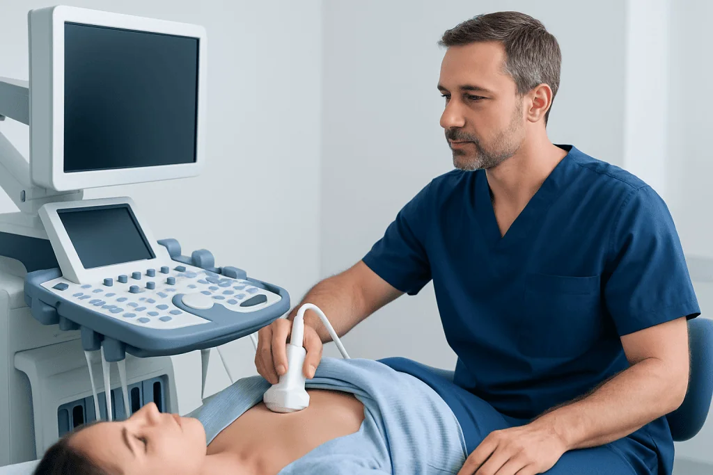
Abdominal ultrasound is an imaging test that uses high-frequency sound waves to create pictures of structures inside the abdomen. A handheld device called a transducer is moved over the skin after a small amount of gel is applied. The sound waves bounce off internal organs and are turned into images on a screen.
For people with digestive symptoms, this test is often one of the first imaging studies a doctor may request. It is widely used because it is noninvasive, does not use ionizing radiation, and can often be done quickly. It is especially helpful when symptoms suggest a problem involving the liver, gallbladder, bile ducts, pancreas, kidneys, or fluid in the abdomen.
Although it is called an abdominal ultrasound, it does not show every part of the digestive system equally well. Some organs and tissues are easier to see than others. The test is particularly useful for solid organs and for detecting stones or enlarged ducts, but it is less detailed for the inside lining of the stomach or intestines.
When it is used for digestive symptoms
Doctors may recommend abdominal ultrasound when a person has symptoms such as upper abdominal pain, right-sided abdominal pain, nausea, vomiting, bloating, unexplained fullness, jaundice, or abnormal liver blood tests. It may also be used when there is concern about gallstones, inflammation of the gallbladder, bile flow problems, liver enlargement, or fluid buildup.
In some situations, ultrasound is used to help evaluate a specific symptom pattern. For example, pain after fatty meals may raise concern for gallbladder disease, while yellowing of the skin or eyes may lead a doctor to look for bile duct blockage. It can also be useful in assessing abdominal swelling or checking whether an organ appears enlarged.
Ultrasound may be part of a broader digestive workup rather than a complete answer on its own. Depending on the symptoms, doctors may also consider blood tests, stool tests, endoscopy, or other imaging studies. If there is concern for conditions such as gallstones or fatty liver disease, ultrasound is often an appropriate first step.
What an abdominal ultrasound can show

One of the main strengths of abdominal ultrasound is its ability to assess the liver and biliary system. It can show whether the liver looks enlarged, whether there are changes consistent with fatty infiltration, and whether bile ducts are widened. It is also commonly used to detect gallstones and signs that may suggest gallbladder inflammation.
The pancreas may sometimes be partly visible, especially if there is not too much bowel gas blocking the view. Ultrasound can occasionally suggest swelling, a mass, or duct changes, but the pancreas is not always seen clearly. The spleen and kidneys are also usually evaluated during the same scan, which can help identify other causes of abdominal discomfort that may overlap with digestive symptoms.
Abdominal ultrasound can also detect free fluid in the abdomen, called ascites, and may identify some cysts, enlarged organs, or masses that need further assessment. Common findings may include:
- Gallstones or gallbladder sludge
- Widened bile ducts
- Fatty changes in the liver
- Liver cysts or some focal liver lesions
- Enlarged liver or spleen
- Fluid in the abdomen
- Kidney stones or kidney swelling that may mimic abdominal pain
If a more detailed look is needed, a doctor may recommend further imaging such as MRI scanning or CT scanning to clarify the finding.
What it may not show clearly
Abdominal ultrasound has important limitations, and understanding them can help set realistic expectations. It does not directly examine the inside lining of the esophagus, stomach, or colon the way endoscopy or colonoscopy can. It is also less reliable for structures hidden behind bone or obscured by bowel gas.
For this reason, ultrasound may not fully explain symptoms such as heartburn, difficulty swallowing, many causes of chronic diarrhea, or small ulcers in the stomach or small intestine. It also may not detect every pancreatic problem, especially if the pancreas cannot be seen well. A normal ultrasound does not always mean there is no digestive condition.
If symptoms point to a problem within the digestive tract itself, a doctor may advise other tests. These may include endoscopy for upper digestive symptoms or other targeted studies depending on the person’s age, history, examination, and blood test results.
How to prepare and what happens during the test
Preparation depends on why the ultrasound is being performed, but many abdominal ultrasounds require fasting for several hours beforehand. Fasting can reduce gas in the intestines and help the gallbladder fill, making it easier to evaluate. The imaging center or doctor will usually give specific instructions in advance.
During the test, the person typically lies on an examination bed while gel is placed on the abdomen. The sonographer moves the transducer across the skin and may ask the person to change position or hold their breath briefly. These steps can improve the view of certain organs.
The scan is usually painless, though there may be mild pressure if the abdomen is tender. Most appointments are relatively short, and people can usually return to their normal activities afterward unless told otherwise. The images are then reviewed by a radiologist or another qualified specialist, who sends a report to the referring doctor.
How doctors interpret the results
Ultrasound findings are interpreted in the context of the person’s symptoms, medical history, physical examination, and laboratory results. For example, a small liver cyst may be an incidental finding that is not causing symptoms, while a widened bile duct in someone with jaundice may require prompt follow-up.
Sometimes the report clearly identifies a likely cause of symptoms, such as gallstones or a fatty liver pattern. In other cases, the report may say that the exam is limited or that a finding is nonspecific. This does not necessarily mean something serious is present; it usually means the image alone cannot provide a final diagnosis.
Depending on the result, a doctor may recommend monitoring, lifestyle changes, medication, or further testing. In some cases, additional imaging or specialist evaluation is needed to confirm the cause. If treatment is required, options may range from diet-based management to procedures such as gallbladder surgery when clinically appropriate.
After the scan: next steps, self-care, and when to seek medical advice
After an abdominal ultrasound, most people can eat, drink, and resume their usual routine unless they have been told otherwise. The most important next step is to review the result with the doctor who ordered the test. That discussion helps connect the imaging findings with symptoms and decide whether any treatment or further evaluation is needed.
Self-care depends on the symptom pattern and the underlying cause. For some people, practical steps such as eating smaller meals, staying hydrated, limiting alcohol, avoiding foods that trigger pain, and following advice for weight management may support digestive health. These measures are helpful but should not replace medical assessment when symptoms are persistent or unexplained.
A person should seek medical advice sooner if abdominal pain is severe, symptoms are worsening, there is fever, repeated vomiting, black stools, blood in vomit or stool, fainting, unexplained weight loss, or yellowing of the skin or eyes. In these situations, timely evaluation is important. Near the end of the care pathway, some patients may choose centers such as Acibadem International, where multidisciplinary specialists in JCI-accredited hospitals evaluate digestive conditions for international patients.
Frequently asked questions
Can abdominal ultrasound diagnose the cause of stomach pain?
It can help identify some causes of abdominal pain, especially problems involving the gallbladder, liver, bile ducts, kidneys, or fluid in the abdomen. However, it may not show every cause of stomach or intestinal pain, so additional tests are sometimes needed.
Does abdominal ultrasound show the stomach and intestines well?
Not usually in detail. Ultrasound is less effective for looking at the inside lining of the stomach and intestines, especially when bowel gas blocks the view. Tests such as endoscopy or CT may be more informative for certain digestive symptoms.
Is abdominal ultrasound safe?
Yes. Abdominal ultrasound is generally considered very safe because it uses sound waves rather than radiation. It is commonly used in many age groups and is a routine part of evaluating abdominal symptoms.
Do patients need to fast before an abdominal ultrasound?
Often yes, but not always. Fasting for several hours may help improve image quality, particularly when the gallbladder or upper abdomen is being assessed. The exact instructions depend on the reason for the test.
Can an abdominal ultrasound detect gallstones?
Yes, it is one of the most common and useful tests for detecting gallstones. It can also show signs that suggest gallbladder inflammation or changes in the bile ducts.
What happens if the ultrasound result is normal but symptoms continue?
A normal result can be reassuring, but it does not rule out all digestive conditions. If symptoms persist, a doctor may recommend blood tests, stool tests, endoscopy, or other imaging based on the symptom pattern and medical history.
References
- World Health Organization
- National Institute of Diabetes and Digestive and Kidney Diseases
- American College of Radiology
- Radiological Society of North America
- National Health Service
This article is for general information only and is not a substitute for professional medical advice. Please consult a qualified doctor about your individual situation.
Ultrasound in Turkey — costs, top hospitals & a free quote
JCI-accredited · board-certified surgeons · reply within 24h
Add us as a Preferred Source to see more of our trusted health content across Google Search, AI Overviews and Discover.

Check-up & Preventive Medicine
Comprehensive health screenings designed for early detection and prevention.
1 specialists in this unitMore from the Health Library



Related Specialists

Dt. Sinem Uzuner
Oral & Dental Health
Prof. Dr. Vildan Ertekin
Pediatrics
Prof. Dr. Behice Kurtaran
Infectious Diseases & Clinical Microbiology
Asst. Prof. Dr. Harika Bodur Öztürk
Gynecology & Obstetrics