

Abdominal Ultrasound: What It Shows and When Doctors Use It

Abdominal ultrasound uses sound waves, not radiation, to create real-time images of organs and blood flow. It can show the liver, gallbladder, bile ducts, pancreas, spleen, kidneys, bladder, abdominal aorta and fluid in the abdomen.
Key Takeaways
- Abdominal ultrasound uses sound waves, not radiation, to create real-time images of organs and blood flow.
- It can show the liver, gallbladder, bile ducts, pancreas, spleen, kidneys, bladder, abdominal aorta and fluid in the abdomen.
- Doctors often request it for abdominal pain, suspected gallstones, liver or kidney problems, unexplained swelling, or follow-up of known conditions.
- Preparation may include fasting for several hours or arriving with a full bladder, depending on the organs being examined.
- Ultrasound is very safe, but results sometimes need to be clarified with CT, MRI, blood tests, endoscopy or a specialist consultation.
Abdominal ultrasound is a widely used imaging test that helps doctors view organs, blood vessels and some soft tissues inside the abdomen without radiation. It is commonly used to investigate pain, swelling, abnormal blood tests, gallstones, liver conditions and many other concerns.
Overview
Abdominal ultrasound, also called abdominal sonography, is a diagnostic imaging test that uses high-frequency sound waves to produce pictures of structures inside the belly. A small handheld device called a transducer is moved over the skin with the help of a clear gel. The transducer sends sound waves into the body and receives echoes that are converted into images on a screen.
Unlike X-rays or CT scans, ultrasound does not use ionizing radiation. It is non-invasive, generally painless and can provide real-time information about organ movement, fluid collections and blood flow when Doppler ultrasound is used. For these reasons, it is often one of the first tests doctors choose when evaluating abdominal symptoms.
An abdominal ultrasound may be performed in an outpatient imaging department, hospital, emergency setting or specialist clinic. The examination is usually completed within a short visit, although timing can vary depending on the reason for the test and whether additional Doppler measurements or focused views are needed.
What an Abdominal Ultrasound Can Show
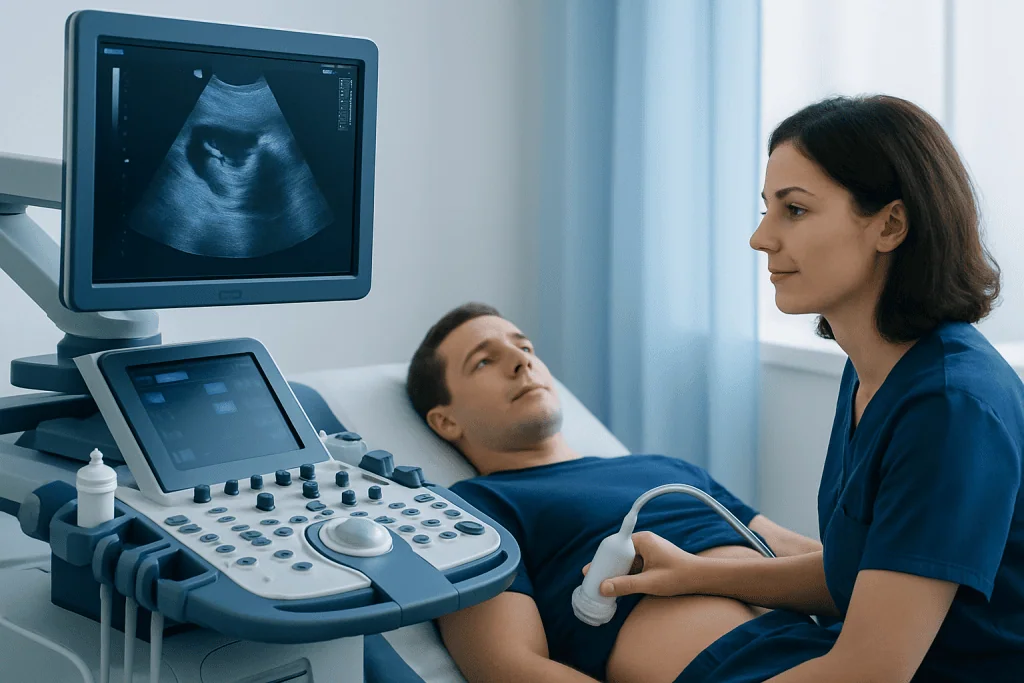
Abdominal ultrasound can show the size, shape, texture and position of several major organs. It is especially useful for evaluating the liver, gallbladder, bile ducts, spleen, kidneys, bladder and abdominal aorta. In many cases it can also assess parts of the pancreas, although bowel gas may sometimes limit the view.
Doctors may use ultrasound to look for gallstones, inflammation of the gallbladder, widening of the bile ducts, liver cysts, fatty liver changes, signs of cirrhosis, kidney stones, kidney swelling, kidney cysts, enlarged organs, abdominal fluid and some masses. Doppler ultrasound can assess blood flow in selected vessels, such as the portal vein, hepatic vessels, renal vessels or abdominal aorta.
Common findings that ultrasound may help identify include:
- Gallstones or sludge in the gallbladder
- Enlargement or texture changes in the liver or spleen
- Fluid in the abdomen, known as ascites
- Blocked urine flow causing kidney swelling
- Aneurysm or enlargement of the abdominal aorta
- Some cysts, abscesses, tumors or inflammatory changes
Ultrasound is helpful, but it does not show every abdominal condition with equal detail. The quality of the images can be affected by body habitus, bowel gas, recent meals and the location of the organ being examined. If ultrasound findings are unclear, the doctor may recommend further testing.
When Doctors Use Abdominal Ultrasound

Doctors request an abdominal ultrasound when symptoms, examination findings or laboratory results suggest a possible problem inside the abdomen. It is commonly used to investigate pain in the right upper abdomen, suspected gallbladder disease, abnormal liver enzymes, jaundice, unexplained swelling, urinary symptoms, blood in the urine or an enlarged abdominal organ felt during an exam.
In emergency or urgent care settings, ultrasound may help assess causes of acute abdominal pain or check for free fluid. It may also be used to monitor known conditions over time, such as liver disease, kidney cysts, gallbladder polyps or an abdominal aortic aneurysm. In some cases, ultrasound guides procedures such as draining fluid or obtaining a tissue sample, although these are separate interventional procedures performed under specific medical indications.
Abdominal ultrasound is often selected because it is accessible, quick and safe for repeated use when clinically appropriate. For example, a patient with recurring gallbladder-type pain may have an ultrasound to look for stones, while a patient with abnormal blood tests may have one to assess liver texture and bile duct size.
How to Prepare for the Test
Preparation depends on the area being examined. For an upper abdominal ultrasound that includes the liver, gallbladder or pancreas, patients are often asked not to eat or drink for several hours before the test. Fasting helps reduce gas and allows the gallbladder to fill, which can make it easier to see.
If the bladder or pelvic area is being assessed as part of the abdominal examination, the patient may be asked to drink water beforehand and avoid urinating until after the scan. A full bladder can create a better window for certain views. The imaging center or doctor’s office should provide specific instructions based on the test request.
Patients should bring previous imaging reports if available, along with a list of current medications and relevant medical history. Most regular medications can usually be taken as directed, but patients with diabetes, kidney disease or special dietary needs should ask their doctor how to manage fasting safely. Jewelry or tight clothing around the abdomen may need to be removed, and a gown may be provided.
What Happens During an Abdominal Ultrasound
During the scan, the patient usually lies on an examination table. The sonographer or doctor applies warm or room-temperature gel to the skin, then moves the transducer gently across the abdomen. The gel helps sound waves travel between the transducer and the body, improving image quality.
The examiner may ask the patient to change position, hold the breath briefly or take a deep breath. These simple steps can move organs into a better viewing position, especially the liver and gallbladder. Mild pressure from the transducer may be felt, particularly over a tender area, but the test should not be painful.
If Doppler ultrasound is included, the machine may produce sounds that represent blood flow. After the examination, the gel is wiped away and the patient can usually return to normal activities immediately, unless the doctor has given different instructions. A radiologist or appropriately trained physician interprets the images and sends a report to the referring doctor.
Understanding Results and Limitations
Ultrasound results are usually described in a written report. The report may state that the organs appear normal, or it may describe findings such as stones, cysts, fluid, organ enlargement, duct widening or changes in tissue appearance. The meaning of these findings depends on the patient’s symptoms, examination, blood tests and medical history.
Some ultrasound findings are incidental, meaning they are discovered while looking for something else and may not be causing symptoms. Others require follow-up imaging, blood tests or specialist evaluation. For example, a simple kidney cyst is often harmless, while a complex cyst may need further assessment. Similarly, fatty changes in the liver may prompt discussion about metabolic health and liver function tests.
It is important to understand that a normal ultrasound does not always rule out every cause of abdominal pain. Conditions affecting the stomach, intestines, appendix or very small structures may be difficult to evaluate with ultrasound alone. Depending on the situation, the doctor may recommend CT, MRI, endoscopy, colonoscopy, urine tests or blood tests to complete the assessment.
Safety, Benefits and Possible Risks
Diagnostic abdominal ultrasound is considered very safe when performed by trained professionals for appropriate medical reasons. It does not expose the patient to ionizing radiation, and it can be repeated when medically necessary. This makes it particularly useful for monitoring certain conditions over time.
The main benefits are that ultrasound is non-invasive, widely available, relatively quick and capable of showing real-time images. It can help guide clinical decisions, reduce uncertainty and identify conditions that may require treatment or monitoring. It is also useful for patients who cannot receive contrast dye used in some CT or MRI studies, although each imaging choice depends on the clinical question.
There are very few risks from the ultrasound itself. Some patients may feel temporary discomfort from pressure on a sore area, and fasting may be inconvenient or require planning for people with medical conditions such as diabetes. The most important limitation is that the test may not provide a complete answer in every case, so follow-up with the referring doctor remains essential.
When to See a Doctor
A doctor should be consulted if abdominal pain is persistent, worsening, recurrent or associated with other symptoms such as fever, vomiting, yellowing of the skin or eyes, unexplained weight loss, swelling of the abdomen, blood in the urine or changes in bowel habits. These symptoms do not always indicate a serious condition, but they deserve proper medical evaluation.
Patients who already have liver, kidney, gallbladder or vascular conditions should follow their doctor’s recommended imaging schedule. If an ultrasound report contains unclear or unexpected findings, the next step is to review the results with the referring physician, who can explain the significance and whether further testing is needed.
For international patients who need coordinated evaluation, Acibadem International’s multidisciplinary specialists and JCI-accredited hospitals diagnose and treat abdominal and digestive conditions using appropriate imaging and specialist consultation. Patients should always seek individualized advice from a qualified healthcare professional, as imaging decisions depend on symptoms, medical history and examination findings.
Frequently asked questions
Is an abdominal ultrasound painful?
Abdominal ultrasound is usually painless. The patient may feel mild pressure from the transducer, especially if the area is already tender, but this is generally brief and well tolerated. If discomfort occurs, the patient should tell the examiner.
How long does an abdominal ultrasound take?
Many abdominal ultrasound examinations take about 15 to 30 minutes, although the time can vary. More complex studies, Doppler measurements or difficult viewing conditions may take longer. The total appointment time may also include registration and preparation.
Can abdominal ultrasound detect cancer?
Ultrasound can sometimes detect masses or suspicious changes in abdominal organs, but it cannot always determine whether a finding is cancer. If a concerning area is seen, the doctor may recommend additional imaging such as CT or MRI, laboratory tests or a biopsy. Diagnosis depends on the full clinical evaluation.
Why do patients need to fast before some abdominal ultrasounds?
Fasting helps reduce bowel gas and allows the gallbladder to fill, which can improve the quality of the images. This is especially important when the liver, gallbladder, bile ducts or pancreas are being examined. Patients should follow the specific instructions provided by their doctor or imaging center.
Is abdominal ultrasound safe during pregnancy?
Ultrasound uses sound waves and does not use ionizing radiation. It is widely used in pregnancy when medically indicated, including for obstetric and some abdominal concerns. Pregnant patients should tell the healthcare team before any imaging test so the most appropriate examination can be selected.
What happens if the ultrasound result is normal but symptoms continue?
A normal abdominal ultrasound is reassuring, but it does not rule out every possible cause of symptoms. The doctor may review blood tests, urine tests, medications, diet and symptom patterns, and may recommend other imaging or procedures if needed. Persistent or worsening symptoms should be discussed with a qualified clinician.
References
- American College of Radiology
- Radiological Society of North America
- World Health Organization
- National Institute of Biomedical Imaging and Bioengineering
- European Society of Radiology
This article is for general information only and is not a substitute for professional medical advice. Please consult a qualified doctor about your individual situation.
Ultrasound in Turkey — costs, top hospitals & a free quote
JCI-accredited · board-certified surgeons · reply within 24h
Add us as a Preferred Source to see more of our trusted health content across Google Search, AI Overviews and Discover.

Check-up & Preventive Medicine
Comprehensive health screenings designed for early detection and prevention.
3 specialists in this unitMore from the Health Library



Related Specialists

Prof. Dr. Ercan Karaarslan
Radiology
Prof. Dr. Bora Peynircioğlu
Interventional Radiology
Prof. Dr. Koray Güven
Interventional Radiology
Assoc. Prof. Dr. Deniz Can Aliş
Radiology