Acral Lentiginous Melanoma: Early Signs, Risk Factors, and How It Is Treated

Acral lentiginous melanoma most often affects the palms, soles, and nail units. It can be mistaken for a bruise, wart, fungal infection, or minor injury.
Key Takeaways
- Acral lentiginous melanoma most often affects the palms, soles, and nail units.
- It can be mistaken for a bruise, wart, fungal infection, or minor injury.
- Diagnosis usually requires a skin examination and biopsy.
- Treatment depends on the stage and often includes surgery.
- Prompt medical review is important for any changing or unexplained lesion on the hands, feet, or nails.
Acral lentiginous melanoma is a type of skin cancer that appears on the palms, soles, or under the nails. Early signs can be subtle, so any new, changing, or non-healing dark spot in these areas should be checked by a qualified doctor.
Overview
Acral lentiginous melanoma is a form of melanoma that develops on acral skin, meaning the skin of the palms of the hands, soles of the feet, and the area under or around the nails. Although it is less common than other melanoma subtypes, it is clinically important because it can be harder to recognize early. The lesion may look like a persistent dark patch, a streak in the nail, or an area that seems bruised or irritated but does not resolve.
This type of melanoma is not defined by sun exposure in the same way as many other skin cancers. Because it occurs in places people may not examine closely, diagnosis is sometimes delayed. Early detection matters, as treatment is generally more straightforward and outcomes are usually better when the cancer is found before it has grown deeply or spread.
Acral lentiginous melanoma can affect people of any skin tone. It is especially important to understand that darker skin does not eliminate melanoma risk. Regular self-checks of the feet, palms, and nails can help people notice suspicious changes that deserve professional assessment.
Early signs and symptoms
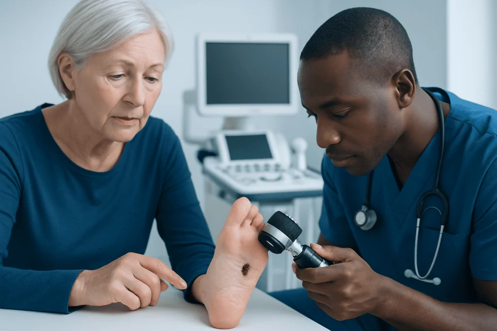
The earliest signs of acral lentiginous melanoma are often subtle. On the sole or palm, it may begin as a flat brown, black, gray, or mixed-color patch with an irregular border. Over time, it may enlarge, become darker, develop several shades, or start to thicken. Some lesions may ulcerate, bleed, or become tender, but pain is not always present.
When it affects the nail unit, it may appear as a dark vertical band running from the cuticle toward the nail tip. This is sometimes called subungual melanoma. Warning signs include a band that becomes wider, darker, or more uneven, pigment spreading onto the nearby skin, nail splitting, or a nail that becomes distorted without a clear cause.
These lesions can be confused with harmless conditions, especially in active people or those with repeated friction on the feet. Common look-alikes include bruising, warts, fungal nail changes, calluses, or a chronic wound. A lesion should be checked if it does not heal, keeps changing, or returns after seeming to improve.
- A new dark spot on the sole, palm, or around a nail
- A streak in the nail that changes in width or color
- Irregular edges or several colors within one lesion
- Bleeding, crusting, ulceration, or persistent tenderness
- A lesion mistaken for trauma that does not resolve over time
Causes and risk factors

The exact cause of acral lentiginous melanoma is not fully understood. Like other melanomas, it begins when pigment-producing cells called melanocytes grow abnormally. Unlike many melanomas on sun-exposed skin, this subtype is less clearly linked to ultraviolet exposure. That difference can make it less expected by patients and sometimes even less obvious in routine skin checks focused mainly on sun-exposed areas.
Researchers continue to study the genetic and molecular changes involved. What is known is that acral lentiginous melanoma can occur in people with no obvious risk factors and in areas that receive little sunlight. Repeated trauma has been discussed in research, but it is not considered a proven direct cause. More likely, injury draws attention to a lesion that was already present or makes an abnormal area easier to notice.
Risk rises with age, and a personal history of melanoma or certain other skin cancers may also increase concern for suspicious lesions. Even so, many people diagnosed with acral lentiginous melanoma do not fit a classic skin cancer profile. That is why unexplained changes on the feet, hands, or nails should not be ignored simply because a person has darker skin or limited sun exposure history.
How doctors diagnose it
Diagnosis starts with a careful medical history and skin examination. A dermatologist or other experienced clinician will ask when the lesion first appeared, whether it has changed, and whether there has been bleeding, pain, injury, or previous treatment. The doctor will also examine the lesion’s color pattern, border, symmetry, and location and may use dermoscopy, a magnifying tool that helps evaluate pigment structures in more detail.
If acral lentiginous melanoma is suspected, a biopsy is needed to confirm the diagnosis. This involves removing part or all of the lesion so a pathologist can examine the tissue under a microscope. A biopsy is the only reliable way to distinguish melanoma from other conditions such as a mole, wart, hematoma, or fungal nail problem. If the lesion is beneath a nail, sampling may involve the nail unit and should be performed by clinicians familiar with this area.
Once melanoma is confirmed, the pathology report helps guide treatment. Important details include tumor thickness, ulceration, and whether cancer cells are present at the edges of the sample. Some patients may need further staging tests depending on how advanced the tumor appears. In broader melanoma care, evaluation may be connected with melanoma pathways and imaging or lymph node assessment when clinically appropriate.
Treatment options
Treatment for acral lentiginous melanoma depends on the stage, location, tumor thickness, and whether the cancer has spread. Surgery is usually the main treatment for localized disease. The goal is to remove the melanoma completely with a margin of healthy tissue around it. Depending on the exact site, this can be challenging on the sole, palm, or nail unit, so planning aims to balance complete cancer removal with function and wound healing.
For some patients, doctors may recommend a sentinel lymph node biopsy to check whether melanoma cells have reached nearby lymph nodes. This is more often considered when the primary tumor is thicker or has other higher-risk features. If the disease is more advanced, treatment may also involve medical oncology approaches such as medical oncology care, which can include immunotherapy or targeted therapy when suitable.
In selected cases, surgical oncology teams and reconstructive specialists may work together, especially if the lesion is large or located in a weight-bearing part of the foot. Some patients with advanced or complex disease may also need cancer treatment planning across several specialties. At Acibadem International, multidisciplinary specialists in JCI-accredited hospitals diagnose and treat melanoma for international patients, with care plans tailored to the person’s stage and overall health.
Living with the diagnosis and self-care
After treatment, follow-up is an important part of care. Melanoma surveillance may include regular skin examinations, checks of the lymph nodes, and sometimes imaging depending on the original stage. Patients are often advised to monitor their own skin and nails between visits and to report any new or changing lesions promptly. Ongoing review helps detect recurrence early and also supports identification of other skin cancers.
Self-care also includes protecting the skin in general, even though acral lentiginous melanoma is not mainly sun-driven. Good foot and nail awareness is particularly useful. People can examine the soles, between the toes, palms, and nail beds regularly, using a mirror or asking for help if needed. Any pigmented streak under the nail or dark patch on the foot that does not match a clear injury should be assessed.
It can be emotionally difficult to receive a melanoma diagnosis, particularly when it was mistaken for a minor problem at first. Clear communication with the healthcare team, practical wound care instructions, and support from family or counseling services can all help during recovery. Patients should follow medical advice closely and ask before using over-the-counter treatments on suspicious or healing areas.
When to seek medical care
Medical care should be sought promptly for any dark or unusual lesion on the palms, soles, or nails that is new, changing, or not healing. This is especially important if the spot has irregular borders, more than one color, bleeding, crusting, nail distortion, or pigment spreading onto the surrounding skin. Persistent lesions should not be repeatedly treated as a bruise, fungal infection, or wart without a proper examination.
Urgent assessment is also appropriate if a known lesion starts growing more quickly, becomes painful, or begins to ulcerate. People with a history of melanoma, many atypical moles, or previous skin cancer should have a lower threshold for arranging review. A doctor can decide whether specialist referral, biopsy, or additional tests are needed.
While not every dark mark on the foot or nail is cancer, it is safer to have uncertain lesions examined than to wait for them to declare themselves. Early assessment supports early diagnosis, and early diagnosis offers the best chance of simpler treatment and better long-term control.
Frequently asked questions
What is acral lentiginous melanoma?
Acral lentiginous melanoma is a subtype of melanoma that develops on the palms, soles, or nail unit. It is different from many other melanomas because it often appears in areas that receive little sun exposure.
What does acral lentiginous melanoma look like?
It may look like a dark patch with uneven borders on the sole or palm, or a brown-to-black streak under a nail. The color can be irregular, and the lesion may gradually enlarge, thicken, bleed, or fail to heal.
Is acral lentiginous melanoma caused by sun exposure?
It is not as strongly linked to ultraviolet exposure as many other melanomas. Even so, the exact causes are not fully understood, and anyone with a suspicious lesion should seek medical evaluation.
How is acral lentiginous melanoma diagnosed?
Diagnosis requires a clinical examination and a biopsy. The biopsy allows a pathologist to confirm whether melanoma is present and provides details that help guide treatment.
Can acral lentiginous melanoma be mistaken for something else?
Yes. It is often confused with bruises, warts, fungal nail disease, calluses, or minor trauma, especially on the feet. A spot that persists or changes despite simple care should be checked by a doctor.
How is acral lentiginous melanoma treated?
Treatment usually begins with surgery to remove the melanoma completely. Depending on the stage, some patients also need lymph node assessment, immunotherapy, targeted therapy, or coordinated oncology care.
References
- American Academy of Dermatology
- National Cancer Institute
- American Cancer Society
- National Comprehensive Cancer Network
- World Health Organization
This article is for general information only and is not a substitute for professional medical advice. Please consult a qualified doctor about your individual situation.
Melanoma in Turkey — costs, top hospitals & a free quote
JCI-accredited · board-certified surgeons · reply within 24h
Add us as a Preferred Source to see more of our trusted health content across Google Search, AI Overviews and Discover.

Medical Oncology Department
Medical treatment of cancer with chemotherapy, immunotherapy and targeted therapies under a multidisciplinary tumor board.
60 specialists in this unitMore from the Health Library



Related Specialists

Dr. Nu Nu Win
Radiology
Assoc. Prof. Dr. Bahadir Osman Bozkirli
General Surgery
Dr. Hudheifa Yussuf Haji
Inpatient Clinic Physicians Clinical Service
Prof. Dr. Gül Esen İçten
Breast Clinic Clinical Service