Adem MRI: Preparation, Procedure and Results
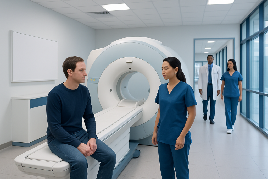
ADEM MRI commonly shows multiple areas of inflammation in the brain’s white matter and may also show spinal cord involvement. MRI does not diagnose ADEM on its own; clinicians interpret it together with symptoms, timing, blood tests and sometimes spinal fluid tests.
Key Takeaways
- ADEM MRI commonly shows multiple areas of inflammation in the brain’s white matter and may also show spinal cord involvement.
- MRI does not diagnose ADEM on its own; clinicians interpret it together with symptoms, timing, blood tests and sometimes spinal fluid tests.
- The scan is painless and does not use ionizing radiation, but contrast dye may be recommended for more detailed images.
- Many people, especially children, recover well from ADEM, although follow-up is important to monitor symptoms and MRI changes.
- New or rapidly worsening weakness, confusion, seizures, vision changes or difficulty walking needs urgent medical assessment.
An ADEM MRI is an MRI scan of the brain and sometimes the spinal cord used to look for inflammation and changes in myelin, the protective covering around nerves. It is a key part of assessing acute disseminated encephalomyelitis (ADEM), alongside symptoms, neurological examination and laboratory tests.
ADEM MRI: What It Is and Why It Is Used
An ADEM MRI is a magnetic resonance imaging scan performed when acute disseminated encephalomyelitis (ADEM) is suspected. ADEM is an immune-mediated inflammatory condition that affects the brain and sometimes the spinal cord. MRI uses strong magnets and radio waves to produce detailed pictures of nervous system tissues, without using X-rays.
The scan helps clinicians identify areas where inflammation has affected myelin, the insulating layer around nerve fibers. It can also help rule out other possible explanations for neurological symptoms, including infection, stroke, tumors and other inflammatory conditions. A brain MRI is usually requested first, while an MRI of the spine may be added when there is weakness, numbness, bladder changes or other symptoms suggesting spinal cord involvement.
ADEM is a neurological disease because it involves the central nervous system: the brain, spinal cord and optic nerves. It often develops after a viral illness, though not every person has a clear preceding infection. MRI findings are important, but they are only one part of the assessment for acute disseminated encephalomyelitis (ADEM).
How an ADEM MRI Works
MRI measures signals from hydrogen atoms in the body after they are influenced by a powerful magnetic field and radiofrequency pulses. A computer converts these signals into cross-sectional images. Different MRI sequences highlight different tissue features, helping radiologists distinguish inflammation, swelling, older tissue changes and active lesions.
In ADEM, radiologists commonly look for broad, poorly defined areas of increased signal on T2-weighted and FLAIR images. These often involve the brain’s white matter, but may also appear in deeper brain structures, the brainstem, cerebellum or spinal cord. The appearance, location and timing of lesions can help the neurology team consider ADEM in the appropriate clinical setting.
A contrast agent containing gadolinium may be given through a small vein cannula when it is clinically appropriate. Contrast can show areas where inflammation has temporarily altered the blood-brain barrier. Not everyone needs contrast, and the imaging team will review kidney health, prior contrast reactions, pregnancy status and other relevant factors before its use.
Who May Need an ADEM MRI and How to Prepare

An ADEM MRI may be recommended for a person who develops sudden or subacute neurological symptoms, particularly after a recent infection. Possible symptoms include confusion or unusual sleepiness, headache, fever, weakness, balance problems, numbness, visual disturbance, seizures or difficulty speaking. In children, changes in alertness or behavior can be especially important clues.
Preparation is usually straightforward. Patients should tell the MRI team about implanted devices, previous surgery, metal fragments from work or injury, hearing implants, medication patches, pregnancy, kidney disease and any history of contrast reaction. Many modern implants can be scanned safely, but their exact type and MRI compatibility must be checked before the appointment.
Most patients can eat, drink and take usual medicines unless the care team gives different instructions. Comfortable clothing without metal fasteners is helpful, although a gown may be provided. People with claustrophobia, significant pain or difficulty staying still should mention this in advance; practical support, a wider scanner where available, or clinician-prescribed sedation may be considered.
- Bring prior MRI scans or reports if they were performed elsewhere.
- Remove jewelry, watches, hearing aids, hairpins and other metal items before entering the scan room.
- Arrange an accompanying adult if sedation is planned, as driving afterward may not be safe.
ADEM MRI Procedure: Step by Step
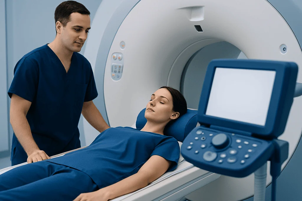
Before the scan, a radiographer reviews the safety checklist and explains what will happen. If contrast is planned, a small intravenous line is placed in the hand or arm. The patient lies on a padded table, usually on their back, with a head support or head coil placed around the head to improve image quality.
The table moves slowly into the MRI scanner. The machine makes loud tapping and knocking sounds during image acquisition, so earplugs or headphones are provided. The patient can communicate with the radiographer through an intercom and is usually given an alarm button to use if they need assistance. Remaining as still as possible is important because movement can blur the images.
The duration varies according to whether the brain alone or both brain and spine are scanned, and whether contrast is needed. A brain MRI commonly takes between 30 and 60 minutes, while combined studies can take longer. The patient does not feel the magnetic field; however, some people find the noise or enclosed space uncomfortable.
After images are obtained, a neuroradiologist or radiologist reviews them and sends a report to the referring clinician. If ADEM is strongly suspected or symptoms are severe, the neurology team may act on preliminary findings while the full report and additional test results are being completed.
Understanding ADEM MRI Results
A typical ADEM MRI may show multiple areas of inflammation, often affecting both sides of the brain but not always in a perfectly symmetrical pattern. Lesions can be large and may involve white matter, deep gray matter, the brainstem or cerebellum. Some people also have abnormalities in the spinal cord. The pattern can be different from person to person, and a normal early MRI does not always exclude an inflammatory neurological condition.
What does encephalopathy look like on MRI? Encephalopathy describes altered brain function, such as confusion, reduced alertness or behavior change, rather than one specific MRI appearance. In ADEM, encephalopathy may occur alongside inflammatory lesions visible on MRI; however, some causes of encephalopathy have normal or nonspecific MRI findings. The scan must therefore be interpreted with the clinical examination, blood tests, infection testing and, in selected cases, cerebrospinal fluid analysis.
Radiologists also assess whether lesions enhance after contrast, whether there is swelling, and whether the distribution resembles another condition. Follow-up MRI may be arranged weeks or months later to check whether inflammatory areas have improved and to look for any new changes. This comparison can support diagnosis and guide ongoing care.
Because symptoms and scans may overlap with other disorders, clinicians may consider conditions such as multiple sclerosis and other forms of central nervous system inflammation. An experienced neurology and neuroradiology review is especially valuable when the diagnosis remains uncertain.
Benefits, Risks and Recovery After an ADEM MRI
The main benefit of an ADEM MRI is detailed, noninvasive imaging of the brain and spinal cord. It can show the extent and location of inflammation, help exclude alternative causes of symptoms and provide a baseline for future comparison. MRI does not expose patients to ionizing radiation, making it particularly useful when repeat imaging is needed.
For most people, the scan itself has no recovery period. They can return to normal activities immediately after a standard MRI. If sedation was used, they should follow the discharge instructions, avoid driving or operating machinery for the recommended period, and have someone accompany them home.
MRI is generally very safe when screening procedures are followed. Possible issues include anxiety in confined spaces, temporary discomfort from lying still, noise exposure without proper hearing protection, and rare reactions to contrast. Gadolinium contrast is used carefully, particularly in people with severe kidney impairment. The imaging team is prepared to manage unexpected reactions.
Recovery from ADEM refers to recovery from the neurological illness rather than from the MRI scan. Treatment may include hospital monitoring and medicines to reduce inflammation, followed by rehabilitation support when needed. Depending on symptoms, care may include neurological rehabilitation to support mobility, daily activities, cognition or speech.
Does ADEM Turn Into MS?
ADEM and multiple sclerosis (MS) are different inflammatory conditions, although they can sometimes look similar at first. Most people diagnosed with ADEM do not go on to develop MS. ADEM is generally a single episode of widespread inflammation, while MS is characterized by inflammatory episodes or new MRI lesions occurring over time.
Doctors do not rely on one scan alone to make this distinction. They consider the person’s age, symptoms, presence of encephalopathy, MRI lesion pattern, spinal fluid findings and whether new symptoms or lesions develop on follow-up. Follow-up with a neurologist is important when there are features that make the diagnosis less clear.
If recurrent neurological symptoms occur after apparent recovery, prompt reassessment is needed. This does not automatically mean MS, but it helps the clinical team determine whether another inflammatory or neurological condition should be investigated.
Can Someone Recover From ADEM? When to Seek Medical Care
Yes, many people recover substantially or completely from ADEM, particularly with timely assessment and treatment. Recovery can take days, weeks or months depending on the severity of inflammation and the symptoms involved. Some people experience lingering fatigue, weakness, attention difficulties, mood changes or balance problems, which may improve gradually with rehabilitation and follow-up care.
Medical follow-up may include repeat neurological examinations, MRI monitoring and support from physiotherapy, occupational therapy, speech and language therapy, psychology or pediatric specialists when appropriate. Treatment plans are individualized and may include neurology care for diagnosis, monitoring and management of inflammatory nervous system conditions.
Urgent medical care is needed for new or worsening confusion, loss of consciousness, seizures, severe headache, vision loss, new weakness, trouble walking, difficulty speaking, loss of bladder or bowel control, or breathing problems. These symptoms can have several causes and should be assessed without delay rather than managed at home.
Acibadem International’s multidisciplinary specialists and JCI-accredited hospitals provide assessment and treatment planning for international patients with complex neurological conditions, including suspected ADEM.
Frequently asked questions
What is an ADEM MRI?
An ADEM MRI is an MRI scan of the brain, and sometimes the spinal cord, performed to look for inflammation associated with acute disseminated encephalomyelitis. It helps clinicians assess the location and extent of changes in the central nervous system and exclude other possible causes of symptoms.
Does ADEM turn into MS?
Most people with ADEM do not develop multiple sclerosis. However, ADEM and MS can overlap in their early features, so neurologists may recommend follow-up examinations and MRI scans to clarify the diagnosis over time.
Can someone recover from ADEM?
Many people recover well from ADEM, especially children, although the pace and completeness of recovery vary. Some symptoms can persist for a period and may benefit from rehabilitation, neurological follow-up and supportive care.
Is ADEM a neurological disease?
Yes. ADEM is a neurological disease because it causes inflammation in the central nervous system, which includes the brain and spinal cord. It can affect movement, sensation, vision, balance, thinking and alertness.
What does encephalopathy look like on MRI?
Encephalopathy does not have one single MRI appearance because it is a clinical term for altered brain function. In ADEM, MRI may show inflammatory lesions in the brain, but some forms of encephalopathy can have normal or nonspecific imaging findings.
How long does an ADEM MRI take?
A brain MRI often takes about 30 to 60 minutes, although the exact time depends on the imaging protocol and whether contrast is used. An MRI that includes the spine can take longer.
References
- National Institute of Neurological Disorders and Stroke
- American Academy of Neurology
- Radiological Society of North America
- National Multiple Sclerosis Society
- Mayo Clinic
This article is for general information only and is not a substitute for professional medical advice. Please consult a qualified doctor about your individual situation.
Joint and spine care in Turkey — expert assessment & treatment
JCI-accredited · board-certified surgeons · reply within 24h
Add us as a Preferred Source to see more of our trusted health content across Google Search, AI Overviews and Discover.