How an Arthrogram Shows the Inside of a Joint
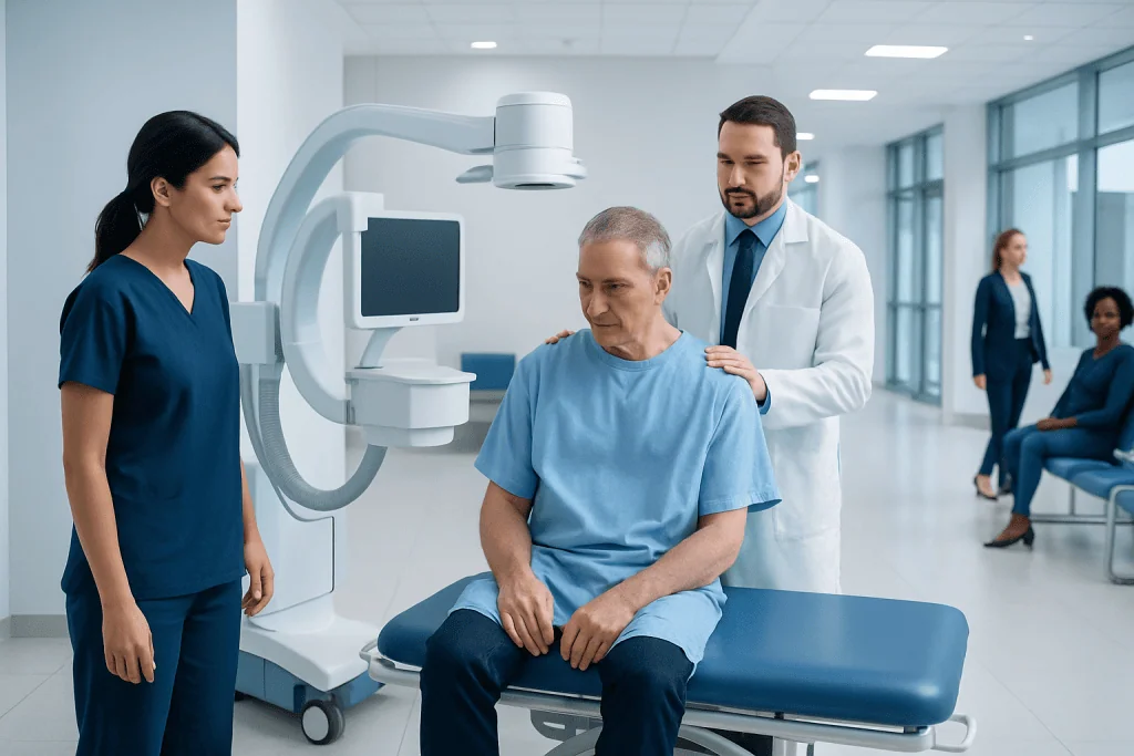
An arthrogram involves injecting contrast dye into a joint, then imaging it with MRI, CT, or X-ray to show cartilage, ligaments, and the labrum in more detail. It is usually recommended when joint pain, clicking, or instability is not explained by a physical exam or routine X-rays. It is an outpatient procedure done under local anesthetic.
Key Takeaways
- An arthrogram helps doctors see joint structures in greater detail than standard imaging alone.
- It may be performed on the shoulder, knee, hip, wrist, elbow, or ankle.
- The test usually involves injecting contrast dye into the joint, followed by X-ray, CT, or MRI imaging.
- Most people can go home the same day and resume light activity soon after the procedure.
- Mild temporary soreness is common, but worsening pain, fever, or significant swelling should be assessed by a doctor.
Your shoulder clicks every time you reach overhead, or your knee catches when you stand up — and the X-ray came back looking fine. That gap between what you feel and what the pictures show is exactly where an arthrogram comes in.
An arthrogram is an imaging test that uses contrast dye placed inside the joint so structures like cartilage, ligaments, and the joint lining show up more clearly. Doctors turn to it when standard imaging doesn’t fully explain joint pain, stiffness, clicking, or a sense that the joint isn’t stable.
Overview: what an arthrogram is
An arthrogram is a diagnostic imaging test used to look closely at a joint. During the procedure, a radiologist or other trained specialist injects contrast dye into the joint space. The contrast outlines the inside of the joint, making structures such as cartilage, ligaments, the joint capsule, and labrum easier to evaluate on imaging.
This test is often recommended when a person has joint symptoms that are not fully explained by a physical exam or routine X-rays. It can help identify tears, inflammation, looseness in the joint, cartilage injury, or other problems that may not be as visible on standard imaging alone. The test may be combined with fluoroscopy, CT, or MRI, depending on the question the doctor needs answered.
Arthrograms are commonly performed in the shoulder, knee, hip, wrist, elbow, and ankle. In many cases, the goal is to clarify whether symptoms are related to a sports injury, overuse, a degenerative condition, or a structural abnormality. It’s a focused test: it answers specific questions about one joint rather than screening your whole body.
Why doctors recommend an arthrogram
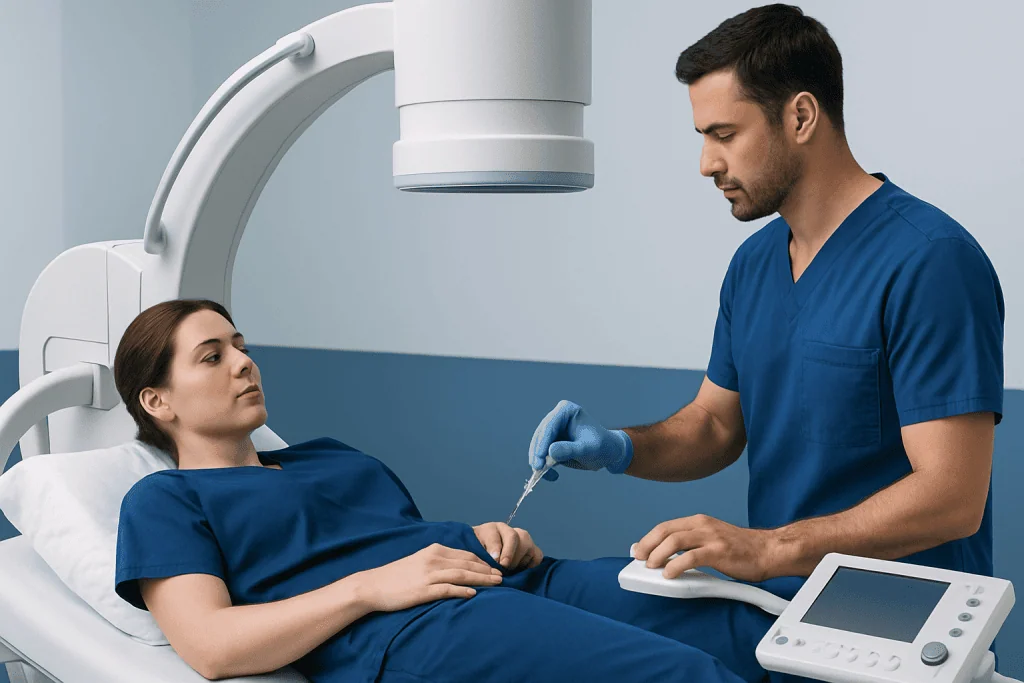
Doctors may suggest an arthrogram when joint pain, catching, locking, weakness, or instability continues despite rest and initial evaluation. The test is particularly useful when there is concern about soft-tissue structures inside the joint. These structures can be difficult to assess on plain X-rays because X-rays mainly show bone.
For example, an arthrogram may help detect a shoulder labral tear, subtle cartilage damage in the hip, or a ligament-related problem in the wrist. In some situations, MRI without contrast is enough. In others, the added contrast inside the joint improves detail and gives the care team clearer information for planning treatment.
It may also be used after a previous injury or surgery if symptoms return or healing is uncertain. If a person has ongoing joint pain, the doctor may also consider related conditions such as osteoarthritis or a meniscus problem. The choice of test depends on the joint involved, the symptoms, age, activity level, and whether surgery is being considered.
- Unexplained joint pain
- Clicking, catching, or locking
- Suspected cartilage or labral tears
- Joint instability or repeated dislocation
- Persistent symptoms after injury
How the arthrogram procedure is performed
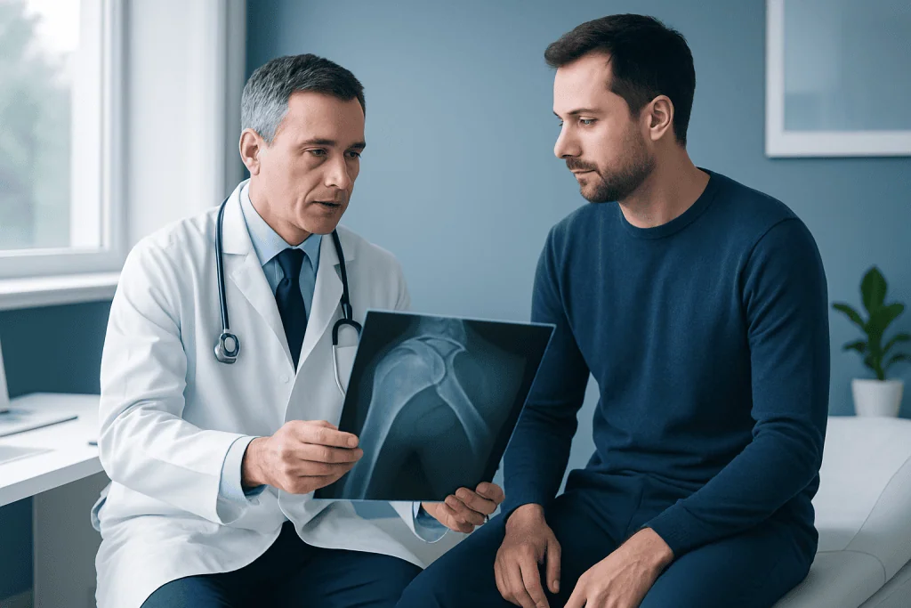
Before the test, the medical team usually reviews the person’s symptoms, medications, allergies, and any possibility of pregnancy. The skin over the joint is cleaned carefully to reduce the risk of infection. A local anesthetic is then used to numb the area, so the injection is more comfortable.
Using imaging guidance, often fluoroscopy or ultrasound, the doctor places a small needle into the joint space and injects contrast dye. Some people feel pressure, fullness, or a brief stinging sensation, but the procedure is generally well tolerated. Once the contrast is in place, images are taken right away using X-ray, CT, or most commonly MRI, depending on what the doctor needs to see.
The entire visit may take longer than the injection itself because positioning, image planning, and follow-up scanning are part of the process. If MRI is used after the injection, patients then move to the MRI scanner for additional imaging. In centers offering advanced MRI scanning, this combination can provide detailed information about internal joint structures without the need for immediate surgery.
After the images are completed, the person is usually observed briefly and then allowed to go home. Most arthrograms are outpatient procedures. Written aftercare instructions are typically provided, including guidance about activity, soreness, and when to call a doctor.
What an arthrogram can show
An arthrogram can reveal problems that may be difficult to identify with routine imaging alone. It is especially helpful for showing tears or defects in soft tissues that line or stabilize a joint. In the shoulder, for instance, it may help identify labral injury or damage related to repeated dislocation. In the knee, it may add information about cartilage surfaces or post-surgical changes.
In the hip, an arthrogram can help assess the labrum and cartilage when symptoms suggest a structural problem. In the wrist or elbow, it may help detect small ligament injuries that affect stability and cause pain with movement. Results are interpreted in the context of symptoms and examination, because not every imaging finding explains the person’s discomfort.
The test can support decisions about conservative care, physical therapy, injections, or surgery. Depending on the findings, treatment may involve specialist evaluation in areas such as orthopedics or, when severe wear-and-tear damage is present, discussion of options including knee replacement. An arthrogram doesn’t treat anything. It simply tells your team, with more confidence, what the next best step should be.
Preparing for the test and recovering afterward
Preparation is usually simple. Patients are often able to eat and drink normally unless they are given different instructions. It is important to tell the care team about allergies, especially prior reactions to contrast material, bleeding disorders, use of blood thinners, infection near the joint, or any implanted devices if MRI will be performed afterward.
After the arthrogram, mild soreness, a sense of fullness in the joint, or temporary discomfort with movement can happen for a day or two. Some people find it helpful to rest the joint, use ice, and avoid strenuous activity for a short time. The exact advice may vary depending on the joint involved and whether MRI or CT followed the injection.
Most people can return to routine daily activities fairly quickly, but heavy lifting, intense exercise, or sports may need to wait briefly. If a sedating medication was used, the person may need someone else to drive them home, though many arthrograms are done with local anesthetic only. The care team will explain when results are expected and who will review them.
People who are already being assessed for related issues, such as meniscus tear, may find that the arthrogram helps confirm whether symptoms match the suspected diagnosis. Usually the test is one step in a bigger plan, not the whole answer on its own.
Risks, side effects, and limitations
Arthrograms are safe for most people, though no procedure is completely without risk. Mild temporary discomfort at the injection site is the most common side effect. There may also be short-lived swelling, bruising, or a popping sensation in the joint as the contrast disperses.
Less commonly, people can have an allergic reaction to the contrast material or develop infection or bleeding in the joint. These complications are uncommon, especially when the procedure is performed using sterile technique and proper imaging guidance. Patients should always tell the medical team about previous contrast reactions, medications, or conditions that may increase risk.
An arthrogram also has limitations. It does not replace a full clinical assessment, and imaging findings do not always explain symptoms completely. In some cases, another imaging study, follow-up examination, or referral to a specialist is still needed. You get the most out of an arthrogram when it’s ordered for a clear reason and read alongside your history and examination.
When to seek medical care
Medical advice should be sought if joint pain is persistent, worsening, or limiting daily activities, exercise, work, or sleep. A person should also speak with a doctor if the joint repeatedly gives way, locks, clicks painfully, or becomes difficult to move after an injury. These symptoms do not always require an arthrogram, but they do deserve proper assessment.
After an arthrogram, prompt medical attention is important if there is fever, increasing redness, significant swelling, drainage from the injection site, severe pain that does not improve, or numbness that persists. Don’t wait these out — they can point to a complication.
If a joint condition needs specialist evaluation, multidisciplinary teams can help determine whether imaging, therapy, medication, or surgery is most appropriate. Acibadem International’s multidisciplinary specialists and JCI-accredited hospitals diagnose and treat joint conditions for international patients, including advanced imaging and orthopedic care when needed.
Frequently asked questions
Is an arthrogram painful?
Most people feel some pressure or brief discomfort when the needle and contrast are placed into the joint, but local anesthetic is usually used to numb the area. Mild soreness for a day or two afterward is common and usually manageable with rest and simple aftercare.
How long does an arthrogram take?
The injection part of the procedure is often fairly short, but the full appointment can take longer because of preparation, imaging guidance, and follow-up scans such as MRI or CT. The exact timing depends on which joint is being examined and which imaging method is used.
What is the difference between an MRI and an arthrogram?
An MRI creates detailed images using magnetic fields and radio waves, while an arthrogram involves placing contrast dye into the joint first to improve visualization of certain structures. Some arthrograms are followed by MRI, which combines both techniques for more detailed joint assessment.
Do patients need to rest after an arthrogram?
Light activity is usually possible soon after the test, but strenuous exercise and heavy use of the joint may need to be avoided briefly. The care team will give specific instructions based on the joint examined and the person’s overall condition.
Why would a doctor order an arthrogram instead of a regular X-ray?
A regular X-ray is very good for showing bones, but it does not show soft tissues inside the joint as clearly. An arthrogram can provide more information about cartilage, ligaments, the labrum, and the joint lining when these are the suspected source of symptoms.
Can someone drive home after an arthrogram?
Many people can drive home if only local anesthetic was used and they feel comfortable moving the joint. If sedating medication was given, or if the joint is too sore to use safely, another driver may be needed.
This article is for general information only and is not a substitute for professional medical advice. Please consult a qualified doctor about your individual situation.
Joint and spine care in Turkey — expert assessment & treatment
7 JCI-accredited hospitals · board-certified surgeons · reply within 24h
Add us as a Preferred Source to see more of our trusted health content across Google Search, AI Overviews and Discover.

Medically reviewed by the Acıbadem International Medical Board — August 24, 2026
See our medical review board →
Update history
- PublishedAugust 6, 2026
- Medical review approvedAugust 24, 2026
- Last content updateAugust 24, 2026
References2
- Arthrogram – MedlinePlus — medlineplus.gov
- Osteoarthritis – MedlinePlus — medlineplus.gov
Radiology
Advanced imaging and image-guided interventions supporting diagnosis across every specialty.
124 specialists in this unitRadiology Specialists at Acibadem





More from the Health Library
Private mri scan Risks & Side Effects Explained by Surgeons

Radioactive Iodine Pills Treat Thyroid Disease, Not Oral Cancer
Liver Ultrasound: Uses, Preparation and Results

Radiation Precautions After Pluvicto Prostate Cancer Therapy

PSMA Therapy Delivers Radiation to Prostate Cancer Cells