

Biopsy: Types, Image Guidance, and What Pathology Results Mean

A biopsy helps doctors make a tissue diagnosis when imaging, blood tests, or examination cannot provide enough certainty. Biopsies can be done with a needle, endoscope, surgical technique, skin punch, or image guidance such as ultrasound, CT, MRI, or mammography.
Key Takeaways
- A biopsy helps doctors make a tissue diagnosis when imaging, blood tests, or examination cannot provide enough certainty.
- Biopsies can be done with a needle, endoscope, surgical technique, skin punch, or image guidance such as ultrasound, CT, MRI, or mammography.
- Most biopsies are performed with local anesthesia and are planned to collect the safest and most useful sample possible.
- Pathology reports may include terms such as benign, malignant, atypical, inflammatory, grade, margins, and special test results.
- Results should always be reviewed with a qualified doctor, because meaning and next steps depend on the patient’s full clinical picture.
A biopsy is a procedure that removes a small sample of tissue or cells so a pathologist can examine it under a microscope and often perform additional tests. It is one of the most reliable ways to understand whether a suspicious area is benign, inflammatory, precancerous, or cancerous.
Overview
A biopsy is a medical procedure in which a doctor removes a small piece of tissue, a group of cells, or fluid from the body for laboratory examination. The sample is sent to a pathology laboratory, where a specialist doctor called a pathologist studies it under a microscope. In many cases, the pathologist can also use special stains, immunohistochemistry, genetic testing, or molecular tests to gain more detail.
Doctors recommend a biopsy when they need a clearer answer than imaging or physical examination can provide. For example, a scan may show a lump, thickening, enlarged lymph node, or abnormal area, but imaging alone cannot always confirm exactly what it is. A biopsy can help determine whether the finding is benign, due to infection or inflammation, precancerous, or cancerous.
Although the word biopsy can sound worrying, it is a common diagnostic tool used across many medical specialties. Many biopsies show non-cancerous conditions. When a serious condition is found, the biopsy provides information that helps doctors choose the most appropriate treatment plan.
Why a Biopsy May Be Recommended
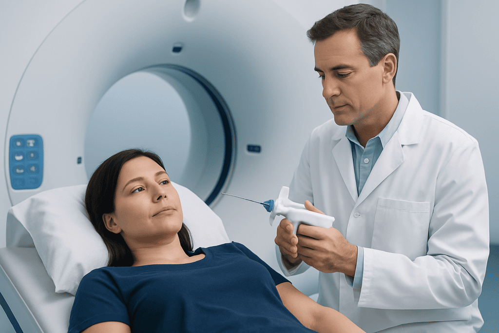
A biopsy may be advised after an abnormal imaging test, such as an ultrasound, mammogram, CT scan, MRI, PET scan, or X-ray. It may also be needed when a doctor feels a lump during examination, sees an unusual skin lesion, finds abnormal cells on a screening test, or needs to investigate persistent symptoms. The goal is to obtain enough tissue to answer a specific clinical question.
Common reasons for biopsy include checking a breast lump, thyroid nodule, prostate abnormality, enlarged lymph node, lung nodule, liver lesion, gastrointestinal growth, skin mole, bone lesion, or uterine lining change. Biopsies are also used to diagnose inflammatory or autoimmune conditions, infections, and organ diseases, such as kidney, liver, or muscle disorders.
A biopsy is usually recommended only when the expected benefit is greater than the small risks of the procedure. Before the biopsy, the medical team considers the location of the abnormality, the safest path to reach it, the patient’s medical history, medications, bleeding risk, and whether image guidance would improve accuracy.
Common Types of Biopsy

There are several biopsy techniques, and the best choice depends on the body area, the size of the abnormality, and the amount of tissue needed. Some biopsies remove only a few cells, while others remove a core of tissue or the entire suspicious area. The doctor will explain which method is planned and why.
- Fine needle aspiration: A very thin needle removes cells or fluid. It is often used for thyroid nodules, lymph nodes, cysts, or superficial lumps.
- Core needle biopsy: A slightly larger hollow needle removes small cylinders of tissue. This is commonly used for breast, liver, kidney, prostate, lymph node, and soft tissue lesions.
- Vacuum-assisted biopsy: A specialized needle device collects multiple tissue samples, often in breast diagnosis, and may reduce the need for repeated needle passes.
- Endoscopic biopsy: Tissue is taken through a flexible camera scope, such as during gastroscopy, colonoscopy, bronchoscopy, or cystoscopy.
- Skin biopsy: A shave, punch, or excisional technique removes part or all of a skin lesion.
- Surgical biopsy: An incision is made to remove part of a lesion or the entire abnormal area when a needle biopsy is not suitable or more tissue is required.
Some biopsies are performed in an outpatient clinic, while others take place in an imaging suite, endoscopy unit, operating room, or interventional radiology department. Anesthesia may be local, sedation-based, or general, depending on the procedure and the patient’s needs.
Image-Guided Biopsy: Ultrasound, CT, MRI, and Mammography
Image-guided biopsy means the doctor uses real-time or planned imaging to direct the needle to the correct spot. This is especially helpful when the abnormal area is small, deep inside the body, close to important structures, or not easy to feel by hand. Image guidance can improve sampling accuracy and help avoid blood vessels, nerves, bowel, or other organs.
Ultrasound-guided biopsy is widely used because it provides live images, involves no ionizing radiation, and is useful for many soft tissue, breast, thyroid, liver, kidney, and lymph node biopsies. CT-guided biopsy is helpful for deeper areas such as lung nodules, pelvic lesions, bone lesions, or abdominal masses. MRI-guided biopsy may be used for abnormalities best seen on MRI, particularly in selected breast, prostate, or soft tissue cases.
Mammography-guided, or stereotactic, biopsy is often used for breast calcifications or small findings visible on mammograms but not clearly seen on ultrasound. In some cases, a tiny marker clip may be placed at the biopsy site so the area can be identified on future imaging or during surgery if needed.
During image-guided biopsy, the skin is cleaned, local anesthetic is given, and the doctor advances the needle while monitoring its position. The patient may be asked to hold still or briefly hold their breath. After sampling, pressure and a small dressing are usually applied, and the patient receives instructions about activity, wound care, and signs to watch for.
What to Expect Before, During, and After
Before a biopsy, patients are usually asked about allergies, pregnancy possibility, bleeding disorders, previous reactions to anesthesia, and current medications. Blood thinners, antiplatelet medicines, and some supplements may need special instructions, but patients should never stop prescribed medication without guidance from the doctor who manages it. Some biopsies require fasting, blood tests, or a companion to drive the patient home if sedation is used.
During the procedure, the medical team will aim to keep the patient comfortable and informed. Local anesthetic may sting briefly and then numb the area. Patients may feel pressure, pushing, or vibration, but sharp pain should be reported. Several samples may be taken to increase the chance of a clear diagnosis.
After the biopsy, mild soreness, bruising, or a small amount of swelling can occur. Most patients return to normal light activities soon, although heavy lifting, strenuous exercise, swimming, or certain medications may need to be avoided for a short time depending on the biopsy site. Written aftercare instructions should be followed carefully.
Possible risks vary by biopsy type and location. They may include bleeding, infection, bruising, discomfort, and, rarely, injury to a nearby structure. Lung biopsy can sometimes cause a pneumothorax, which means air around the lung; this is monitored and treated if necessary. The medical team explains relevant risks before the procedure.
Understanding Pathology Results
After the sample reaches the laboratory, it is processed, placed on slides, stained, and examined by a pathologist. Some results are available within a few days, while more complex testing can take longer. Timing depends on the tissue type, the tests required, and whether expert review or additional staining is needed.
A pathology report may use several important terms. Benign means non-cancerous. Malignant means cancerous. Atypia means cells look unusual, but this does not always mean cancer. Dysplasia refers to abnormal cell growth that may be precancerous in some tissues. Inflammation or infection may suggest a non-cancer cause of symptoms or imaging changes.
If cancer is diagnosed, the report may include the cancer type, tumor grade, receptor status, molecular markers, and whether invasion is present. Grade describes how abnormal the cells look and how they may behave; it is not the same as stage. Stage usually requires additional information, such as imaging, surgical findings, lymph node assessment, or other tests.
Sometimes a biopsy result is described as indeterminate, insufficient, non-diagnostic, or discordant with imaging. This does not necessarily mean something serious; it means the sample did not fully answer the question or does not match the clinical picture. The doctor may recommend repeat biopsy, different image guidance, short-interval follow-up imaging, or surgery to obtain more tissue.
Questions to Ask and When to See a Doctor
Patients are encouraged to ask why the biopsy is needed, what type will be performed, how image guidance will be used, what anesthesia is planned, and how results will be communicated. It is also useful to ask whether a marker clip, stitches, or follow-up imaging will be needed. Understanding the plan can reduce uncertainty and help patients prepare.
After a biopsy, patients should contact their healthcare team if they develop increasing pain, heavy or persistent bleeding, spreading redness, fever, drainage from the biopsy site, shortness of breath after a chest procedure, fainting, or any symptom listed in their discharge instructions. Most post-biopsy symptoms are mild, but timely advice is important if symptoms change or worsen.
Results should be discussed with the referring doctor or specialist, who can connect the pathology findings with imaging, examination, symptoms, and medical history. If a diagnosis is complex, a multidisciplinary team may review the case. Acibadem International’s multidisciplinary specialists and JCI-accredited hospitals diagnose and treat conditions requiring biopsy for international patients, including coordination of imaging, pathology, and specialty care.
Frequently asked questions
Is a biopsy always done to check for cancer?
No. While biopsies are often used to confirm or rule out cancer, they also help diagnose infections, inflammation, autoimmune conditions, and organ diseases. Many biopsy results are benign or show non-cancerous causes.
Does a biopsy hurt?
Most biopsies are performed with local anesthesia, which numbs the area. Patients may feel pressure, pushing, or brief discomfort, but sharp pain should be reported during the procedure. Sedation or general anesthesia may be used for some deeper or surgical biopsies.
How long do biopsy results take?
Some biopsy results are ready within a few days, but timing varies. If special stains, molecular testing, or a second pathology review is needed, results may take longer. The healthcare team should explain when and how the patient will receive results.
Can imaging replace a biopsy?
Imaging can provide very important information about size, location, and appearance, but it cannot always determine the exact cell type or disease process. A biopsy provides tissue for microscopic and laboratory analysis. In some situations, imaging follow-up is appropriate instead of immediate biopsy, depending on risk and clinical judgment.
What does a non-diagnostic biopsy mean?
A non-diagnostic biopsy means the sample did not provide enough information to make a clear diagnosis. This can happen if the abnormal area was difficult to reach, the sample was too small, or the tissue changes were not specific. The doctor may recommend repeat biopsy, different imaging guidance, follow-up imaging, or another procedure.
What should patients do before a biopsy?
Patients should tell the medical team about all medications, allergies, bleeding problems, implanted devices, and any possibility of pregnancy. They should follow instructions about fasting, transportation, and medication changes. Blood thinners or prescribed medicines should only be adjusted under medical guidance.
References
- World Health Organization
- National Cancer Institute
- Radiological Society of North America
- College of American Pathologists
- American College of Radiology
This article is for general information only and is not a substitute for professional medical advice. Please consult a qualified doctor about your individual situation.
Oncology care in Turkey — second opinion and treatment plan
JCI-accredited · board-certified surgeons · reply within 24h
Add us as a Preferred Source to see more of our trusted health content across Google Search, AI Overviews and Discover.

More from the Health Library



Related Specialists

Prof. Dr. Ercan Karaarslan
Radiology
Prof. Dr. Bora Peynircioğlu
Interventional Radiology
Prof. Dr. Koray Güven
Interventional Radiology
Assoc. Prof. Dr. Deniz Can Aliş
Radiology