Bone and Joint MRI: What It Shows and When You May Need One
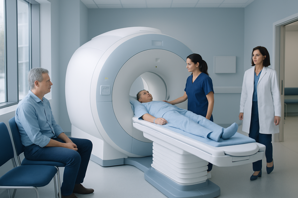
Bone and joint MRI can show soft tissues, cartilage, bone marrow, and joint structures in great detail. It is commonly used to assess injuries, persistent pain, inflammation, infection, and some tumors.
Key Takeaways
- Bone and joint MRI can show soft tissues, cartilage, bone marrow, and joint structures in great detail.
- It is commonly used to assess injuries, persistent pain, inflammation, infection, and some tumors.
- MRI does not use ionizing radiation, but it may not be suitable for everyone with certain metal implants or devices.
- Some MRI exams use contrast dye to improve detail, depending on what the doctor needs to evaluate.
- Preparation is usually simple, but patients should tell the imaging team about implants, pregnancy, kidney problems, or claustrophobia.
Bone and joint MRI is an imaging test that creates detailed pictures of bones, joints, cartilage, ligaments, tendons, muscles, and surrounding soft tissues. It is often used when pain, swelling, injury, or reduced movement needs a closer look than an X-ray can provide.
Overview of Bone and Joint MRI
Bone and joint MRI, also called musculoskeletal MRI, is a scan that uses a strong magnetic field and radio waves to create very detailed images of the body. Unlike X-rays and CT scans, MRI is especially good at showing soft tissues as well as changes inside the bones. This makes it particularly helpful for evaluating joints such as the knee, shoulder, hip, ankle, wrist, and spine-related structures.
An MRI can show cartilage, ligaments, tendons, muscles, nerves, joint lining, bone marrow, and fluid in and around a joint. Because many causes of pain or reduced movement begin in these tissues, MRI can help explain symptoms that may not be visible on standard X-rays. It is often used after an injury, when symptoms persist, or when a doctor needs to clarify the cause of swelling, stiffness, or instability.
Doctors may request a bone and joint MRI to investigate sports injuries, overuse problems, suspected tears, arthritis, infection, stress injuries, and some bone or soft tissue masses. The scan can also help guide treatment planning and monitor recovery after surgery or other therapies.
What a Bone and Joint MRI Can Show
One of the main strengths of MRI is that it can look beyond the outer shape of a bone or joint. It can reveal damage to cartilage, tears in ligaments or tendons, inflammation of the joint lining, fluid collections, and swelling in the bone marrow. These details can help doctors understand whether symptoms are related to injury, wear and tear, inflammation, or another process.
In bones, MRI can detect bone marrow edema, occult fractures that may not appear clearly on X-ray, stress reactions, infection, and some tumors. In joints, it can show cartilage loss, meniscus tears in the knee, labral tears in the shoulder or hip, bursitis, tendon injuries, and synovitis. MRI can also assess muscles for strains or tears and can help evaluate nerve compression around joints and limbs.
Because it provides high-contrast images of soft tissues, MRI is often valuable when there is a need to distinguish between several possible causes of pain. For example, a painful shoulder may involve the rotator cuff, bursae, labrum, or joint surfaces. A painful knee may involve cartilage, ligaments, menisci, or the underlying bone. MRI helps localize the source more precisely.
- Ligament injuries, such as ACL or ankle ligament tears
- Tendon problems, including tears or inflammation
- Cartilage damage and degenerative joint disease
- Bone marrow changes, stress injuries, or hidden fractures
- Inflammatory or infectious conditions affecting bone or joints
When a Doctor May Recommend One

A doctor may recommend bone and joint MRI when symptoms continue despite rest or initial treatment, or when the physical examination suggests damage that needs closer evaluation. Common reasons include persistent joint pain, swelling, stiffness, clicking, instability, weakness, limited range of motion, or symptoms after a fall or sports injury.
MRI is often used when X-rays are normal but symptoms remain unexplained. X-rays are very good for showing fractures, alignment, and advanced arthritis, but they do not provide the same level of detail for soft tissues. If there is concern for a ligament tear, tendon injury, cartilage defect, bone marrow problem, or early inflammatory disease, MRI may offer important answers.
The scan may also be requested before surgery to define the extent of injury, or after treatment to assess healing or complications. In some cases, doctors use MRI to investigate suspected arthritis or other inflammatory joint conditions, or to evaluate symptoms related to bone weakness when stress injury or fracture is suspected. The exact reason depends on the body part involved and the person’s symptoms and medical history.
How the Scan Is Performed and How to Prepare
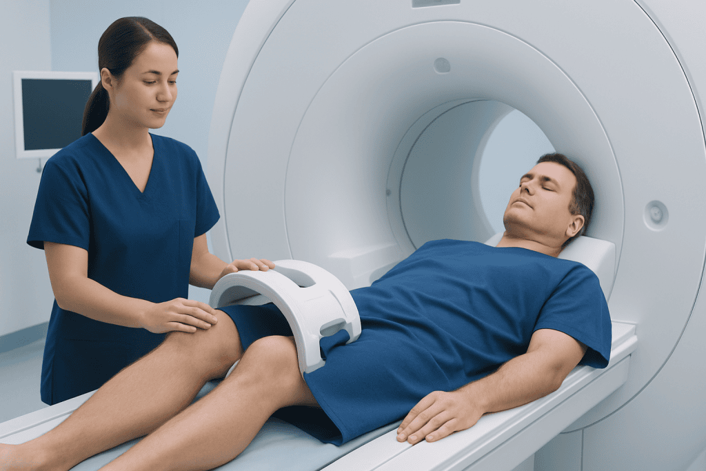
During a bone and joint MRI, the patient lies on a table that slides into the MRI scanner. Depending on the area being examined, a special coil may be placed around the knee, shoulder, wrist, or other body part to improve image quality. It is important to stay as still as possible, because movement can blur the pictures and make the scan harder to interpret.
The test is painless, but the machine can be noisy, often producing knocking or thumping sounds. Ear protection is usually provided. Some MRI studies are done without contrast, while others may require a contrast agent to highlight inflammation, tumors, infection, or blood supply to tissues. If contrast is needed, it is typically given through a vein.
Preparation is usually straightforward. Patients are asked to remove metal objects such as jewelry, watches, hairpins, and hearing aids. They should tell the imaging team about pacemakers, joint replacements, metal fragments, aneurysm clips, insulin pumps, cochlear implants, pregnancy, allergies, or kidney problems. People who feel anxious in enclosed spaces should mention this in advance, as comfort measures or sedation may sometimes be arranged.
In many centers, MRI is part of a wider imaging workup that may also include radiology services and, when needed, a dedicated MRI examination tailored to the joint or bone being studied.
MRI Compared With X-ray, CT, and Ultrasound
Each imaging method has different strengths. X-rays are usually the first test for bone and joint symptoms because they are quick and effective for showing fractures, dislocations, alignment problems, and advanced arthritis. However, they do not show tendons, ligaments, cartilage, or bone marrow in detail. MRI is often the next step when those structures need closer assessment.
CT scans are very good for showing complex bone anatomy and certain fractures, especially when surgery is being planned. They are faster than MRI but use ionizing radiation and generally provide less soft tissue detail. Ultrasound can be excellent for evaluating some tendons, bursae, fluid collections, and guided injections, especially around the shoulder or ankle, but it is more limited for looking deep inside a joint or inside the bone.
MRI is often chosen when the clinical question involves soft tissue injury, bone marrow changes, or early disease that may not yet have altered the bone surface. In some cases, the best diagnosis comes from combining MRI findings with other tests, a physical examination, and specialist review by orthopedics and traumatology teams.
Benefits, Limits, and Possible Risks
A major benefit of bone and joint MRI is that it provides highly detailed images without using ionizing radiation. This makes it useful for many adults and children when repeated imaging may be needed. It can help detect disease earlier, refine a diagnosis, and support treatment decisions such as physical therapy, medication, injections, or surgery.
At the same time, MRI has some limitations. Not everyone can undergo the scan safely, especially if they have certain implanted medical devices or metal fragments in the body. In addition, MRI may show abnormalities that are not actually causing symptoms, so results must always be interpreted in the context of the person’s examination and history.
Some people find the enclosed space uncomfortable, and the need to remain still can be difficult for those in pain. When contrast is used, there is a small risk of allergic reaction or concerns related to reduced kidney function. The imaging team reviews these factors carefully to make sure the test is appropriate and safe.
What Happens After the MRI
After the scan, most people can return to normal activities right away unless they received sedation. A radiologist reviews the images and prepares a report for the referring doctor, who then explains what the findings mean in relation to the patient’s symptoms. MRI results are most useful when combined with an examination, medical history, and sometimes blood tests or other imaging.
Treatment depends on what the MRI shows. Some conditions improve with rest, activity modification, ice, bracing, or physical therapy. Others may require medication, joint injection, or surgery. For example, a major ligament tear, advanced cartilage damage, or mechanical joint problem may lead to referral for orthopedic surgery evaluation, while inflammation may be managed non-surgically.
Patients should feel comfortable asking what the MRI found, whether the findings explain their symptoms, and what the next steps are. Near the end of the care pathway, if specialist assessment is needed, Acibadem International’s multidisciplinary specialists and JCI-accredited hospitals evaluate and treat bone and joint conditions for international patients.
When to Seek Medical Advice
Medical advice is recommended for joint or bone pain that lasts more than a few days, keeps returning, or interferes with walking, lifting, exercise, work, or sleep. A person should also seek evaluation if there is swelling, warmth, redness, joint locking, repeated instability, or reduced range of motion that does not improve.
Prompt assessment is especially important after trauma if the limb cannot bear weight, the joint looks deformed, or there is significant weakness or numbness. Fever with bone or joint pain, unexplained night pain, or rapidly worsening symptoms also deserves timely attention. These symptoms do not always mean a serious problem, but they should be checked by a qualified clinician.
Seeing a doctor early can help clarify whether MRI is needed or whether another test would be more appropriate first. The right imaging choice depends on symptoms, examination findings, age, medical history, and the body area involved.
Frequently asked questions
Is bone and joint MRI painful?
The scan itself is not painful. The main challenge for some people is lying still, especially if the affected joint is already sore. The machine can also be loud, but ear protection is usually provided.
How long does a bone and joint MRI take?
The exact time depends on the body part and whether contrast is needed. Many exams take between about 20 and 60 minutes. More detailed or multi-part studies may take longer.
Do all bone and joint MRIs need contrast dye?
No. Many bone and joint MRI scans are done without contrast. A doctor may request contrast when extra detail is needed to assess inflammation, infection, a tumor, or postoperative changes.
Can MRI show arthritis?
Yes, MRI can show signs of arthritis, including cartilage damage, inflammation, fluid, and changes in the bone marrow. It may be especially helpful when early inflammatory disease is suspected or when symptoms are not fully explained by X-rays.
What should a person do before the scan?
Patients should follow the instructions given by the imaging center and tell the team about any implants, metal in the body, kidney disease, pregnancy, or claustrophobia. In most cases, they will need to remove metal objects before the exam. If sedation is planned, there may be additional instructions.
Is MRI safe if someone has a joint replacement or metal implant?
Many people with orthopedic implants can still have an MRI, but the imaging team must know exactly what type of implant is present. Some devices are MRI-compatible, while others need special precautions or may make MRI unsuitable. Safety is checked carefully before the scan.
References
- American College of Radiology
- Radiological Society of North America
- National Institute of Arthritis and Musculoskeletal and Skin Diseases
- MedlinePlus
- NHS
This article is for general information only and is not a substitute for professional medical advice. Please consult a qualified doctor about your individual situation.
Joint and spine care in Turkey — expert assessment & treatment
JCI-accredited · board-certified surgeons · reply within 24h
Add us as a Preferred Source to see more of our trusted health content across Google Search, AI Overviews and Discover.