Bone Tumors on Xray: Preparation, Procedure and Results

An X-ray is often the first imaging test used to assess persistent bone pain, swelling, or an unexplained bone lesion. Many bone tumors are benign, and an abnormal X-ray finding does not automatically mean cancer.
Key Takeaways
- An X-ray is often the first imaging test used to assess persistent bone pain, swelling, or an unexplained bone lesion.
- Many bone tumors are benign, and an abnormal X-ray finding does not automatically mean cancer.
- The lesion’s location, margins, bone reaction, and surrounding tissues help guide the next diagnostic steps.
- MRI, CT, blood tests, and biopsy may be needed to establish a precise diagnosis.
- New or worsening bone pain, a growing lump, an unexplained fracture, or persistent symptoms should be medically assessed.
Bone tumors on xray can sometimes be recognized by characteristic changes in the bone, such as an unusual area of bone loss, extra bone formation, expansion, or a break in the outer bone layer. An X-ray is usually an important first test, but it cannot confirm whether a tumor is benign or cancerous on its own; specialists may recommend further imaging and, in some cases, a biopsy.
Bone tumors on Xray: what the examination can show
Bone tumors on xray may look different depending on the type of lesion, its location, and whether it is growing slowly or more aggressively. An X-ray uses a small amount of radiation to create images of dense structures such as bone. It can show an area that is less dense than usual, more dense than usual, enlarged, weakened, or altered in shape.
Radiologists evaluate several details: which bone is involved, whether the lesion is in the center or edge of the bone, its borders, the pattern of bone destruction or formation, and whether the outer layer of bone is intact. These features can suggest whether a finding is more likely to be benign or needs urgent further assessment. However, no image feature alone can reliably diagnose every lesion.
Some abnormalities found on an X-ray are not tumors. Infection, a healing injury, arthritis-related changes, bone cysts, and normal developmental variations can sometimes resemble a tumor. For this reason, X-ray results are interpreted alongside symptoms, age, medical history, examination findings, and additional tests when appropriate.
How an X-ray for a suspected bone tumor works
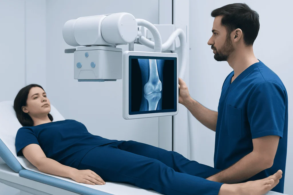
An X-ray machine directs a controlled beam of radiation through the body part being examined. Bone absorbs more radiation than soft tissue, producing a detailed two-dimensional image of the skeleton. Several views are usually taken from different angles so the radiologist can assess the lesion’s size, position, and effect on the surrounding bone.
People may be referred for an X-ray after reporting ongoing localized bone pain, swelling, reduced movement, or a suspected injury. It may also be ordered after an incidental finding on another scan. X-rays are suitable for many people, including children when clinically necessary, because the radiation exposure is kept as low as reasonably achievable.
Pregnant people, or those who think they could be pregnant, should tell the imaging team before the examination. The team can decide whether the X-ray should proceed, be modified, or be replaced with another test. An X-ray is not usually the only test when a bone tumor is suspected, but it is often the examination that helps plan the next step.
Preparation, procedure and recovery timeline

Preparation for a bone X-ray is usually simple. In most cases, no fasting, injections, or special medication changes are needed. The person may be asked to remove jewelry, belts, clothing with metal fasteners, or other objects near the body part being imaged. Comfortable clothing can make this easier.
During the procedure, a radiographer positions the body part on an X-ray table or in front of an imaging plate. Remaining still for a few seconds is important for sharp images. The radiographer may reposition the person to obtain two or more views. The examination itself commonly takes only a short time, although positioning can take longer if movement is painful or an area is difficult to image.
There is no recovery period after a standard X-ray. People can usually return to usual activities immediately. A radiologist reviews the images and sends a report to the referring clinician, who explains what the findings may mean and whether further imaging, specialist review, or follow-up X-rays are needed.
Understanding results: benign, uncertain and concerning patterns
Many benign bone tumors have relatively well-defined borders and may cause little or no symptom. Examples include osteochondromas, enchondromas, and non-ossifying fibromas. Depending on their appearance and location, some can be safely monitored with repeat imaging, while others may require treatment if they cause pain, weaken a bone, interfere with movement, or grow.
More concerning X-ray patterns can include poorly defined bone destruction, a lesion that extends through the outer bone layer, an aggressive reaction in nearby bone, or an associated soft-tissue mass. These findings do not by themselves prove cancer, but they require prompt assessment by a specialist team. Further evaluation may include MRI to examine bone marrow and soft tissues, CT for detailed bone anatomy, or a bone scan or PET scan in selected cases.
If a biopsy is needed, it should generally be planned by the orthopedic oncology or sarcoma team that would manage any treatment. This approach helps ensure that the biopsy route does not complicate later surgery. Bone tumor treatment may involve observation, surgery, medicines, radiotherapy, chemotherapy, or a combination, depending on the confirmed diagnosis.
Where does bone cancer usually show up first?
Primary bone cancer begins in bone rather than spreading there from another organ. It often develops in long bones, especially around the knee in the lower end of the thighbone or upper end of the shinbone. It can also occur in the upper arm, pelvis, shoulder region, ribs, or spine, depending on the cancer type and a person’s age.
Bone cancer does not always produce an obvious lump at first. Persistent pain in one area, particularly pain that becomes more frequent, occurs at rest, or disturbs sleep, may be an early symptom. These symptoms are common in many non-cancerous conditions, so they should be assessed without assuming the cause.
It is important to distinguish primary bone cancer from bone metastases, which occur when a cancer that started elsewhere spreads to bone. The diagnostic process and treatment plan differ, which is why accurate imaging and pathology are essential. Bone cancer care commonly involves orthopedic oncology, medical oncology, radiology, pathology, rehabilitation, and other relevant specialties.
Are bone tumors curable?
Many bone tumors are curable or can be managed successfully. Benign tumors may need no treatment at all, while others can often be removed or treated when they cause symptoms or place the bone at risk of fracture. The outlook depends on the exact tumor type, its size and location, and whether it has affected nearby structures.
For malignant bone tumors, treatment and outlook vary widely. Some primary bone cancers can be treated with curative intent, particularly when identified before spread and when complete local treatment is possible. Treatment may include surgery, chemotherapy, radiotherapy, or carefully planned combinations of these options.
Specialist evaluation is important because outcomes and treatment choices are influenced by the tumor’s pathology and stage. At Acibadem International, multidisciplinary specialists at JCI-accredited hospitals assess and treat bone tumors for international patients, coordinating imaging, pathology, orthopedic oncology, and cancer care where needed.
What is the most common benign bone tumor?
Osteochondroma is generally considered the most common benign bone tumor. It is an outgrowth of bone covered by cartilage and often develops near the ends of long bones, particularly around the knee or shoulder. It may be noticed during childhood or adolescence, or found incidentally when an X-ray is performed for another reason.
Many osteochondromas do not require treatment. A clinician may recommend observation when the lesion is painless and does not affect function. Surgery may be considered if it causes pain, limits movement, presses on a nerve or blood vessel, creates a significant deformity, or has concerning changes after skeletal maturity.
Other common benign lesions include enchondromas, which form within cartilage in bone, and non-ossifying fibromas, which are often discovered in children and adolescents. A specialist can explain whether a particular X-ray finding is consistent with a benign lesion and whether follow-up imaging is appropriate.
What are the 7 warning signs of a bone tumor?
Possible warning signs of a bone tumor include persistent localized bone pain; pain that worsens over time or occurs at rest or during the night; a lump or swelling near a bone; reduced movement of a nearby joint; limping or difficulty using a limb; an unexplained fracture after minor force; and ongoing fatigue, fever, or unintentional weight loss without a clear cause.
These signs do not necessarily indicate a bone tumor. Sports injuries, infections, inflammatory conditions, and common musculoskeletal problems can cause similar symptoms. Still, persistent or progressive symptoms deserve clinical evaluation, particularly when pain is focused in one area and does not improve as expected.
For children and teenagers, recurring limb pain should not automatically be attributed to growth. A healthcare professional can determine whether an examination and imaging are needed. Early assessment can clarify the cause and help guide appropriate care.
Benefits, limitations and risks of bone X-rays
The main benefit of an X-ray is that it is quick, widely available, and especially useful for showing the structure of bone. It can help distinguish a fracture from a lesion, reveal features that suggest a slow-growing process, and identify cases that need urgent referral. It also provides a baseline image for monitoring certain known benign lesions.
The principal limitation is that an X-ray cannot show every detail. It may not detect early changes within bone marrow or accurately define involvement of nearby muscles, nerves, blood vessels, or cartilage. MRI, CT, and other tests can add information that is not visible on plain X-rays.
X-rays involve a low dose of ionizing radiation. For a medically necessary single examination, the risk is considered small, and imaging teams use the lowest appropriate exposure. The greater risk may be delaying evaluation of significant symptoms, so the benefits and limitations are weighed by the treating clinician.
When to seek medical care
Medical advice is appropriate for bone pain that persists for several weeks, steadily worsens, occurs at night, or is associated with a new lump or swelling. Assessment is also important after an unexplained fracture, new difficulty walking or using an arm, or pain that does not improve with usual rest and conservative care.
Urgent medical assessment is needed for severe pain after an injury, a limb that appears deformed, sudden inability to bear weight, numbness or weakness, or symptoms suggesting infection such as fever with marked redness and swelling. These symptoms may have causes other than a tumor but should not be ignored.
When an X-ray identifies a possible lesion, the next step should be guided by the referring doctor and appropriate specialists. Avoid trying to interpret an image without the formal radiology report and clinical context. Timely follow-up allows benign conditions to be reassured and monitored appropriately, while more serious findings can be investigated without unnecessary delay.
Frequently asked questions
Can an X-ray tell if a bone tumor is cancerous?
An X-ray can show patterns that may suggest a benign or more aggressive lesion, but it cannot confirm cancer in every case. MRI, CT, specialist review, and sometimes a biopsy are needed to establish the diagnosis.
Do bone tumors always cause pain?
No. Some benign bone tumors cause no symptoms and are discovered incidentally during imaging for an injury or another concern. Pain may occur when a lesion weakens bone, irritates nearby tissues, affects a joint, or causes a fracture.
What happens if an X-ray shows a spot on the bone?
A spot can have many explanations, including a benign lesion, healing change, infection, or less commonly a malignant tumor. The clinician will consider the X-ray appearance, symptoms, age, and medical history to decide whether follow-up imaging or specialist referral is needed.
Is MRI better than X-ray for bone tumors?
The tests provide different information. X-ray is often the first examination because it shows bone structure clearly, while MRI gives more detail about bone marrow, soft tissues, and the extent of a lesion.
Can a benign bone tumor become cancerous?
Most benign bone tumors do not become cancerous. A small number of particular tumor types or inherited conditions can carry a higher risk, which is why recommended monitoring and specialist advice should be followed.
How long does it take to get bone X-ray results?
The images are obtained immediately, but the timing of the formal report varies by facility and clinical urgency. A referring clinician can explain when and how results will be discussed, as well as whether any further tests are needed.
References
- American Cancer Society
- National Cancer Institute
- RadiologyInfo.org
- American Academy of Orthopaedic Surgeons
- World Health Organization
This article is for general information only and is not a substitute for professional medical advice. Please consult a qualified doctor about your individual situation.
Oncology care in Turkey — second opinion and treatment plan
JCI-accredited · board-certified surgeons · reply within 24h
Add us as a Preferred Source to see more of our trusted health content across Google Search, AI Overviews and Discover.