Bowens Disease: Early Signs, Risk Factors, and How It Is Treated
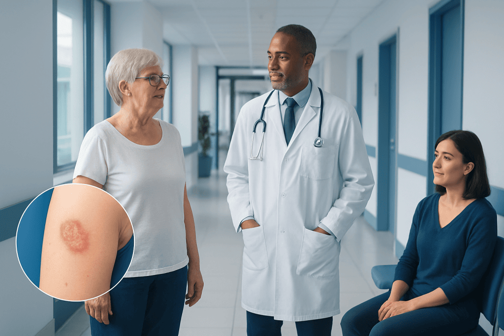
Bowens disease is squamous cell carcinoma in situ, meaning abnormal cells are limited to the epidermis, the outermost skin layer. A persistent red, pink, brown, or scaly patch that does not heal should be checked by a qualified clinician.
Key Takeaways
- Bowens disease is squamous cell carcinoma in situ, meaning abnormal cells are limited to the epidermis, the outermost skin layer.
- A persistent red, pink, brown, or scaly patch that does not heal should be checked by a qualified clinician.
- Sun exposure, older age, light skin, immune suppression, and previous skin damage can increase risk.
- A skin biopsy is usually needed to confirm the diagnosis and rule out invasive skin cancer.
- Treatment may include creams, freezing, light-based treatment, curettage, or surgical removal, depending on the lesion and the person’s health.
Bowens disease, also called squamous cell carcinoma in situ, is a very early form of skin cancer confined to the outer layer of the skin. It often appears as a persistent scaly patch and can usually be treated effectively before it grows deeper into the skin.
What Is Bowens Disease?
Bowens disease is an early form of squamous cell carcinoma, a common type of skin cancer. It is also known as squamous cell carcinoma in situ. “In situ” means that the abnormal cells are present only in the epidermis, the thin outer layer of the skin, and have not grown into deeper tissues.
Because it is confined to the surface layer, Bowens disease is generally very treatable. However, it should not be ignored. Without treatment, a lesion can sometimes develop into invasive squamous cell carcinoma, in which cancer cells extend deeper into the skin. Early assessment and appropriate treatment help reduce this possibility.
Bowens disease is not contagious, and it is not the same as melanoma. It can occur on sun-exposed skin, such as the lower legs, head, neck, hands, and forearms, but it may also develop in areas that receive little sun exposure. When it affects the genital or anal area, it may be described using site-specific terms and requires careful specialist assessment.
How Bowens Disease May Look and Feel
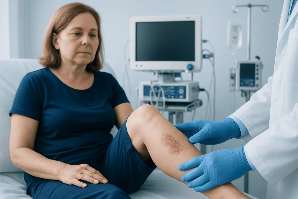
Bowens disease often starts as a slowly enlarging patch that remains in the same place for months or longer. The patch may be red, pink, brown, or skin-colored. It commonly has a dry, rough, crusted, or scaly surface and may resemble eczema, psoriasis, a fungal rash, or a healing injury.
The edges can be well defined, although this is not always the case. Some lesions are flat, while others are slightly raised. They are often painless, which can make them easy to overlook. A lesion may occasionally itch, feel tender, bleed after rubbing, or form a crust that repeatedly returns.
On darker skin tones, the area may appear darker than surrounding skin rather than red. A persistent patch that changes in appearance, becomes thicker, develops an ulcer, or does not respond to usual skin care deserves medical review. Photographs taken over time can be helpful for documenting change, but they cannot replace an in-person skin examination.
- A scaly patch that persists despite moisturizers or prescribed rash treatments
- A slowly growing red, brown, or pink area with a rough surface
- Repeated crusting, minor bleeding, tenderness, or itching in one spot
- A non-healing lesion, especially on previously sun-damaged skin
Why Bowens Disease Develops and Who Is at Risk
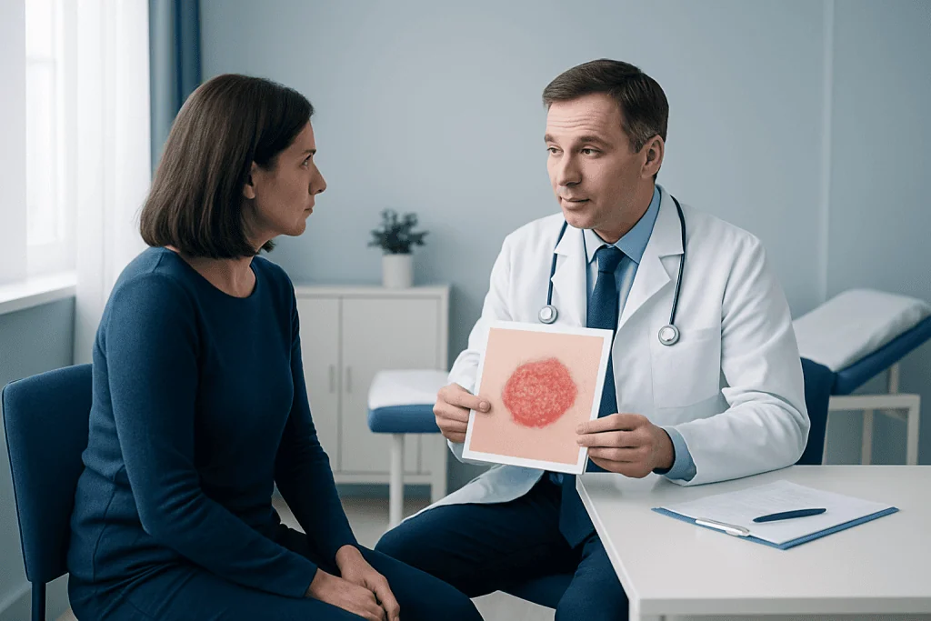
Long-term exposure to ultraviolet, or UV, radiation is an important risk factor for Bowens disease. UV radiation comes mainly from sunlight and can also come from tanning beds. It can damage skin-cell DNA over time, especially in areas that have received years of intermittent or regular sun exposure.
Risk increases with age because UV damage and other cellular changes accumulate over many years. People with fair skin, light-colored eyes, freckles, a history of frequent sunburns, or extensive outdoor sun exposure may be more likely to develop sun-related skin changes. Still, Bowens disease can occur in people of every skin tone and should not be dismissed based on appearance alone.
Other factors may include a weakened immune system due to certain medicines or medical conditions, prior radiation treatment, chronic wounds or inflammation, and past exposure to arsenic. Some lesions in genital or anal skin are associated with certain types of human papillomavirus, or HPV. Having one lesion also means it is sensible to have the rest of the skin checked regularly, as sun damage can affect multiple areas.
How the Diagnosis Is Confirmed
A dermatologist or another qualified clinician will begin by asking about the lesion, including when it appeared, whether it has changed, and any symptoms such as bleeding or itching. They will examine the area and may assess other parts of the skin for signs of sun damage or additional suspicious lesions.
Although the appearance can suggest Bowens disease, many skin conditions look similar. For this reason, diagnosis is usually confirmed with a skin biopsy. After numbing the skin with local anesthetic, the clinician removes a small sample or the entire lesion and sends it to a laboratory for examination under a microscope.
The biopsy helps distinguish Bowens disease from inflammatory conditions, benign growths, fungal infection, and invasive squamous cell carcinoma. It also helps guide treatment planning. The location, size, thickness, borders, number of lesions, immune status, and any previous treatment all influence which approach is most appropriate.
Treatment Options for Bowens Disease
Treatment is individualized. The best option depends on where the lesion is located, how large it is, whether it has been treated before, and the person’s general health, skin-healing ability, and preferences. In some situations, a clinician may recommend a short period of monitoring before treatment planning, but confirmed lesions are commonly treated because of the possibility of progression.
Surgical excision removes the lesion along with a small margin of surrounding skin and allows the tissue to be examined. This approach may be especially useful when the diagnosis is uncertain, the area is thickened, the borders are unclear, or there is concern for invasion. Curettage and cautery, in which the lesion is scraped and treated with heat or electricity, may be suitable for selected superficial lesions.
Non-surgical choices include cryotherapy, which destroys abnormal cells by freezing them; topical medicines such as 5-fluorouracil or imiquimod; and photodynamic therapy, which combines a light-sensitive treatment with a specific light source. These methods can be useful for superficial or larger areas and may avoid surgery in some locations. They can cause temporary redness, soreness, crusting, or inflammation as the skin responds to treatment.
Radiotherapy is used less often but may be considered when surgery is not suitable or for selected difficult-to-treat areas. Whatever method is chosen, follow-up is important to confirm healing and identify recurrence or new lesions early. A specialist may also recommend regular full-skin examinations for people with significant sun damage or other risk factors.
Protecting the Skin and Supporting Follow-Up
Sun protection cannot reverse all existing sun damage, but it can help reduce further UV injury and lower the risk of additional skin cancers. Protection is useful throughout the year, including on cloudy days, because UV radiation can still reach the skin. It is particularly important for people who have had Bowens disease or other types of skin cancer.
Practical measures include seeking shade when possible, wearing a broad-brimmed hat and protective clothing, avoiding tanning beds, and using a broad-spectrum sunscreen with SPF 30 or higher on exposed skin. Sunscreen should be applied generously and reapplied according to the product instructions, especially after swimming, sweating, or towel drying.
Regular self-checks can help a person notice a changing patch, sore, or growth. It is useful to examine commonly exposed areas as well as the scalp, backs of the legs, feet, and nails. A mirror or a trusted family member can help check hard-to-see areas. Any persistent or changing lesion should be assessed rather than repeatedly treated at home as a presumed rash.
When to Seek Medical Care
A person should arrange a medical appointment for a skin patch that lasts longer than several weeks, especially if it is rough, scaly, crusted, enlarging, bleeding, or repeatedly failing to heal. Prompt review is also advisable for new changes in a previous scar, chronic wound, or area of skin that has received radiation treatment.
More timely assessment is appropriate if a lesion becomes painful, rapidly increases in size, forms an open sore, or develops a firm raised area. People with reduced immunity, a personal history of skin cancer, or numerous sun-damaged spots should have a lower threshold for seeking advice.
Bowens disease is often manageable with outpatient treatment, and early evaluation can make treatment more straightforward. Acibadem International’s multidisciplinary specialists and JCI-accredited hospitals diagnose and treat skin conditions, including Bowens disease, for international patients. A dermatologist can explain the diagnosis, discuss suitable options, and create an appropriate follow-up plan.
Frequently asked questions
Is Bowens disease cancer?
Bowens disease is an early form of squamous cell skin cancer called squamous cell carcinoma in situ. The abnormal cells are confined to the outermost layer of skin and have not invaded deeper tissue. It is generally very treatable, but medical assessment and treatment are important.
Can Bowens disease go away on its own?
A persistent lesion should not be expected to resolve on its own. Some skin conditions that resemble Bowens disease may improve, but a confirmed diagnosis usually needs treatment or specialist-directed monitoring. This helps reduce the chance of progression to invasive squamous cell carcinoma.
What does Bowens disease look like?
It often looks like a persistent dry, scaly, red, pink, brown, or skin-colored patch. It may be flat or slightly raised and can resemble eczema, psoriasis, or a fungal infection. Its appearance varies between individuals and across skin tones, so a biopsy may be needed for confirmation.
Is Bowens disease caused by sun exposure?
Long-term UV exposure is a major risk factor, particularly for lesions on sun-exposed skin. However, Bowens disease may also occur in areas with little sun exposure. Other possible factors include immune suppression, previous radiation, chronic skin injury, and certain HPV-related changes in genital or anal skin.
How is Bowens disease treated?
Treatment can include surgical removal, curettage and cautery, freezing with cryotherapy, topical prescription medicines, or photodynamic therapy. The choice depends on the lesion’s site, size, features, and the individual’s medical circumstances. A clinician will discuss expected healing, possible side effects, and follow-up needs.
Can Bowens disease come back after treatment?
A treated lesion can sometimes recur, and people who have had Bowens disease may also develop new lesions elsewhere. Follow-up appointments and regular skin checks help detect changes early. Ongoing sun protection is an important part of reducing additional UV-related skin damage.
References
- American Academy of Dermatology Association
- National Cancer Institute
- NHS
- American Cancer Society
- Skin Cancer Foundation
This article is for general information only and is not a substitute for professional medical advice. Please consult a qualified doctor about your individual situation.
Oncology care in Turkey — second opinion and treatment plan
JCI-accredited · board-certified surgeons · reply within 24h
Add us as a Preferred Source to see more of our trusted health content across Google Search, AI Overviews and Discover.