

Brain MRI or Lumbar Puncture for CNS Infection: Why Both May Be Needed
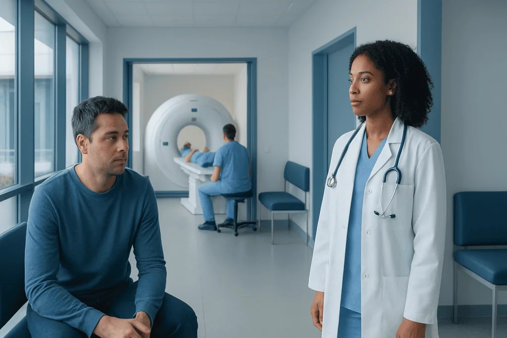
Brain MRI and lumbar puncture are complementary tests, not competing alternatives. MRI helps doctors look for brain swelling, inflammation, abscesses, or pressure-related risks before lumbar puncture in selected patients.
Key Takeaways
- Brain MRI and lumbar puncture are complementary tests, not competing alternatives.
- MRI helps doctors look for brain swelling, inflammation, abscesses, or pressure-related risks before lumbar puncture in selected patients.
- Lumbar puncture analyzes cerebrospinal fluid and is often essential for diagnosing meningitis, encephalitis, and other CNS infections.
- The order of testing depends on symptoms, examination findings, and how urgently treatment must begin.
- Doctors may start treatment before all test results return if a serious CNS infection is strongly suspected.
When a central nervous system (CNS) infection is suspected, brain MRI and lumbar puncture often answer different but equally important questions. MRI can reveal swelling, inflammation, or complications in the brain, while lumbar puncture examines cerebrospinal fluid to help confirm infection and identify its cause.
Overview: Why both tests may be needed
A central nervous system infection affects the brain, spinal cord, or the tissues and fluid around them. Common examples include meningitis, which involves inflammation of the protective membranes around the brain and spinal cord, and encephalitis, which affects brain tissue itself. Because these conditions can become serious quickly, doctors aim to diagnose them as early and accurately as possible.
Brain MRI and lumbar puncture play different roles in that process. A brain MRI creates detailed images of the brain and nearby structures. It can show inflammation, swelling, abscesses, bleeding, or other changes that support the diagnosis and help explain a person’s symptoms. A lumbar puncture, also called a spinal tap, collects cerebrospinal fluid so it can be tested for signs of infection, inflammation, and the likely organism causing the illness.
In many cases, one test does not replace the other. MRI is excellent for showing structure, but it cannot directly analyze the fluid that surrounds the brain and spinal cord. Lumbar puncture provides that laboratory information, but it does not give the same visual detail about the brain itself. For this reason, doctors often use both tests together when evaluating suspected meningitis or encephalitis.
The exact order depends on the individual situation. Some people need urgent lumbar puncture right away, while others should have brain imaging first if there are signs that pressure in the skull may be increased or if another brain problem needs to be ruled out. The goal is always to balance speed, safety, and diagnostic accuracy.
What symptoms can suggest a CNS infection?
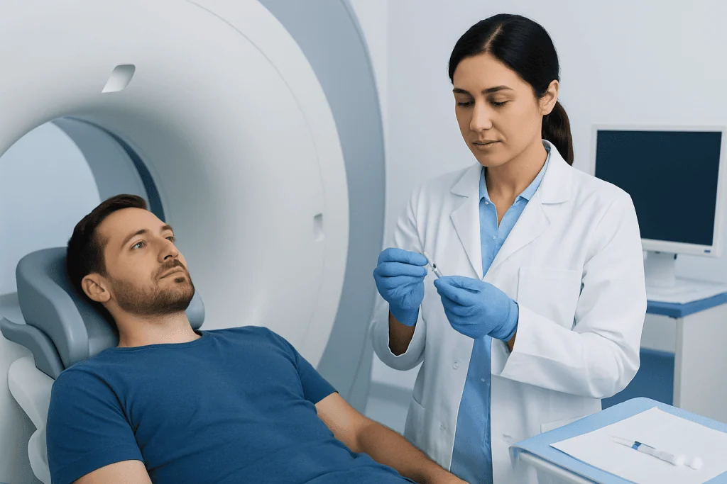
Symptoms of a CNS infection can vary widely depending on the person’s age, the area affected, and the cause of the infection. Common symptoms include fever, headache, neck stiffness, confusion, sensitivity to light, nausea, vomiting, drowsiness, and seizures. Some people may also develop weakness, speech difficulty, or changes in behavior or personality.
Not every patient has the classic symptom pattern. For example, older adults may mainly show confusion or reduced alertness. Young children and infants may have poor feeding, irritability, unusual sleepiness, or a bulging soft spot on the head. In people with weakened immune systems, symptoms can be less typical and may develop more gradually.
These symptoms can overlap with other neurological problems, including stroke, migraine, autoimmune inflammation, tumors, or metabolic disorders. That is one reason doctors often use imaging and fluid testing together. The combination helps separate infection from other causes and guides urgent treatment decisions.
When symptoms suggest a possible infection of the brain or its coverings, time matters. Medical teams usually assess vital signs, perform a neurological examination, and order blood tests while deciding whether MRI, lumbar puncture, or both should happen first.
What a brain MRI shows in suspected infection
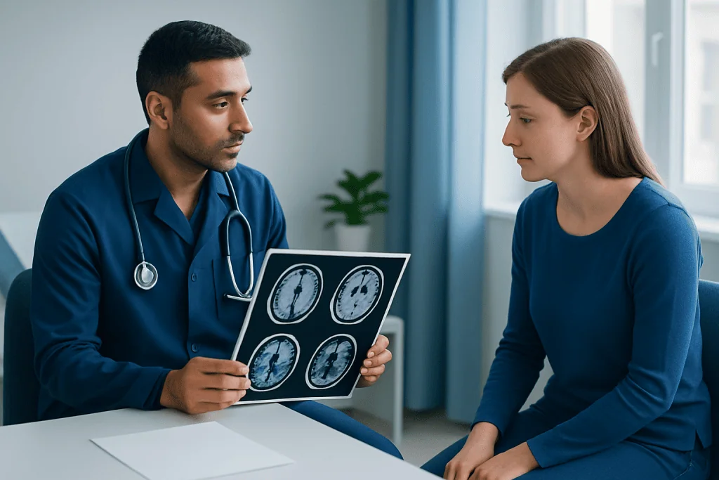
Brain MRI is one of the most detailed imaging tests for the brain. In suspected CNS infection, it can reveal signs such as brain swelling, inflammation, abnormal enhancement after contrast, collections of pus such as abscesses, involvement of specific brain regions, and complications like hydrocephalus. MRI may also help identify patterns that suggest certain infections more strongly than others.
For example, doctors may see findings that support viral encephalitis, fungal infection, abscess formation, or infection spreading from the sinuses or ear. MRI can also show whether there are mass-like lesions or marked swelling that could make lumbar puncture less safe until the situation is better understood. In selected cases, MRI of the spine may be needed as well if spinal cord infection is suspected.
Another important role of MRI is to look for alternative diagnoses. Symptoms such as fever and confusion do not always come from infection. MRI can help distinguish infection from stroke, tumor, autoimmune inflammation, demyelinating disease, or bleeding. This can be especially helpful when clinical symptoms are unclear.
If imaging is needed, the care team may arrange a brain MRI scan early in the evaluation. Even so, a normal MRI does not completely rule out CNS infection, especially in the early stages. That is why doctors often still recommend lumbar puncture when it is safe and appropriate.
Why lumbar puncture is often essential
Lumbar puncture allows doctors to collect cerebrospinal fluid, the clear fluid that surrounds the brain and spinal cord. This sample can provide critical information that imaging alone cannot. The fluid is checked for white blood cells, protein, glucose, pressure, and other markers that help show whether infection or inflammation is present.
Laboratories may also test the fluid for bacteria, viruses, fungi, or other organisms using culture, antigen tests, and molecular methods such as PCR. These results can help identify the likely cause of conditions such as bacterial meningitis, viral encephalitis, or other neuroinfections. Knowing the cause helps doctors tailor treatment rather than relying only on broad initial therapy.
In some situations, lumbar puncture also helps monitor how a disease is evolving or whether treatment is working. For instance, repeat cerebrospinal fluid testing may be considered in selected cases if symptoms continue, if the diagnosis remains uncertain, or if the infection is caused by a hard-to-treat organism.
Many patients worry that lumbar puncture is unusually painful or dangerous. In practice, it is a commonly performed procedure done with careful sterile technique. The care team explains positioning, numbing medication, and aftercare to improve comfort. When needed, lumbar puncture can be a key step in diagnosing and managing a CNS infection safely.
Why doctors sometimes do MRI before lumbar puncture
Although lumbar puncture is central to diagnosis, it is not always the first test performed. Doctors may order brain imaging first if there are warning signs suggesting increased pressure inside the skull, a mass lesion, or another structural problem. Examples include markedly reduced consciousness, new focal neurological deficits, recent seizures, severe immunosuppression, or signs of significant brain swelling on examination.
The concern is not that lumbar puncture is routinely unsafe, but that in some patients, removing cerebrospinal fluid before assessing the brain could increase the risk of complications. Imaging helps the medical team judge whether it is safe to proceed immediately or whether treatment and monitoring should come first.
It is also important to understand that imaging should not cause harmful delays in urgent care. If doctors strongly suspect a serious CNS infection, they usually obtain blood cultures and start appropriate treatment promptly, even if MRI or lumbar puncture must wait. Early treatment can be lifesaving, especially in bacterial infections.
In hospitals with advanced neurological services, specialists from neurology, infectious diseases, neuroradiology, and critical care may work together to decide the safest sequence of tests. In some patients, neuroradiology-guided imaging assessment helps clarify the next step when the diagnosis is complex.
How diagnosis and treatment work together
Diagnosis of a CNS infection rarely relies on one finding alone. Doctors combine the medical history, physical and neurological examination, blood tests, imaging, and cerebrospinal fluid results. Sometimes additional tests such as electroencephalography, chest imaging, or immune testing are also useful, depending on the suspected cause and the person’s health background.
Treatment may begin before a final cause is confirmed. This is common when bacterial meningitis, viral encephalitis such as herpes simplex encephalitis, or another severe infection is suspected. Initial treatment may include antibiotics, antiviral medicines, and supportive hospital care while test results are pending. If later testing points to a different cause, treatment can be adjusted.
MRI findings may also influence treatment decisions by showing complications that need special management, such as abscess drainage, seizure risk, or swelling that requires close monitoring. Likewise, lumbar puncture results may help determine whether a patient needs isolation precautions, prolonged antimicrobial therapy, or evaluation for a noninfectious inflammatory condition instead.
In more complex cases, patients may benefit from centers experienced in neurological infections and advanced imaging. Acibadem International’s multidisciplinary specialists in JCI-accredited hospitals diagnose and treat CNS infections for international patients, including conditions that may require coordinated imaging, laboratory analysis, and neurological care.
What patients can expect during testing and recovery
For a brain MRI, the patient lies still on a table that moves into the scanner. The machine is noisy, but staff usually provide hearing protection, and the test itself is painless. Some people receive contrast dye to improve image detail. If a person feels claustrophobic, anxious, or unable to remain still, the team may discuss options to help them complete the scan comfortably and safely.
During a lumbar puncture, the patient is usually asked to lie on their side or sit leaning forward. After the skin is cleaned, a local anesthetic numbs the area in the lower back. A thin needle is then used to collect cerebrospinal fluid. The procedure usually takes only a short time, although preparation and observation add to the overall visit.
After lumbar puncture, mild back soreness or headache can occur. Patients are usually advised to rest, drink fluids as recommended, and report severe headache, fever, new numbness, weakness, or worsening symptoms. The healthcare team explains what is normal and when to seek help.
Most importantly, patients and families should know that these tests are ordered to answer different questions. When both are recommended, it usually means the medical team wants the clearest and safest picture possible rather than performing duplicate testing.
When to seek urgent medical care
Possible CNS infections need prompt medical evaluation. Urgent care is especially important for symptoms such as high fever with severe headache, neck stiffness, confusion, unusual drowsiness, seizures, new weakness, difficulty speaking, or sudden behavior changes. In infants and older adults, signs may be less typical, so any rapid change in alertness or function deserves attention.
People with weakened immune systems, recent neurosurgery, head injury, cancer treatment, organ transplantation, or implanted neurological devices may have a higher risk of certain serious infections. For them, even milder neurological symptoms may require early assessment. Delaying care can make diagnosis and treatment more difficult.
Emergency evaluation does not always mean every patient will need both MRI and lumbar puncture immediately. The care team chooses the safest path based on the person’s condition. However, if a doctor recommends one or both tests, it is usually because each can add important information that could affect treatment.
Anyone recovering from a known CNS infection should also seek follow-up care if symptoms return, new neurological problems appear, or recovery is slower than expected. Ongoing communication with a qualified doctor helps ensure complications are recognized and treated promptly.
Frequently asked questions
Is brain MRI better than lumbar puncture for diagnosing a CNS infection?
Neither test is better in every situation because they answer different questions. MRI shows structural changes in the brain, while lumbar puncture analyzes cerebrospinal fluid to look for infection and identify the cause. Doctors often need both to make the most accurate diagnosis.
Why can’t doctors just do one test?
A single test may miss important information. MRI may show inflammation or complications, but it cannot directly test spinal fluid for bacteria, viruses, fungi, or inflammatory markers. Lumbar puncture provides that fluid analysis, but it does not replace detailed brain imaging.
Is it dangerous to do a lumbar puncture before imaging?
For many patients, lumbar puncture can be done safely without prior imaging. However, if there are signs of increased pressure in the skull, a mass lesion, or certain neurological deficits, doctors may order imaging first to reduce risk. The decision is based on the patient’s symptoms and examination.
Can treatment start before MRI or lumbar puncture results are back?
Yes. If doctors strongly suspect a serious CNS infection, they often begin treatment right away after initial assessment and blood tests. This is done because waiting for full results could be unsafe, especially in severe bacterial or viral infections.
Does a normal brain MRI rule out meningitis or encephalitis?
No. Early in the illness, MRI may be normal or may not show all forms of infection clearly. If symptoms and clinical findings still suggest a CNS infection, lumbar puncture and other tests may still be needed.
What does cerebrospinal fluid testing look for?
Cerebrospinal fluid testing checks cell counts, protein, glucose, opening pressure, and signs of inflammation. It can also be tested for bacteria, viruses, fungi, and other organisms using culture and molecular techniques. These findings help confirm infection and guide treatment.
References
- World Health Organization
- Centers for Disease Control and Prevention
- National Institute of Neurological Disorders and Stroke
- Infectious Diseases Society of America
- American College of Radiology
This article is for general information only and is not a substitute for professional medical advice. Please consult a qualified doctor about your individual situation.
Joint and spine care in Turkey — expert assessment & treatment
JCI-accredited · board-certified surgeons · reply within 24h
Add us as a Preferred Source to see more of our trusted health content across Google Search, AI Overviews and Discover.

More from the Health Library



Related Specialists

Prof. Dr. Elif Akpek
Anesthesiology
Dt. Ceren Ayözger
Dentistry
Assoc. Prof. Dr. Levent Aydemir
Otorhinolaryngology
Prof. Dr. Salih Marangoz
Orthopedic Surgery & Traumatology