Brain PET vs MRI: When Different Neuroimaging Tests Are Used
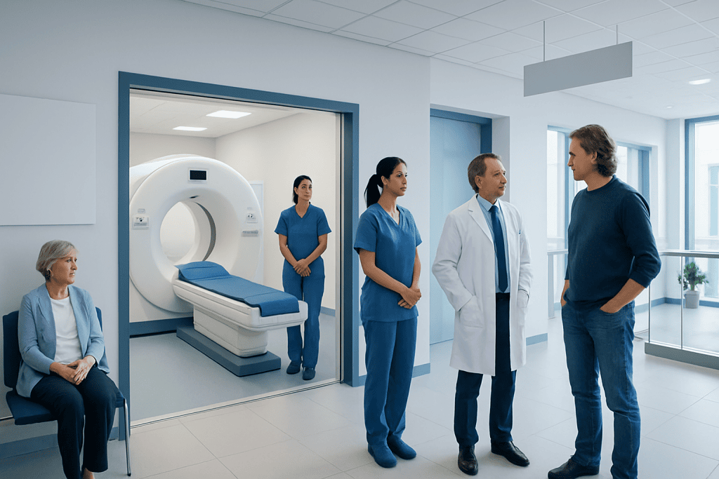
MRI is usually used to show detailed brain anatomy and structural changes. PET helps show how brain tissue is functioning, including metabolism and activity patterns.
Key Takeaways
- MRI is usually used to show detailed brain anatomy and structural changes.
- PET helps show how brain tissue is functioning, including metabolism and activity patterns.
- The best test depends on the reason for imaging, such as stroke, tumor, memory loss, seizures, or infection.
- Some people need both tests because they provide complementary information.
- MRI does not use ionizing radiation, while PET uses a small amount of radioactive tracer.
- A doctor interprets imaging results together with symptoms, examination findings, and other tests.
Brain PET and MRI are different neuroimaging tests that answer different clinical questions. MRI mainly shows detailed brain structure, while PET highlights brain function and metabolism, so doctors choose one or both depending on symptoms and the condition being evaluated.
Overview: how Brain PET and MRI differ
Brain PET and brain MRI are both important imaging tools, but they are not interchangeable. They look at the brain in different ways. MRI, or magnetic resonance imaging, creates highly detailed pictures of brain structures such as the cortex, white matter, blood vessels, and nearby tissues. PET, or positron emission tomography, shows how brain tissue is working by tracking a small amount of radioactive tracer.
In simple terms, MRI is often best for anatomy, while PET is often best for function. MRI can show whether there is a mass, swelling, bleeding, tissue injury, or changes in brain structure. PET can show patterns of metabolism, blood flow, or specific molecular activity that may help explain symptoms even when structure looks relatively preserved.
Doctors do not usually choose one test because it is generally “better” than the other. Instead, they choose the test that best answers the clinical question. In some situations, MRI is the first-line examination. In others, PET adds useful information after MRI, especially when symptoms suggest a problem with brain function, tumor activity, dementia pattern, or seizure focus.
What a brain MRI shows
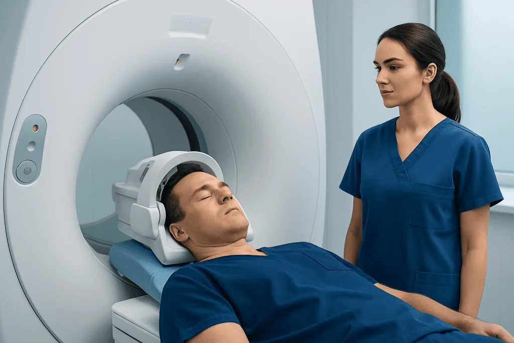
Brain MRI uses a strong magnetic field and radio waves to produce detailed images of the brain. It is especially useful for showing structural abnormalities, including stroke-related changes, tumors, multiple sclerosis plaques, inflammation, bleeding, hydrocephalus, and abnormalities of the brainstem or cranial nerves. Different MRI sequences highlight different tissue properties, which helps radiologists characterize what they see.
MRI is often the preferred test when doctors need precise anatomical detail. For example, it is commonly used to investigate headaches with warning signs, weakness, numbness, seizures, balance problems, sudden neurologic symptoms, and unexplained changes in vision or speech. It can also help guide treatment planning before surgery, radiation therapy, or ongoing neurologic care.
In some cases, a contrast agent is given through a vein to improve visualization of inflammation, tumors, blood vessels, or breakdown of the blood-brain barrier. MRI does not use ionizing radiation, which is an important advantage. However, not everyone can have MRI safely, especially people with certain implanted devices or metal fragments, although many modern implants are MRI-compatible.
What a brain PET scan shows
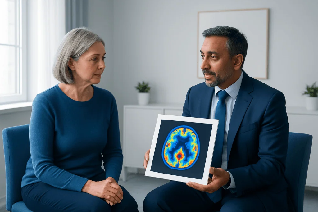
A brain PET scan shows how active different parts of the brain are. The most commonly used brain PET study measures glucose metabolism, which reflects how brain cells are using energy. Areas of unusually low or high activity can point to disease processes that may not be obvious on structural imaging alone.
PET can be helpful in evaluating dementia, epilepsy, some brain tumors, and certain inflammatory or infectious conditions. In memory disorders, PET may show characteristic patterns of reduced activity in specific brain regions that support the diagnostic picture. In epilepsy, PET can sometimes help identify an area of abnormal brain function between seizures, which may be useful when surgery is being considered.
PET involves a small amount of radioactive tracer injected into a vein. After a short uptake period, images are taken while the patient lies still on the scanner. Because PET reflects function rather than detailed anatomy, it is often interpreted together with MRI or CT. In many centers, hybrid imaging or side-by-side review helps specialists compare function with structure more precisely.
When doctors choose MRI, PET, or both
The choice between Brain PET vs MRI depends on the reason for testing. MRI is usually the first choice when a doctor suspects a structural problem, such as a recent stroke, bleeding, tumor, demyelinating disease, infection, or injury. It is also widely used when symptoms are new, focal, or urgent because it can quickly show whether there is a visible anatomical cause.
PET is often used when doctors need more information about how the brain is functioning. This may happen in complex cases of memory loss, seizure disorders, or brain tumors. For example, MRI may show a lesion, but PET may help estimate whether tissue is metabolically active, whether a tumor appears more aggressive, or whether treatment has changed the biology of the lesion.
Both tests may be used together in several situations. A person with suspected Alzheimer’s disease may have MRI to assess brain volume loss and exclude other causes, while PET may help reveal a typical metabolic pattern. A person with a brain tumor may have MRI for precise structural mapping and PET to better understand tumor activity. Someone with difficult-to-control seizures may need MRI plus functional studies as part of an advanced epilepsy workup.
Doctors also consider practical factors such as urgency, availability, prior test results, implanted devices, the need for radiation avoidance, and the person’s ability to lie still. The decision is individualized, and imaging results are always interpreted in the context of symptoms, medical history, physical examination, and laboratory findings.
Common conditions where these tests are used
In stroke care, MRI is especially valuable for showing acute ischemic injury, older infarcts, small-vessel disease, and other structural changes. PET is not usually the first-line emergency test for stroke, but in selected research or specialized settings it may contribute information about tissue metabolism or viability. In routine practice, MRI generally plays the larger role for brain stroke assessment.
For tumors, MRI is central because it shows location, size, edema, relation to surrounding structures, and contrast enhancement. PET may complement this by helping distinguish active tumor from treatment-related change in some cases. This can support planning for brain tumor surgery or further oncologic treatment, though the exact approach depends on the tumor type and the imaging tracer used.
In neurodegenerative disease, MRI may show generalized or regional atrophy and help exclude other causes of cognitive decline. PET may identify characteristic functional patterns seen in conditions such as Alzheimer’s disease or frontotemporal disorders. This can be useful when symptoms are atypical or when the diagnosis remains uncertain after clinical evaluation.
In epilepsy, MRI looks for structural causes such as cortical malformations, scarring, tumors, or prior injury. PET may help localize an area of altered metabolism when MRI is normal or unclear, especially before epilepsy surgery. In movement disorders or inflammatory diseases, doctors may also use MRI first and add PET selectively if it may change diagnosis or treatment decisions.
What to expect before, during, and after the test
Before a brain MRI, patients are asked about metal implants, pacemakers, prior surgeries, and possible metal exposure. They may need to remove jewelry, hearing aids, and other metallic items. The scan itself is painless, but it can feel noisy and enclosed. Some people receive contrast, and some may need sedation if claustrophobia or inability to stay still is a concern.
Before a brain PET scan, instructions may include fasting for several hours, depending on the tracer and the reason for the test. Blood sugar control can be important, especially for glucose-based PET imaging. A small amount of tracer is injected into a vein, followed by a waiting period to allow distribution. The scan is then performed while the person lies still.
After MRI, most people can return to normal activities right away unless they were given sedation. After PET, patients can usually resume normal routines as advised by the care team. The amount of radiation used in PET is generally low, but doctors still order it only when the expected benefit is meaningful. Results are reviewed by a radiologist or nuclear medicine specialist and then discussed with the treating physician.
In advanced centers, brain imaging may be part of coordinated care that includes neurology, neurosurgery, oncology, neuroradiology, and rehabilitation. Near the end of the care pathway, some international patients may seek evaluation at institutions such as Acibadem International, where multidisciplinary specialists and JCI-accredited hospitals assess neurologic conditions and provide treatments including Gamma Knife radiosurgery or brain MRI when appropriate.
Benefits, limits, and safety considerations
MRI offers excellent soft-tissue detail and does not expose the patient to ionizing radiation. It is particularly strong for diagnosing structural brain problems and following them over time. Its main limitations include sensitivity to movement, the need to lie still in a confined space, and safety restrictions for some metal implants or devices. Certain findings also may still need further evaluation because MRI shows structure more clearly than metabolism.
PET provides a different kind of information by showing physiologic activity. That can be a major advantage when symptoms are related to altered brain function rather than a visible structural lesion. PET may help answer questions that MRI cannot answer alone, but it is not usually the first test in many neurologic situations. It also has lower anatomical detail and uses a radioactive tracer, so it is selected thoughtfully.
Neither test should be interpreted in isolation. An abnormal scan does not automatically mean serious disease, and a normal scan does not always exclude a condition at an early stage. Doctors combine imaging with examination findings, cognitive testing, blood work, EEG, spinal fluid studies, or biopsy when needed. This step-by-step approach helps avoid overdiagnosis and improves the chance of reaching the most accurate explanation for symptoms.
When to speak with a doctor about neuroimaging
A person should speak with a doctor promptly if there are neurologic symptoms such as sudden weakness, facial drooping, new trouble speaking, confusion, severe sudden headache, seizure, unexplained loss of consciousness, or sudden vision changes. These symptoms may require urgent assessment, and imaging is often part of that evaluation. Timing matters, especially when stroke or bleeding is possible.
Medical review is also important for ongoing concerns such as progressive memory loss, persistent balance problems, repeated headaches with red-flag features, new personality or behavior changes, or seizures that are not well controlled. In these situations, the doctor can decide whether MRI, PET, another imaging test, or no imaging at all is most appropriate.
It is helpful for patients to bring prior scan reports, a list of medications, and information about implants, allergies, kidney disease, pregnancy, or diabetes. Asking why a test is being recommended can also clarify expectations. Often the most useful question is not “Which test is better?” but “Which test is best for this specific problem?”
Frequently asked questions
Is brain PET better than MRI?
Neither test is better in every situation. MRI is generally better for detailed brain structure, while PET is better for showing patterns of brain function or metabolism. Doctors choose based on the clinical question.
Why would someone need both a brain MRI and a PET scan?
Some conditions require both structural and functional information. MRI may show where a problem is located, while PET may show how active or abnormal that tissue is. Using both can improve diagnostic confidence in selected cases.
Does a brain PET scan show dementia?
A brain PET scan does not diagnose dementia by itself, but it can show activity patterns that support certain types of dementia. Doctors interpret PET findings together with symptoms, examination, cognitive testing, and often MRI.
Which scan is used more often for seizures?
MRI is commonly used first to look for a structural cause of seizures. PET may be added when seizures are difficult to control or when doctors are trying to localize the seizure focus more precisely, especially before surgery.
Is MRI safer than PET?
MRI does not use ionizing radiation, which is an important safety advantage. PET uses a small amount of radioactive tracer, but it is generally considered safe when medically indicated. Each test also has its own precautions, such as metal-device screening for MRI.
Can a brain MRI miss problems that PET can find?
Yes. MRI can sometimes appear normal or only mildly abnormal even when brain function is altered. PET may reveal abnormal metabolic patterns that help explain symptoms in selected conditions.
References
- Radiological Society of North America
- American College of Radiology
- National Institute of Neurological Disorders and Stroke
- Alzheimer's Association
- Society of Nuclear Medicine and Molecular Imaging
This article is for general information only and is not a substitute for professional medical advice. Please consult a qualified doctor about your individual situation.
Medical weight-loss options in Turkey — costs & eligibility
JCI-accredited · board-certified surgeons · reply within 24h
Add us as a Preferred Source to see more of our trusted health content across Google Search, AI Overviews and Discover.