Brain Vessel Imaging: MRA vs CTA vs Cerebral Angiography

MRA, CTA, and cerebral angiography all visualize brain blood vessels, but they differ in detail, speed, and invasiveness. MRA usually does not use ionizing radiation and is often useful for planned, non-emergency evaluation.
Key Takeaways
- MRA, CTA, and cerebral angiography all visualize brain blood vessels, but they differ in detail, speed, and invasiveness.
- MRA usually does not use ionizing radiation and is often useful for planned, non-emergency evaluation.
- CTA is fast and widely available, making it especially helpful in urgent settings such as suspected stroke or bleeding.
- Cerebral angiography is an invasive catheter-based test, but it provides the most detailed images and can also allow treatment during the same procedure.
- The safest and most useful imaging test depends on symptoms, kidney function, allergies, metal implants, and the need for possible intervention.
Brain vessel imaging helps doctors examine the arteries and veins of the brain to look for problems such as aneurysms, narrowing, malformations, or blocked blood flow. MRA, CTA, and cerebral angiography each have different strengths, and the best choice depends on the clinical question, urgency, and the person’s overall health.
Overview: what brain vessel imaging shows
Brain vessel imaging refers to tests that show the arteries and veins supplying the brain. These tests help doctors evaluate blood flow and identify structural problems that may increase the risk of stroke, bleeding, headaches, or other neurological symptoms. The three commonly compared methods are magnetic resonance angiography (MRA), computed tomography angiography (CTA), and cerebral angiography, which is also called catheter angiography or digital subtraction angiography.
Although all three tests look at blood vessels, they do so in different ways. MRA uses magnetic fields and radio waves. CTA uses X-rays together with contrast dye to create detailed images quickly. Cerebral angiography is a minimally invasive procedure in which a thin catheter is guided through a blood vessel and contrast dye is injected directly to produce very precise images.
Doctors may request brain vessel imaging for many reasons. Common examples include suspected brain aneurysm, blood vessel narrowing, arteriovenous malformation, vascular causes of headache, transient ischemic attack, or emergency evaluation of stroke. In some situations, imaging is also used to plan surgery or endovascular treatment, or to follow a known condition over time.
MRA: how it works, benefits, and limits
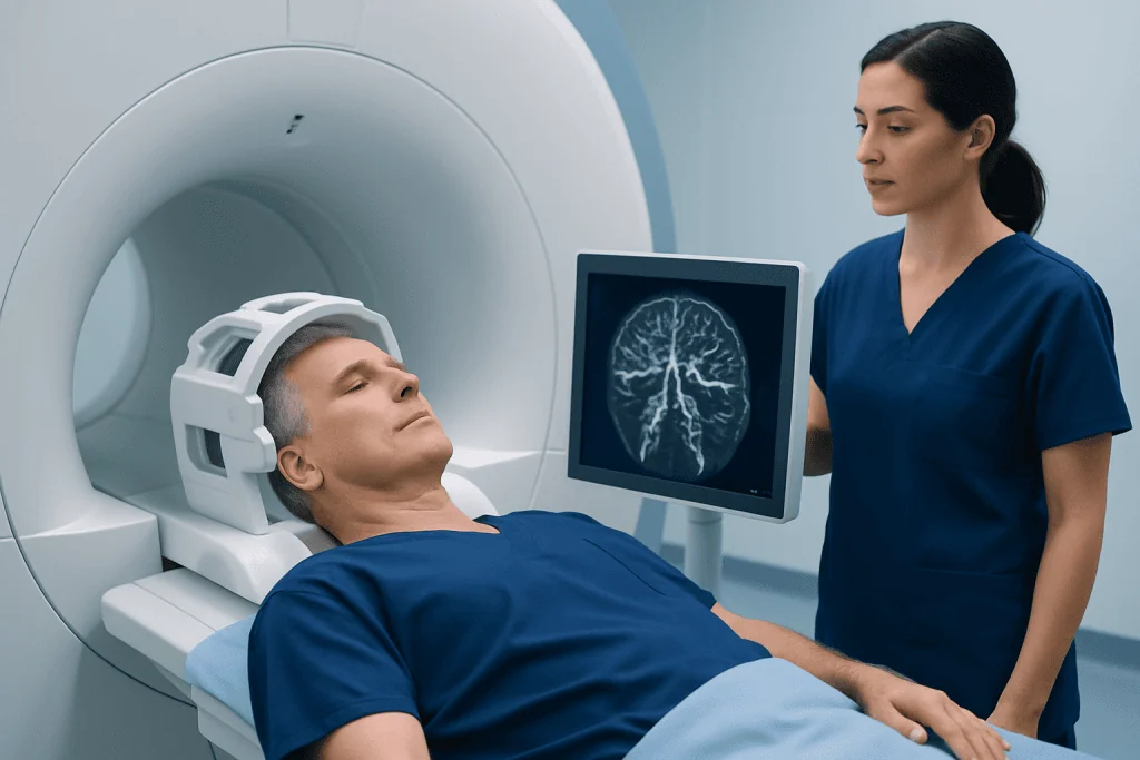
MRA is a special type of MRI designed to highlight blood vessels. It can often be performed without contrast, though some cases require gadolinium-based contrast to improve visualization. Because MRA does not use ionizing radiation, it is often preferred when repeated imaging may be needed or when avoiding radiation is especially important.
One of the main advantages of MRA is safety and convenience in selected patients. It can show many abnormalities of the brain’s arteries, including aneurysms and areas of narrowing. It is often helpful for outpatient assessment, follow-up imaging, and situations where doctors need a noninvasive overview of the vascular system.
MRA does have limits. It generally takes longer than CTA, and some people find the MRI scanner uncomfortable because of noise or a confined space. Motion can reduce image quality, and certain implanted devices or metal fragments may make MRI unsafe or unsuitable. MRA may also be less detailed than catheter angiography for very small vessels or complex abnormalities.
When a doctor recommends MRA, the goal is usually to answer a focused question while minimizing risk. In many people, it provides enough information to guide treatment decisions without the need for an invasive procedure.
CTA: when speed and accessibility matter

CTA combines a CT scan with contrast dye injected into a vein, usually in the arm. The scan is fast, often completed within minutes, and creates detailed cross-sectional images of the brain’s blood vessels. Because it is quick and widely available, CTA is frequently used in emergency departments and stroke centers.
CTA is especially valuable when doctors need rapid answers. It can help detect large-vessel blockage, aneurysm, dissection, bleeding-related vascular changes, and significant arterial narrowing. In acute neurological symptoms, CTA may be part of the workup alongside standard brain CT and other imaging to determine the cause and guide urgent care, including stroke treatment.
The main limitations of CTA are radiation exposure and the need for iodine-based contrast dye. Some people cannot receive this contrast safely because of severe allergy history or reduced kidney function. CTA also can be affected by calcification or nearby bone, which may occasionally make interpretation more difficult in certain areas.
Even with these limits, CTA remains one of the most practical and informative first-line tests when timing is critical. It often provides an excellent balance of speed, detail, and availability.
Cerebral angiography: the most detailed test
Cerebral angiography is considered the reference standard for the most detailed evaluation of brain blood vessels. During the procedure, a specialist inserts a thin catheter, usually through an artery in the wrist or groin, and carefully guides it to the vessels supplying the brain. Contrast dye is then injected while X-ray images are taken in real time.
This test can reveal very fine details that may not be visible on MRA or CTA. It is particularly useful when doctors need to fully define an aneurysm before treatment, assess a complex arteriovenous malformation, investigate unusual vascular anatomy, or confirm uncertain findings from other scans. In some cases, the same session can move directly from diagnosis to treatment, such as brain aneurysm treatment or endovascular therapy for a blocked vessel.
Because cerebral angiography is invasive, it carries more risk than MRA or CTA. Possible complications include bruising at the puncture site, bleeding, reaction to contrast dye, kidney stress, or rarely stroke from the procedure itself. For that reason, it is usually reserved for situations where the extra detail will clearly affect care or when a therapeutic procedure may be needed.
Despite being more involved, cerebral angiography plays a central role in interventional neuroradiology. It does not simply diagnose disease; it can also open the door to image-guided treatment in carefully selected patients.
How doctors choose between MRA, CTA, and angiography
There is no single “best” test for everyone. The choice depends on the person’s symptoms, how urgent the situation is, and what problem the doctor is trying to confirm or rule out. A patient with sudden weakness or speech difficulty may need urgent CTA because of its speed, while someone being monitored for a stable vascular condition may be better suited to MRA.
Doctors also consider medical factors such as kidney function, pregnancy status, contrast allergies, and implanted devices. MRA may be favored when avoiding radiation is important. CTA may be preferred when the patient cannot have MRI or when emergency access is essential. Cerebral angiography is often chosen when the diagnosis remains unclear or when there is a high likelihood that an endovascular procedure may be performed during the same session, such as embolization.
Another consideration is the level of detail needed. Small aneurysms, subtle vessel abnormalities, or treatment planning for complex lesions may require catheter angiography. In contrast, a broader screening or follow-up question may be answered well by MRA or CTA alone.
In practice, these tests are often complementary rather than competitive. A doctor may begin with one imaging method and then recommend another if more detail is needed to guide safe and effective care.
What patients can expect before, during, and after each test
Preparation depends on the test being performed. Before MRA or CTA, patients are usually asked about kidney disease, allergies, pregnancy, and previous reactions to contrast. Before MRI-based tests, they are also screened for pacemakers, cochlear implants, clips, metal fragments, and other devices. Fasting may be recommended in some cases, especially if contrast will be used.
During MRA, the patient lies still on a table that moves into the scanner. During CTA, contrast is injected through a vein and the CT scanner captures images very quickly. For cerebral angiography, the care team cleans the access site, gives local anesthesia and sometimes light sedation, and then performs the catheter procedure under imaging guidance. The person remains monitored throughout.
After MRA or CTA, most people can return to normal activities soon unless they received medication that causes drowsiness. After cerebral angiography, a longer observation period is needed. The access site must be monitored for bleeding, and patients are usually advised to rest, drink fluids if appropriate, and follow discharge instructions carefully.
Many people feel less anxious when they know what to expect. Asking questions in advance about preparation, contrast use, recovery, and when results will be available can make the process easier and more comfortable.
Risks, safety considerations, and special situations
All medical imaging decisions balance benefit and risk. MRA avoids radiation, but not every patient can undergo MRI safely. CTA involves radiation and iodine-based contrast, though for many people the risk is low and acceptable when the test is medically necessary. Cerebral angiography provides unmatched detail but has the highest procedure-related risk because it requires catheter access to the arteries.
Kidney function is an important consideration for contrast-enhanced CTA and angiography, and sometimes for contrast-enhanced MRA. People with previous contrast reactions should inform their doctors well before the scan. Claustrophobia, inability to lie flat, or severe agitation may also affect which test is most practical. In children, older adults, or medically fragile patients, the team may adapt the imaging plan to reduce stress and maximize safety.
Some patients need follow-up over time, for example after treatment of an aneurysm or vascular malformation. In that setting, doctors may choose different imaging methods at different stages. A noninvasive test may be used for surveillance, while angiography may be reserved for cases where results are uncertain or treatment planning is needed, including for conditions such as arteriovenous malformation.
Near the end of the care journey, many patients benefit from discussing results with both the referring neurologist or neurosurgeon and the imaging specialist. At centers such as Acibadem International, multidisciplinary specialists and JCI-accredited hospitals evaluate these conditions and provide diagnostic and interventional care for international patients.
When to seek medical attention
Brain vessel imaging is usually arranged by a doctor after symptoms, examination findings, or another scan suggest a vascular problem. People should seek urgent medical attention for sudden weakness, facial droop, trouble speaking, severe unexplained headache, sudden vision loss, new confusion, seizures, or loss of balance. These symptoms may have many causes, but some require immediate evaluation.
Non-urgent symptoms still deserve medical review if they are persistent or unexplained. Recurrent brief neurological symptoms, pulsating noises in the head, unexplained dizziness with other neurological signs, or a family history of certain vascular conditions may lead a doctor to consider imaging. The timing and type of scan depend on the full clinical picture.
It is helpful for patients to bring a list of medications, allergies, previous imaging results, and details about any implants or kidney problems. This information helps the care team choose the safest and most informative test.
Brain vessel imaging can sound complex, but the purpose is straightforward: to understand blood flow in the brain clearly enough to make informed decisions. With the right test for the right situation, doctors can often diagnose problems accurately and plan treatment with confidence.
Frequently asked questions
Which is better, MRA or CTA for brain vessels?
Neither is always better in every situation. MRA is noninvasive and avoids radiation, while CTA is faster and often preferred in emergencies. The best choice depends on the symptoms, urgency, medical history, and what the doctor needs to see.
Is cerebral angiography the same as MRA or CTA?
No. Cerebral angiography is a catheter-based procedure, while MRA and CTA are noninvasive imaging tests. Angiography is more detailed but also more invasive, so it is usually used when precise definition or possible treatment is needed.
Can brain vessel imaging detect an aneurysm?
Yes. MRA, CTA, and cerebral angiography can all help detect a brain aneurysm. Small aneurysms or complex anatomy may sometimes require catheter angiography for the clearest assessment.
Does brain vessel imaging hurt?
MRA and CTA are generally painless, aside from the brief discomfort of an IV if contrast is used. Cerebral angiography may cause some pressure or soreness at the catheter entry site, but local anesthesia is used and patients are monitored closely.
Are there risks from the contrast dye?
Contrast dye is commonly used safely, but it can cause problems in some people, especially those with kidney disease or a history of contrast allergy. Doctors review these risks before the test and may choose a different imaging method if needed.
How long does it take to get results?
The timing varies by hospital and by how urgent the situation is. In emergency care, results may be reviewed immediately, while non-urgent outpatient scans may take longer. The treating doctor or radiologist usually explains the findings and next steps.
References
- American Heart Association
- American Stroke Association
- Radiological Society of North America
- National Institute of Neurological Disorders and Stroke
- Society of NeuroInterventional Surgery
This article is for general information only and is not a substitute for professional medical advice. Please consult a qualified doctor about your individual situation.
Explore treatments in Turkey — costs, top hospitals & a free quote
JCI-accredited · board-certified surgeons · reply within 24h
Add us as a Preferred Source to see more of our trusted health content across Google Search, AI Overviews and Discover.

Neurosurgery
Surgical treatment of the brain, spine and nervous system, including minimally invasive and functional procedures.
72 specialists in this unit