Breast Ultrasound: When Women Need It and What the Results Can Show
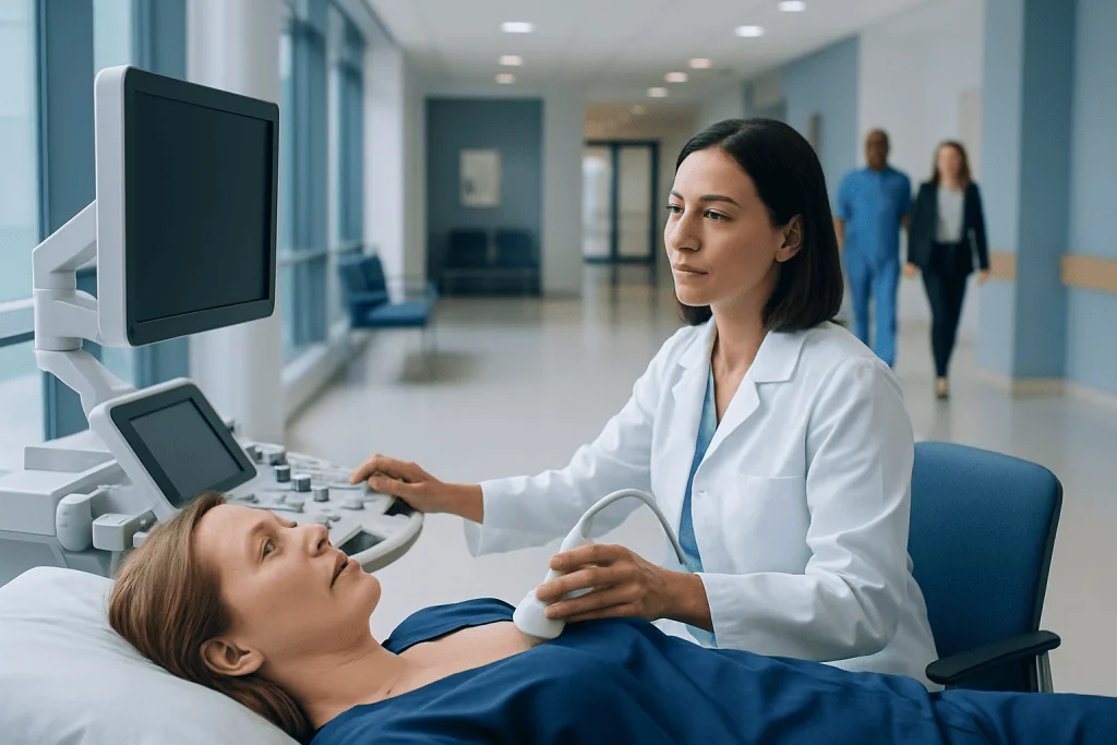
Breast ultrasound uses sound waves, not radiation, to create images of breast tissue. It is often used to evaluate a lump, focal pain, nipple discharge, or an abnormal mammogram.
Key Takeaways
- Breast ultrasound uses sound waves, not radiation, to create images of breast tissue.
- It is often used to evaluate a lump, focal pain, nipple discharge, or an abnormal mammogram.
- Ultrasound can help distinguish a simple cyst from a solid mass, but sometimes more tests are still needed.
- Results may be described as benign, probably benign, suspicious, or needing follow-up imaging or biopsy.
- A breast ultrasound is usually quick, noninvasive, and does not require special preparation.
Breast ultrasound is a common imaging test that helps doctors examine breast tissue, especially when there is a lump, pain, nipple discharge, or an unclear mammogram finding. It does not use radiation and can help show whether a breast change is fluid-filled, solid, likely benign, or in need of further testing.
Overview: What a Breast Ultrasound Is
A breast ultrasound is an imaging test that uses high-frequency sound waves to produce pictures of the inside of the breast. A handheld device called a transducer is moved over the skin after gel is applied. The sound waves bounce off tissues and create real-time images that a radiologist can interpret.
This test is commonly used as a diagnostic tool rather than a routine screening test for most women. It is especially helpful when a woman or her doctor feels a breast lump, when there is localized breast pain, or when a mammogram shows an area that needs a closer look. Ultrasound is also often useful in younger women, whose breast tissue tends to be denser.
Unlike mammography, ultrasound does not use ionizing radiation. It is generally painless, although mild pressure from the transducer may be uncomfortable if the breast is tender. In many cases, it provides important added detail when combined with other forms of breast imaging.
When Women May Need a Breast Ultrasound
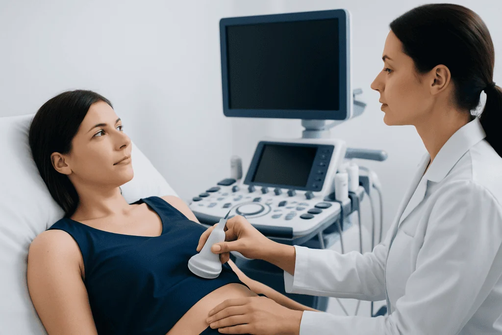
Doctors may recommend a breast ultrasound for several reasons. One of the most common is to evaluate a palpable lump. Although many breast lumps are benign, imaging helps clarify what the lump may be and whether it needs monitoring or biopsy. Ultrasound is also often ordered when a screening mammogram detects an area that is not fully clear.
Other reasons include focal breast pain, nipple discharge, skin thickening, redness, swelling, or changes around the nipple. In women with dense breasts, ultrasound may sometimes be used as a supplemental test, depending on individual risk factors and local practice. It can also help assess findings during pregnancy or breastfeeding, when breast tissue changes can make evaluation more challenging.
Breast ultrasound may be used to guide procedures as well. If a suspicious area needs sampling, the radiologist can use real-time ultrasound to perform a breast biopsy more precisely. Ultrasound guidance may also be used to drain a painful cyst or to mark an area before further treatment.
What the Test Can Show
One of the main strengths of breast ultrasound is its ability to distinguish between a fluid-filled cyst and a solid mass. A simple cyst often appears with features that strongly suggest a benign finding. Solid masses can also be described in detail, including their shape, margins, orientation, and internal appearance.
Radiologists look for imaging characteristics that suggest whether a finding is more likely benign or whether it may need more evaluation. For example, a smooth, oval mass may be more reassuring than one with irregular edges. However, ultrasound alone cannot always determine exactly what a mass is. Some benign findings and some cancers can overlap in appearance.
Ultrasound can also show enlarged lymph nodes in the armpit, areas of inflammation, collections of fluid, and tissue changes related to infection or trauma. In certain cases, it may help identify features seen in conditions such as breast cancer, but the diagnosis itself usually depends on tissue sampling if imaging is suspicious.
How to Prepare and What Happens During the Exam
Preparation for a breast ultrasound is usually simple. Women are often advised to wear a two-piece outfit so the upper body can be uncovered easily. It is helpful to avoid applying lotions, powders, or creams to the chest area on the day of the exam, as these can interfere with the gel or the transducer.
During the exam, the patient lies on an examination table, often with one arm raised above the head. A clear gel is placed on the skin, and the transducer is moved across the breast and sometimes the underarm area. The exam is usually completed within a short visit, though timing can vary depending on the number of areas being assessed.
There is no radiation exposure, and most women can return to normal activities immediately afterward. The sonographer may perform the scan, but a radiologist interprets the images. In some centers, the radiologist may review the findings during the visit and decide whether additional views or tests are needed.
Understanding Breast Ultrasound Results
Breast ultrasound results are usually reported in a structured way. A finding may be described as normal, benign, probably benign, suspicious, or highly suggestive of malignancy. Reports often use the BI-RADS system, which helps standardize how imaging findings are communicated and what next steps may be recommended.
A normal result means no concerning abnormality was seen in the area examined. Benign findings may include simple cysts or other clearly noncancerous changes. A probably benign finding often does not need immediate biopsy, but it may require short-interval follow-up imaging to confirm that it remains stable over time.
If a result is suspicious, the radiologist may recommend further evaluation. This may include additional breast MRI, repeat mammographic views, or a biopsy to obtain a tissue diagnosis. It is important to remember that a suspicious ultrasound does not automatically mean cancer; it means the area needs a closer and more definite assessment.
Results are interpreted in the context of age, symptoms, physical examination, family history, and any prior imaging. Comparing current images with earlier studies can be especially useful in determining whether a finding is new, unchanged, or evolving.
Limits of Breast Ultrasound and When Other Tests Are Needed
Although breast ultrasound is very useful, it has limitations. It is not a replacement for mammography in routine breast cancer screening for most women. Mammography remains better at detecting some findings, such as tiny calcium deposits called microcalcifications, which may be associated with early cancer.
Ultrasound is also operator-dependent, meaning image quality and completeness can vary based on technique and experience. Some areas of the breast may be harder to assess fully, and some abnormalities may still be indeterminate even after careful scanning. For this reason, doctors often combine ultrasound findings with clinical examination and other imaging studies.
Depending on the situation, a woman may need mammography, MRI, or tissue sampling after ultrasound. Additional tests are especially important if there is a persistent lump, a growing lesion, or a result that does not match the physical exam. A coordinated evaluation helps avoid both unnecessary worry and missed diagnoses.
Follow-Up, Self-Care, and When to See a Doctor
After a breast ultrasound, the most important step is to follow the recommended plan. Some women will simply be reassured. Others may be advised to return for repeat imaging after a set interval, especially if a finding appears probably benign. If a biopsy is recommended, arranging it promptly can provide a clearer answer and help guide next steps.
Women should also continue regular breast health care based on their age, symptoms, and risk factors. This may include routine screening, awareness of normal breast changes, and discussing any family history of breast or ovarian cancer with a doctor. New symptoms should not be ignored simply because a previous test was normal, especially if the change persists.
A doctor should be consulted if there is a new breast lump, persistent focal pain, nipple discharge that is bloody or spontaneous, skin dimpling, redness that does not improve, swelling, or changes in breast size or shape. If specialized care is needed, Acibadem International’s multidisciplinary specialists and JCI-accredited hospitals evaluate breast concerns for international patients using modern imaging and coordinated treatment planning.
Frequently asked questions
Is a breast ultrasound painful?
A breast ultrasound is usually not painful. Most women feel only light pressure from the transducer, though the area may feel tender if the breast is already sore or inflamed.
Can a breast ultrasound detect cancer?
A breast ultrasound can show features that may suggest cancer, but it usually cannot confirm the diagnosis on its own. If an area looks suspicious, a biopsy is often needed to determine exactly what it is.
Is breast ultrasound better than mammography?
These tests do different jobs and often complement each other. Ultrasound is especially helpful for evaluating a specific concern such as a lump, while mammography remains the standard screening test for most women.
Why might a doctor order an ultrasound after a mammogram?
A doctor may order ultrasound if a mammogram shows an area that needs closer evaluation or if the breasts are dense. Ultrasound can add detail and help determine whether a finding is a cyst, a solid mass, or normal tissue.
What does it mean if the result says 'probably benign'?
Probably benign means the finding looks very unlikely to be cancer, but follow-up imaging is recommended to confirm that it stays stable. This approach helps avoid unnecessary biopsy while still monitoring the area safely.
Do women need any preparation before a breast ultrasound?
Special preparation is usually not required. Wearing a two-piece outfit can make the visit easier, and it is best to avoid lotions or powders on the chest area before the exam.
References
- American College of Radiology
- Radiological Society of North America
- National Cancer Institute
- Centers for Disease Control and Prevention
- World Health Organization
This article is for general information only and is not a substitute for professional medical advice. Please consult a qualified doctor about your individual situation.
Ultrasound in Turkey — costs, top hospitals & a free quote
JCI-accredited · board-certified surgeons · reply within 24h
Add us as a Preferred Source to see more of our trusted health content across Google Search, AI Overviews and Discover.

Check-up & Preventive Medicine
Comprehensive health screenings designed for early detection and prevention.
3 specialists in this unit