Calcified Pineal Gland — Explained by Medical Evidence, Not Myths

A calcified pineal gland is commonly found on brain imaging and is often a normal age-related change. Calcification itself usually does not cause symptoms; symptoms are more likely related to another neurologic issue or a pineal-region lesion.
Key Takeaways
- A calcified pineal gland is commonly found on brain imaging and is often a normal age-related change.
- Calcification itself usually does not cause symptoms; symptoms are more likely related to another neurologic issue or a pineal-region lesion.
- Doctors interpret pineal calcification in context with age, symptoms, and imaging appearance.
- Further evaluation may be needed if there are headaches with warning signs, vision changes, hydrocephalus, or an unusual mass near the pineal gland.
- Treatment depends on the underlying cause, not on the calcium deposit alone.
A calcified pineal gland usually refers to calcium deposits in the pineal gland that are seen on a CT scan or other brain imaging. In most adults, this is a common incidental finding rather than a disease, but doctors may look more closely if there are symptoms, unusual imaging features, or concern for a nearby pineal-region condition.
What a calcified pineal gland means
A calcified pineal gland means that calcium has built up within the pineal gland, a small structure deep in the brain. This finding is commonly seen on CT scans because calcium appears clearly on that type of imaging. In many adults, it is considered an incidental finding, meaning it is discovered while imaging is being done for another reason.
The pineal gland helps regulate the sleep-wake cycle through melatonin production. Even so, visible calcification on a scan does not automatically mean the gland has stopped working or that a person will develop health problems. Imaging findings and symptoms do not always match, so the result needs to be interpreted carefully rather than assumed to be dangerous.
Medical evidence shows that pineal calcification becomes more common with age. For that reason, doctors often view a small or moderate amount of calcification in an adult as a normal variation. The more important questions are whether there are any neurologic symptoms, whether the appearance is typical, and whether there is any sign of a mass, blockage of cerebrospinal fluid, or another brain condition that needs attention.
Why pineal calcification happens
The exact reason the pineal gland calcifies is not fully understood, but it is generally linked to normal aging and tissue changes over time. Calcium can collect in many tissues in the body, and the pineal gland is one of the places where this is especially visible on imaging.
Researchers have also explored links between calcification, melatonin production, sleep patterns, and environmental or metabolic influences. However, these associations do not prove that calcification directly causes symptoms in most people. A person may have a calcified pineal gland and feel completely well.
Doctors may think about other explanations when calcification appears unusual for the patient’s age, is very irregular, or is associated with a nearby lesion. In some cases, calcification can be seen within or around a pineal cyst or another pineal-region abnormality, which is why the overall imaging pattern matters more than the word calcified alone.
Symptoms: usually none, but context matters

Most people with a calcified pineal gland have no symptoms from the calcification itself. It is often found during imaging done for headaches, trauma, dizziness, or unrelated neurologic complaints, and the calcification turns out not to be the cause.
When symptoms are present, doctors consider whether another condition may be responsible. Important symptoms that need proper assessment include persistent or worsening headaches, nausea and vomiting, double vision, difficulty looking upward, balance problems, unusual sleep disturbances, or signs of increased pressure inside the skull. These symptoms can suggest a pineal-region mass, hydrocephalus, or another neurologic problem rather than simple age-related calcification.
In children and younger adolescents, pineal calcification may prompt a closer look because it is less expected than in adults. Age, symptom pattern, and scan findings together help doctors decide whether reassurance is enough or whether more testing is needed.
- Common incidental finding: no symptoms, no treatment needed
- Symptoms needing evaluation: new severe headache, visual changes, repeated vomiting, imbalance, confusion
- Red flags on imaging: mass effect, enlarged brain ventricles, or atypical calcification pattern
How doctors evaluate a calcified pineal gland
Evaluation begins with the reason imaging was done in the first place. A doctor will ask about headaches, vision changes, sleep issues, hormone concerns, past neurologic conditions, and any recent injuries or infections. The medical history and neurologic examination help determine whether the calcification is likely incidental.
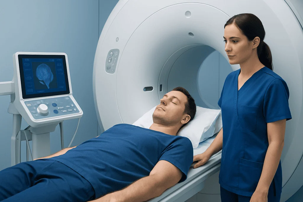
CT scans are especially good at showing calcium, while MRI gives more detail about the surrounding brain tissue, the pineal region, and the presence of a cyst or tumor. If the calcification is typical and there are no concerning symptoms, no further work-up may be needed. If there is uncertainty, the doctor may recommend brain MRI to better define the finding.
Additional tests depend on what the imaging suggests. If a lesion is suspected, doctors may assess the flow of cerebrospinal fluid, look for hydrocephalus, and evaluate whether specialist input from neurology or neurosurgery is needed. In selected situations, referral for neurosurgical evaluation can help determine whether monitoring or treatment is appropriate.
When calcification may be linked to a pineal-region condition
Calcification can sometimes be seen within a benign pineal cyst or, less commonly, in tumors arising in or near the pineal region. The key issue is not the calcium alone but whether there is an abnormal structure causing pressure on nearby parts of the brain. This may affect the normal flow of cerebrospinal fluid and lead to hydrocephalus, a condition that can produce headaches, nausea, drowsiness, or eye movement problems.
Doctors also look at the size, shape, and margins of the pineal area on imaging. A small, typical calcified focus in an adult is very different from a large irregular lesion, a mass with contrast enhancement, or enlarged ventricles. If hydrocephalus is suspected, evaluation for hydrocephalus becomes important because timely treatment can relieve pressure on the brain.
In rare cases, a suspicious pineal-region lesion may need advanced imaging, interval follow-up, or tissue diagnosis. Management then focuses on the underlying diagnosis rather than the calcification. This is why a careful radiology report and specialist review can be more useful than trying to interpret the word calcified in isolation.
Treatment options and what to expect
There is no treatment aimed at removing ordinary age-related pineal calcification, and most people do not need any intervention. If the finding is incidental and the person has no concerning symptoms, reassurance is often the most appropriate approach. Some patients may simply be advised to follow up with their doctor if new neurologic symptoms develop.
When symptoms have another cause, treatment targets that cause. For example, headache management depends on the headache type, sleep concerns may be addressed with lifestyle or medical guidance, and a pineal-region lesion may require monitoring or specialist treatment. If imaging identifies a mass, cyst with pressure effects, or blocked cerebrospinal fluid pathways, management can range from repeat imaging to surgery.
Potential interventions vary by diagnosis and may include specialist monitoring, endoscopic third ventriculostomy for selected cases of obstructed cerebrospinal fluid flow, or other neurosurgical procedures when clearly indicated. Near the end of the care pathway, some international patients may choose centers such as Acibadem International, where multidisciplinary specialists and JCI-accredited hospitals evaluate and treat pineal-region conditions based on imaging findings and clinical need.
Self-care, myths, and when to seek medical care
Many online discussions portray a calcified pineal gland as a hidden cause of major physical or spiritual problems. Medical evidence does not support broad claims that pineal calcification by itself explains complex symptoms or requires detox regimens, supplements, or unproven therapies. Self-treatment based on internet myths can delay proper diagnosis of the real issue.
For general brain and sleep health, people can focus on practical measures: keeping a regular sleep schedule, limiting evening light exposure, managing vascular risk factors, avoiding smoking, and discussing persistent symptoms with a clinician. These habits support overall health, but they do not specifically reverse pineal calcification.
Medical care should be sought promptly for severe or worsening headache, repeated vomiting, double vision, fainting, confusion, seizures, new weakness, difficulty walking, or unusual drowsiness. A doctor should also review the finding if a scan report mentions a mass, hydrocephalus, or an atypical pineal lesion. In most cases, however, a calcified pineal gland on its own is simply an imaging observation that needs context, not fear.
Frequently asked questions
Is a calcified pineal gland normal?
In many adults, yes. Pineal calcification is a common finding on brain imaging and is often considered a normal age-related change rather than a disease.
Can a calcified pineal gland cause headaches?
Usually, no. Headaches often lead to imaging that discovers the calcification incidentally, but the calcium deposit itself is not usually the cause. Doctors look for other explanations if headaches are severe, new, or associated with neurologic symptoms.
Does pineal gland calcification affect sleep or melatonin?
Researchers have studied possible links, but a visible calcification on imaging does not automatically mean a person will have melatonin problems or sleep disorders. Sleep symptoms should be assessed as a separate clinical issue rather than blamed on the scan finding alone.
Should a calcified pineal gland be removed or treated?
Most cases do not need treatment at all. Treatment is considered only if there is an underlying problem, such as a cyst, tumor, or hydrocephalus causing symptoms or pressure on nearby brain structures.
What tests are usually done after this finding?
Often, no further testing is needed if the calcification is typical and there are no concerning symptoms. If the appearance is unusual or symptoms suggest a pineal-region condition, a doctor may recommend MRI and specialist evaluation.
Is a calcified pineal gland more concerning in children?
It can deserve closer attention because pineal calcification is less expected in younger children than in adults. Doctors interpret the finding based on age, symptoms, and the exact imaging appearance.
This article is for general information only and is not a substitute for professional medical advice. Please consult a qualified doctor about your individual situation.
Explore treatments in Turkey — costs, top hospitals & a free quote
JCI-accredited · board-certified surgeons · reply within 24h
Add us as a Preferred Source to see more of our trusted health content across Google Search, AI Overviews and Discover.

More from the Health Library

Mylicon Drops: An Evidence-Based Guide for Patients

Astaxanthin: What Patients Need to Know

Intertrigo Pictures — Explained by Medical Evidence, Not Myths

Hemorrhagic Cyst: A Complete Medical Overview

Pulse Pressure — Explained by Medical Evidence, Not Myths