Can You See Blood Clots in an Xray: Preparation, Procedure and Results

Standard X-rays do not usually show blood clots in veins or arteries directly. Ultrasound is commonly used to look for deep vein thrombosis in the legs or arms.
Key Takeaways
- Standard X-rays do not usually show blood clots in veins or arteries directly.
- Ultrasound is commonly used to look for deep vein thrombosis in the legs or arms.
- CT pulmonary angiography is often used when a pulmonary embolism is suspected.
- Sudden shortness of breath, chest pain, coughing blood, or one-sided leg swelling needs urgent medical assessment.
- Blood clots may cause few or no symptoms, so risk factors and clinical assessment are important.
Can you see blood clots in an Xray? A standard X-ray usually cannot show a clot directly, although it may help doctors identify other causes of symptoms or indirect complications. When a blood clot is suspected, clinicians typically use ultrasound, CT angiography, blood tests, or other targeted imaging based on where the clot may be located.
Overview: Can You See Blood Clots in an Xray?
Can you see blood clots in an Xray? In most cases, no. A standard X-ray uses a small amount of radiation to create images of dense structures such as bones and, to a lesser extent, the lungs and heart silhouette. Blood vessels and the clots inside them generally do not appear clearly enough on a routine X-ray to confirm or exclude a clot.
An X-ray may still be part of an assessment for symptoms such as chest pain, breathlessness, or leg discomfort. For example, a chest X-ray can help identify pneumonia, a collapsed lung, fluid around the lungs, or some heart and lung changes. These findings can guide next steps, but a normal X-ray does not rule out a blood clot in the leg or lungs.
Doctors choose clot-testing based on the suspected location and the person’s symptoms, examination, medical history, and risk factors. A clot in a deep leg vein is often assessed with ultrasound, while a suspected clot in the lungs may require a CT pulmonary angiogram or, in selected situations, a ventilation-perfusion scan.
Will a Regular X-ray Show a Blood Clot?
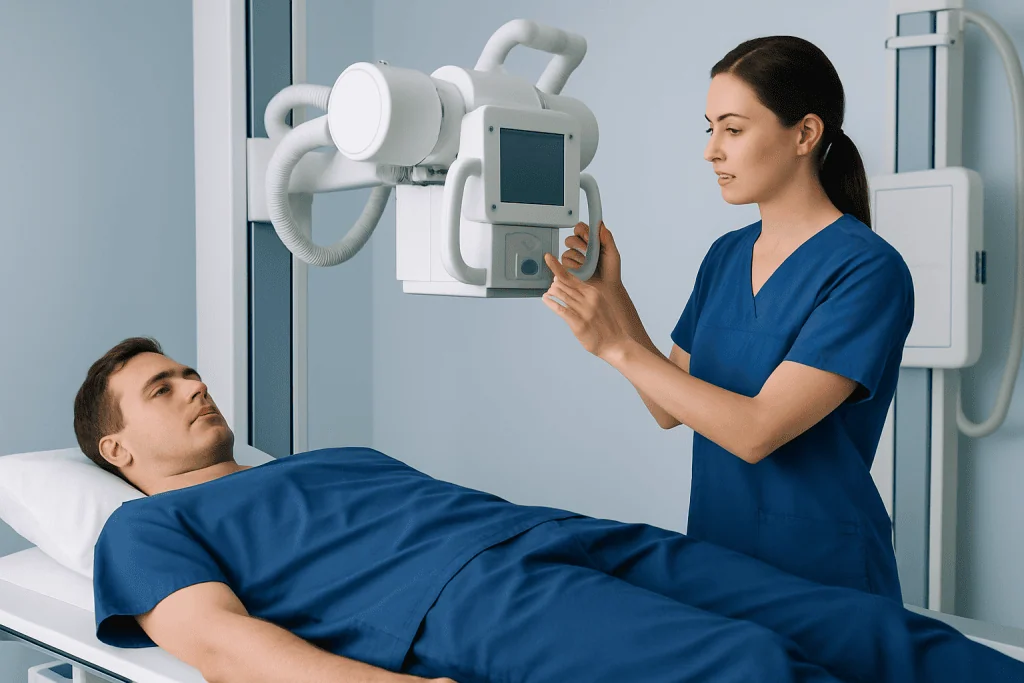
A regular X-ray will not usually show a blood clot in a vein or artery. This is because ordinary X-rays are not designed to distinguish flowing blood from a clot within most blood vessels. They are particularly limited for detecting deep vein thrombosis, also called DVT, which most often affects the deep veins of the leg.
In some situations, an X-ray may show indirect signs that prompt further investigation. A chest X-ray, for example, may reveal an alternative reason for shortness of breath or chest symptoms. However, people with a pulmonary embolism, a clot that has travelled to the lungs, may have a normal chest X-ray.
Specialized X-ray-based procedures using contrast dye can image blood vessels, but they are not the usual first test for most suspected clots. Modern ultrasound and CT-based examinations are often more practical and informative. If symptoms suggest a clot, the right test should be arranged promptly rather than relying on a routine X-ray.
What Are the First Signs of a Blood Clot?

The first signs of a blood clot depend on where it forms. A deep vein clot in the leg may cause new swelling in one leg, pain or tenderness, warmth, redness, or a feeling of heaviness. Symptoms can begin gradually or suddenly, and not every person experiences all of them.
A pulmonary embolism may cause unexplained shortness of breath, chest pain that can worsen with breathing, a fast heartbeat, cough, dizziness, or fainting. Some people may cough up blood, although this is less common. These symptoms can also occur with other conditions, so they require medical evaluation rather than self-diagnosis.
Clots can also occur in other parts of the body. Warning signs may include sudden weakness or difficulty speaking with a stroke, sudden severe abdominal pain with some abdominal clots, or a cold, pale, painful limb with an arterial blockage. Sudden or severe symptoms should be assessed urgently.
- One-sided leg swelling, pain, warmth, or redness may suggest DVT.
- New breathlessness or chest pain may indicate a pulmonary embolism.
- Sudden neurological symptoms, fainting, or severe limb pain require emergency care.
How Long Can You Have a Blood Clot Without Knowing?
A person can have a blood clot without noticing it for days, weeks, or occasionally longer. Some clots cause mild symptoms that may be mistaken for a muscle strain, fatigue, or another common problem. Others produce no clear symptoms until they grow, block blood flow more significantly, or move to another location.
It is not possible to safely estimate how long an individual clot has been present based on symptoms alone. The chance of complications depends on the clot’s location, size, underlying cause, and whether treatment is started. A suspected clot should therefore be evaluated promptly, even if symptoms are mild or intermittent.
Risk may be higher after recent surgery, hospitalization, long periods of limited movement, a long journey, pregnancy or the postpartum period, use of estrogen-containing medicines, cancer, smoking, older age, or a personal or family history of clots. Having a risk factor does not mean a clot is present, but it helps clinicians decide how urgently to investigate symptoms.
What Tests Show Blood Clots?
The most suitable test depends on the part of the body being assessed. Compression ultrasound with Doppler is a common first-line test for suspected DVT in the leg or arm. It uses sound waves rather than radiation and can show how blood moves through a vein and whether the vein can be compressed normally.
For suspected pulmonary embolism, CT pulmonary angiography is commonly used. It combines CT imaging with contrast material injected into a vein to outline the blood vessels in the lungs. A ventilation-perfusion scan may be considered for some people, such as those for whom CT contrast is unsuitable. An echocardiogram may also provide information about heart strain in certain urgent cases, but it does not by itself diagnose every lung clot.
A D-dimer blood test measures a substance that can increase when the body is forming and breaking down clots. It is most useful in people with a low or intermediate clinical likelihood of clotting; a positive result does not confirm a clot because many conditions can raise D-dimer levels. Clinicians combine test results with a structured assessment of symptoms and risk factors.
Depending on the clinical situation, doctors may also use magnetic resonance imaging, catheter angiography, or other specialized imaging. Testing is tailored to the individual, and urgent symptoms should not be delayed while waiting to see whether they improve.
How Blood Clot Imaging Works: Preparation, Procedure and Results
Most imaging for blood clots is straightforward. Ultrasound usually requires no special preparation. The patient lies down while a trained professional applies gel to the skin and moves a handheld probe over the area. Gentle pressure is applied over the veins, and the examination commonly takes less than an hour. Normal activities can generally resume immediately afterward.
For CT pulmonary angiography, staff may ask about kidney disease, thyroid conditions, pregnancy, previous reactions to contrast material, and medicines such as metformin. A small intravenous cannula is placed, contrast is injected, and images are taken while the patient lies still and may briefly hold their breath. The scan itself is quick, and most people return home soon afterward if they are otherwise well.
Imaging specialists interpret the examination alongside the clinical information. Results may identify a clot, show no clot, be inconclusive, or reveal another possible explanation for symptoms. If the first test is negative but suspicion remains high, clinicians may repeat testing or select a different imaging method. The benefit of accurate diagnosis usually outweighs the small risks of radiation or contrast when a serious clot is being considered.
Ultrasound has no ionizing radiation. CT uses radiation and contrast can occasionally cause an allergic-type reaction or affect kidney function, particularly in people with pre-existing kidney impairment. Healthcare teams take steps to reduce these risks and will explain why a particular test is recommended.
Treatment, Prevention and When to Seek Medical Care
If a blood clot is confirmed, treatment often involves anticoagulant medicines, sometimes called blood thinners, which reduce the blood’s ability to form new clots and allow the body to gradually break down the existing clot. The medicine, treatment length, and need for monitoring vary according to the clot location, cause, medical history, pregnancy status, bleeding risk, and other factors. Some people with extensive or life-threatening clots may need hospital-based procedures or medicines to restore blood flow.
Helpful preventive measures include staying as mobile as safely possible after illness or surgery, taking breaks to move during long travel, maintaining hydration, and following any prescribed prevention plan after hospitalization. Compression stockings are appropriate for some individuals but should be used on a clinician’s advice. Anticoagulants should never be started, stopped, or shared without medical guidance because they can cause significant bleeding.
When to seek medical care: Emergency assessment is needed for sudden shortness of breath, chest pain, fainting, coughing blood, a very fast heartbeat, or new severe symptoms in a leg or arm. Prompt medical advice is also appropriate for new one-sided limb swelling, pain, warmth, or redness, especially after surgery, travel, immobility, or another clotting risk.
Acibadem International’s multidisciplinary specialists and JCI-accredited hospitals assess and treat vascular and lung-related conditions for international patients, using individualized diagnostic pathways when a clot is suspected.
Frequently asked questions
Can an X-ray rule out a blood clot in the lungs?
No. A chest X-ray may help doctors look for other causes of symptoms, but it cannot reliably rule out a pulmonary embolism. If a lung clot is suspected, clinicians may use CT pulmonary angiography, a ventilation-perfusion scan, and other tests depending on the situation.
Is ultrasound accurate for a blood clot in the leg?
Ultrasound is commonly used and is highly useful for diagnosing many deep vein clots in the leg. Its accuracy can vary depending on the vein involved, the timing of symptoms, and image quality. If results are unclear or symptoms remain concerning, further or repeat testing may be needed.
Does a D-dimer test mean a person has a blood clot?
No. A raised D-dimer result can occur for many reasons, including infection, inflammation, surgery, pregnancy, increasing age, and cancer. It is interpreted together with symptoms, risk factors, and imaging when appropriate.
Can a blood clot go away on its own?
The body can gradually break down some clots, but an untreated clot can enlarge or travel, creating serious complications. Anyone with possible clot symptoms should seek medical assessment rather than waiting for symptoms to resolve. Treatment decisions should be made by a qualified clinician.
Who is more likely to develop a blood clot?
Risk can increase with recent surgery or hospitalization, reduced mobility, long travel, pregnancy and the postpartum period, estrogen-containing medication, cancer, smoking, and a prior or family history of clots. Many people who have risk factors do not develop a clot, and clots can occasionally occur without an obvious trigger.
What happens if CT contrast is not suitable?
Doctors may consider alternatives such as a ventilation-perfusion scan, ultrasound of the legs, or other imaging based on the suspected clot location and the person’s medical history. The choice is individualized, particularly for people with kidney disease, a prior contrast reaction, or pregnancy.
References
- Centers for Disease Control and Prevention
- National Heart, Lung, and Blood Institute
- National Institute for Health and Care Excellence
- American Heart Association
- Radiological Society of North America
This article is for general information only and is not a substitute for professional medical advice. Please consult a qualified doctor about your individual situation.
Explore treatments in Turkey — costs, top hospitals & a free quote
JCI-accredited · board-certified surgeons · reply within 24h
Add us as a Preferred Source to see more of our trusted health content across Google Search, AI Overviews and Discover.