Cancerous Lung Tumors: How Doctors Tell Benign From Malignant Growths
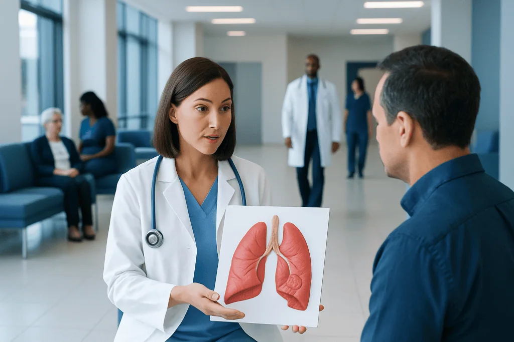
A lung tumor or nodule can be benign or malignant, and imaging alone does not always give a final answer. Doctors assess size, shape, growth pattern, smoking history, age, and other risk factors when judging cancer risk.
Key Takeaways
- A lung tumor or nodule can be benign or malignant, and imaging alone does not always give a final answer.
- Doctors assess size, shape, growth pattern, smoking history, age, and other risk factors when judging cancer risk.
- CT scans, PET scans, bronchoscopy, and biopsy may be used to confirm whether a lung growth is cancerous.
- Early evaluation improves the chance of finding a malignant tumor at a more treatable stage.
- Many small lung nodules are monitored safely over time rather than treated immediately.
Not every lung growth is cancer, and many nodules found on imaging are benign. Doctors tell benign from malignant lung tumors by combining symptoms, personal risk factors, imaging features, growth over time, and sometimes tissue biopsy.
Overview: What Are Cancerous Lung Tumors?
Cancerous lung tumors are malignant growths that begin in the lungs or spread there from another part of the body. A benign lung growth is not cancer and does not invade nearby tissues in the same way. Because both can appear as a spot, mass, or nodule on a chest image, careful evaluation is needed before reaching a diagnosis.
The term “lung nodule” usually refers to a small, round area seen on imaging, while a larger abnormal area may be called a lung mass or tumor. Some nodules are caused by old infections, inflammation, scarring, or noncancerous growths. Others may represent early lung cancer or another malignant condition, which is why doctors look at the full clinical picture rather than one scan finding alone.
In many cases, a suspicious growth is found unexpectedly during imaging done for another reason. This can be worrying, but an abnormal finding does not automatically mean cancer. The next steps usually involve estimating how likely the growth is to be malignant and then choosing follow-up scans, additional tests, or biopsy as appropriate.
Symptoms and Signs to Watch For
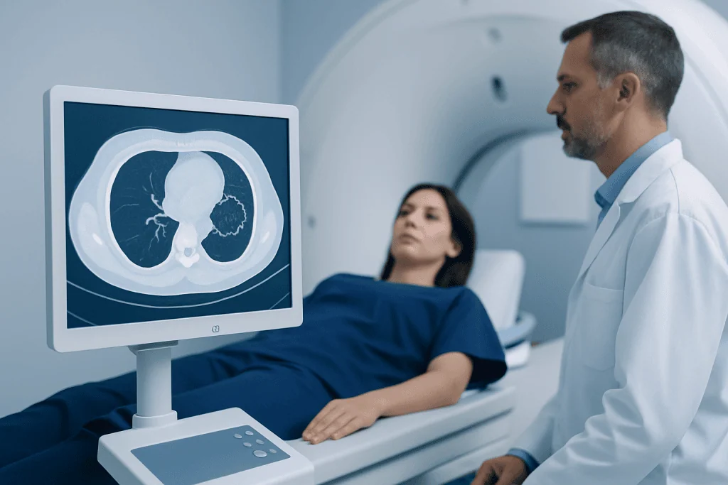
Small benign or malignant lung nodules often cause no symptoms at all. When symptoms do occur, they may include a persistent cough, coughing up blood, chest pain, wheezing, shortness of breath, or repeated chest infections. These symptoms can also happen with noncancerous conditions, so they do not confirm cancer on their own.
Some people notice more general symptoms such as unexplained weight loss, unusual tiredness, reduced appetite, or hoarseness. If a lung tumor affects a larger airway or nearby tissues, symptoms may become more noticeable over time. The pace of change can offer clues, but symptoms alone are not enough to classify a growth as benign or malignant.
Doctors also pay attention to when symptoms began and whether they are getting worse. A growth discovered in someone with no symptoms may be managed differently from one found in a person with ongoing respiratory complaints. If symptoms are significant, evaluation is usually more urgent so that serious causes can be ruled out promptly.
How Doctors Judge Whether a Lung Growth May Be Malignant
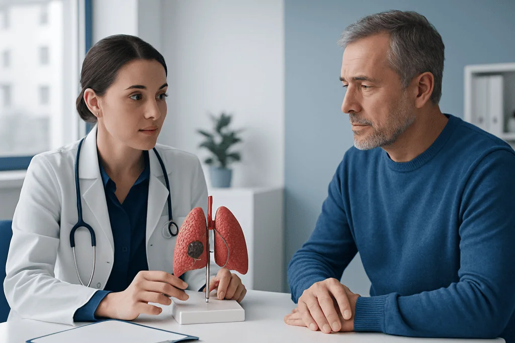
Doctors estimate cancer risk by looking at several factors together. Age, smoking history, exposure to secondhand smoke, radon, asbestos, air pollution, and family history of lung cancer can all increase concern that a growth may be malignant. A previous cancer diagnosis may also matter, because some lung lesions represent spread from another site rather than a new primary tumor.
The appearance of the growth on imaging is equally important. Features that can raise suspicion include a larger size, irregular or spiculated edges, certain patterns of density, and growth over time. In contrast, some nodules have imaging features that are more consistent with a benign cause, such as certain patterns of calcification or long-term stability on repeated scans.
Location can provide useful information as well, but it is rarely decisive on its own. A growth near the airways may be approached differently from one deep in the lung tissue. Rather than labeling a lesion immediately, doctors often place it into a low-, intermediate-, or high-risk category and then choose the safest, most appropriate next step.
- Lower-risk findings may be monitored with repeat imaging.
- Intermediate-risk findings may need additional imaging or biopsy.
- Higher-risk findings may require tissue diagnosis and treatment planning without delay.
Imaging Tests: What Scans Can and Cannot Show
A chest X-ray may detect a larger abnormality, but computed tomography, or CT, gives much more detail and is usually the main imaging test used to evaluate a lung nodule or tumor. CT can show the exact size, borders, density, and location of a growth, and it helps doctors compare current findings with any older scans. The pattern of change over time is one of the most helpful clues in deciding whether a lesion is likely benign or malignant.
Sometimes a PET-CT scan is recommended, especially when a lesion is of uncertain risk or when cancer is strongly suspected. PET imaging can show areas of increased metabolic activity, which may occur in malignant tumors. However, PET is not perfect: inflammation and infection can also appear active, and very small lesions may not be assessed reliably.
This is why scans usually guide the level of suspicion rather than provide an absolute answer. If imaging suggests possible cancer, doctors may recommend PET-CT imaging or further testing to clarify the diagnosis. If a nodule appears low risk, a structured CT follow-up plan may be safer and more appropriate than immediate invasive procedures.
Biopsy and Other Tests That Confirm the Diagnosis
The only way to know for certain whether many lung growths are malignant is to examine cells or tissue under a microscope. This is called a biopsy. Doctors do not biopsy every lung nodule, because some are so small or low risk that careful monitoring is the better choice. When a biopsy is needed, the method depends on where the lesion is located and the person’s overall health.
Common approaches include bronchoscopy, where a doctor passes a thin tube through the airways to collect samples, and needle biopsy through the chest wall guided by CT or ultrasound. A suspicious growth close to the airways may be sampled with bronchoscopy, while more peripheral lesions may be reached with a needle biopsy. If less invasive tests are inconclusive, surgery may sometimes be needed to remove part or all of the lesion for diagnosis.
Additional tests may include sputum analysis, blood tests, breathing tests, and imaging of other body areas if cancer is suspected. If the growth proves to be malignant, the next step is staging, which shows whether the cancer is confined to the lung or has spread. This information helps guide treatment options such as lung cancer treatment planning with a multidisciplinary team.
Treatment Options for Benign and Malignant Lung Tumors
Treatment depends entirely on what the growth turns out to be. Benign nodules often need no treatment at all beyond observation with follow-up scans. If a noncancerous growth is causing symptoms, repeated infections, bleeding, or uncertainty about diagnosis, doctors may recommend removal or another procedure.
Malignant lung tumors are treated based on the type of cancer, its stage, the patient’s lung function, and overall health. Options may include surgery, radiation therapy, chemotherapy, targeted therapy, immunotherapy, or a combination of these. A person with early-stage disease may be treated very differently from someone with more advanced cancer, so individualized planning is essential.
Supportive care is also important. This can include help with breathing symptoms, pain control, nutrition, smoking cessation, and rehabilitation. Near the end of the evaluation pathway, patients may wish to know that Acibadem International’s multidisciplinary specialists in JCI-accredited hospitals diagnose and treat lung tumors for international patients as part of coordinated care.
Prevention, Self-Care, and Follow-Up
The most effective way to lower the risk of malignant lung tumors is to avoid tobacco smoke. Quitting smoking can reduce future risk, even for people who have smoked for many years. It is also helpful to limit exposure to radon, workplace dusts and chemicals, and other known lung irritants when possible.
People with a history of smoking or other major risk factors may benefit from discussing lung cancer screening with their doctor. In selected high-risk adults, low-dose CT screening can help detect cancer earlier, sometimes before symptoms begin. Screening is different from evaluating a known nodule, but both rely on careful imaging and follow-up.
For anyone told they have a lung nodule, keeping follow-up appointments is one of the most important forms of self-care. Stability over time often supports a benign diagnosis, while interval growth may trigger further testing. Patients should also report any new symptoms, especially coughing up blood, worsening shortness of breath, or unexplained weight loss.
When to See a Doctor
A person should see a doctor if they have a cough that does not go away, chest pain, repeated chest infections, wheezing, unexplained weight loss, or blood in the sputum. These symptoms do not always mean cancer, but they do deserve medical assessment. Prompt evaluation is especially important for people with a smoking history or known exposure to lung irritants.
Medical advice is also important after an incidental finding on a chest X-ray or CT scan. Many nodules are harmless, but only a clinician can decide whether a lesion is low risk and suitable for monitoring or whether more testing is needed. Bringing previous imaging studies to the appointment can be very helpful because comparison over time often changes management.
Anyone who feels unsure about the recommended plan can ask about the estimated risk level, the reason for follow-up timing, and whether a second opinion is appropriate. Clear communication helps patients understand why some lesions are watched and others are biopsied quickly. A calm, step-by-step approach is the best way to distinguish benign from malignant lung growths safely.
Frequently asked questions
Can a lung nodule be noncancerous?
Yes. Many lung nodules are benign and may be related to prior infection, inflammation, scarring, or other noncancerous causes. Doctors look at imaging features, risk factors, and growth over time to decide whether further testing is needed.
How do doctors know if a lung tumor is malignant?
Doctors combine information from symptoms, smoking history, exposures, CT findings, and sometimes PET-CT results. If the level of suspicion is high or uncertain, a biopsy may be needed to confirm whether the growth is cancerous.
Does a PET scan always prove lung cancer?
No. A PET scan can show increased activity in a suspicious area, but infection and inflammation can also look active. PET results are helpful, but they are interpreted together with CT findings and, when needed, biopsy results.
Are all cancerous lung tumors treated with surgery?
No. Surgery is one option, mainly for selected early-stage tumors and patients who are fit enough for an operation. Depending on the type and stage of cancer, treatment may also include radiation therapy, chemotherapy, targeted therapy, or immunotherapy.
If a doctor recommends follow-up scans instead of biopsy, is that safe?
Often, yes. Some nodules are small or have low-risk features, so the safest approach is to monitor them with repeat CT scans rather than do an invasive procedure right away. The schedule is based on the nodule’s appearance and the patient’s risk profile.
What symptoms should prompt urgent medical attention?
Coughing up blood, worsening shortness of breath, significant chest pain, or rapid decline in general health should be assessed promptly. Even though these symptoms do not always mean cancer, they warrant timely medical evaluation.
References
- World Health Organization
- National Cancer Institute
- American Cancer Society
- American College of Chest Physicians
- National Comprehensive Cancer Network
This article is for general information only and is not a substitute for professional medical advice. Please consult a qualified doctor about your individual situation.
Oncology care in Turkey — second opinion and treatment plan
JCI-accredited · board-certified surgeons · reply within 24h
Add us as a Preferred Source to see more of our trusted health content across Google Search, AI Overviews and Discover.