

Cardiac MRI: Detailed Imaging for Heart Muscle and Blood Flow

Cardiac MRI provides highly detailed images of heart structure, function, blood flow and tissue health. It is often used when echocardiography, CT or other tests do not provide enough information, or when tissue characterization is important.
Key Takeaways
- Cardiac MRI provides highly detailed images of heart structure, function, blood flow and tissue health.
- It is often used when echocardiography, CT or other tests do not provide enough information, or when tissue characterization is important.
- The test is painless, but patients need to lie still inside the scanner and may receive a contrast agent through a vein.
- People with certain implanted devices, severe kidney disease or metal fragments in the body need careful safety screening before the scan.
- Results are interpreted by specialists and used together with symptoms, examination findings and other cardiac tests.
Cardiac MRI is a non-invasive imaging test that uses a strong magnetic field and radio waves to create detailed pictures of the heart. It helps doctors assess heart muscle, blood flow, valves, congenital heart conditions, inflammation and scarring without using ionizing radiation.
Overview
Cardiac MRI, also called heart MRI or cardiac magnetic resonance imaging, is a specialized scan that produces detailed moving and still images of the heart. It uses a powerful magnet, radio waves and computer processing to show the heart chambers, heart muscle, valves, nearby blood vessels and patterns of blood flow. Unlike X-rays and CT scans, cardiac MRI does not use ionizing radiation.
The test is especially valuable because it can assess both heart anatomy and function. It can show how strongly the heart pumps, whether areas of the heart muscle are thickened or weakened, and whether there are signs of inflammation, swelling, scarring or reduced blood supply. In some cases, cardiac MRI can help clarify findings from an echocardiogram, electrocardiogram, CT scan or coronary angiography.
Cardiac MRI is not needed for every heart concern. Doctors recommend it when the information gained may help guide diagnosis, treatment planning or follow-up. The scan is performed by trained radiology or cardiology imaging teams, and the images are interpreted by specialists with experience in cardiovascular imaging.
What Cardiac MRI Can Show
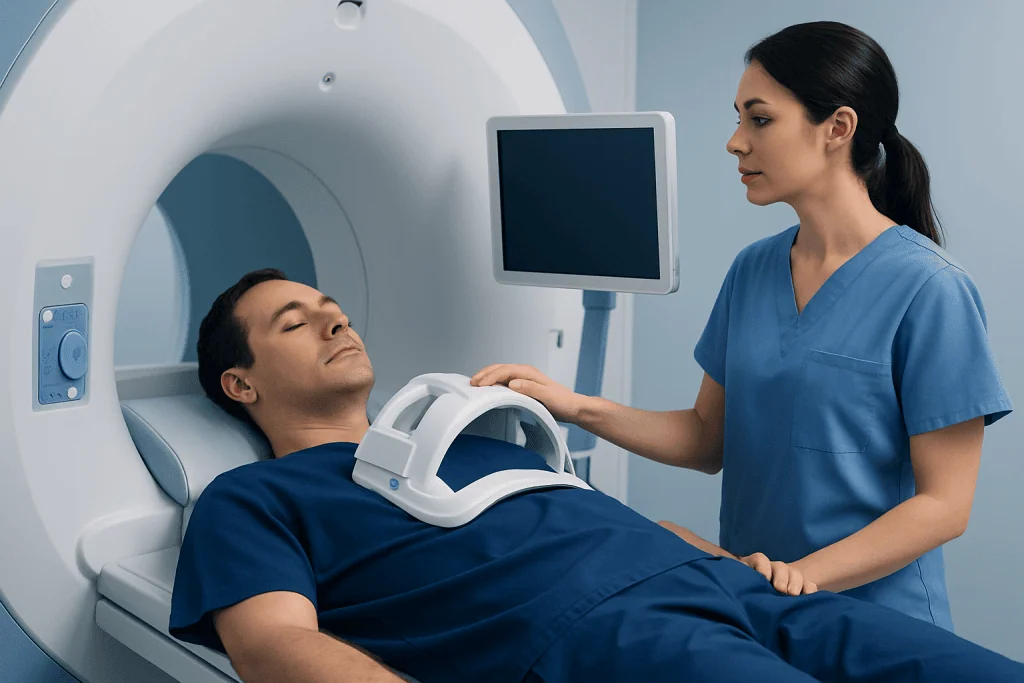
One of the main strengths of cardiac MRI is its ability to measure heart size and pumping function accurately. It can calculate how much blood the left and right ventricles pump with each heartbeat and can show whether the heart muscle moves normally. These measurements may be useful in people with cardiomyopathy, heart failure, previous heart attack, valve disease or congenital heart disease.
Cardiac MRI can also characterize heart tissue. With specific imaging sequences and, when appropriate, a gadolinium-based contrast agent, it can identify patterns of fibrosis, scarring, edema or inflammation. This information can help doctors distinguish between different causes of heart muscle disease, such as myocarditis, prior infarction, infiltrative disease or inherited cardiomyopathies.
Depending on the clinical question, cardiac MRI may be used to evaluate:
- Heart muscle thickness, strength and movement
- Scarring after a heart attack or other injury
- Inflammation such as myocarditis or pericarditis
- Heart valve function and blood flow patterns
- Congenital heart defects and major blood vessels
- Cardiac masses, tumors or blood clots in selected cases
When Doctors Recommend Cardiac MRI

A doctor may recommend cardiac MRI when a detailed view of the heart muscle or blood flow is needed. For example, it can be helpful when echocardiography images are limited or when doctors need a more precise assessment of the right ventricle, complex congenital anatomy or areas of suspected scar tissue. It may also be used to monitor known conditions over time.
In people with chest pain, shortness of breath, fainting, abnormal heart rhythms or unexplained changes on other tests, cardiac MRI may help identify the underlying cause. Stress cardiac MRI, performed with medication that temporarily increases blood flow demand, can assess whether parts of the heart muscle may not be receiving enough blood during stress. This is different from a standard resting cardiac MRI and is chosen only when appropriate.
Common reasons for cardiac MRI include suspected or known cardiomyopathy, myocarditis, previous heart attack, viability assessment before certain treatments, congenital heart disease, aortic disease, pericardial disease and evaluation of selected cardiac masses. The exact scan protocol is tailored to the patient’s symptoms, medical history and the doctor’s referral question.
How the Test Is Performed
Before the scan, the patient completes a safety questionnaire about implanted devices, previous surgery, metal exposure, allergies, kidney disease and pregnancy status if relevant. The care team may check recent kidney function blood tests if contrast is planned. Patients are usually asked to remove metal items, including jewelry, watches, hair clips, hearing aids and some removable dental appliances.
During the scan, the patient lies on a table that moves into the MRI scanner. Small sticky sensors are placed on the chest to monitor the heart rhythm, and a coil may be positioned over the chest to improve image quality. The scanner makes loud knocking or thumping sounds, so ear protection is provided. The technologist communicates with the patient throughout the test.
Many cardiac MRI sequences require short breath-holds, often lasting several seconds at a time. This helps reduce motion blur from breathing. The scan is painless, although some people notice warmth or vibration during imaging. If contrast is used, it is given through a small intravenous line, and most patients can return to normal activities soon afterward unless their doctor advises otherwise.
Contrast, Safety and Who Needs Extra Screening
Some cardiac MRI scans use a gadolinium-based contrast agent to improve the visibility of scar tissue, inflammation, blood flow or masses. Gadolinium contrast is different from iodine-based contrast used in many CT scans. It is generally well tolerated, but it is not suitable for everyone, so the imaging team reviews kidney function, allergy history and other risk factors before use.
MRI safety screening is essential because the magnet is always strong. Many modern pacemakers, defibrillators and other implanted devices are MRI-conditional, meaning they may be scanned only under specific conditions and with appropriate specialist supervision. Older or non-compatible devices, certain aneurysm clips, cochlear implants, metal fragments in the eye and some surgical hardware may require additional evaluation.
Patients should tell the team if they have severe claustrophobia, difficulty lying flat, significant breathing problems or pain that may make it hard to stay still. Options such as careful positioning, reassurance, music, a mirror system or medication prescribed in advance may help some patients. Pregnant patients should discuss the timing and need for MRI with their doctor; MRI may be used in pregnancy when clinically necessary, but contrast is usually avoided unless there is a strong reason.
Benefits and Limitations
Cardiac MRI offers several important benefits. It provides excellent soft-tissue detail, accurate measurements of heart function and the ability to assess scar and inflammation in ways that many other imaging tests cannot. Because it does not use ionizing radiation, it can be particularly useful for patients who need repeated imaging, such as some people with congenital heart disease or cardiomyopathy.
However, cardiac MRI is not the best test for every situation. It may take longer than an echocardiogram or CT scan, and image quality can be affected by irregular heart rhythms, inability to hold the breath or movement during the scan. Some patients cannot undergo MRI because of device or metal safety concerns. In urgent situations, other tests may be faster or more appropriate.
Cardiac MRI also does not usually replace all other heart tests. Echocardiography remains widely used for valve assessment and bedside evaluation, CT can be excellent for coronary artery anatomy in selected patients, and invasive angiography may still be needed for certain coronary artery problems. Doctors choose the test, or combination of tests, based on the clinical question and the patient’s individual situation.
Preparing for Results and Follow-Up
After the scan, the images are reviewed by a radiologist, cardiologist or cardiac imaging specialist. The report may include measurements of chamber size, pumping function, valve flow, tissue characteristics and any abnormal findings. Results are interpreted together with the patient’s symptoms, medical history, physical examination, blood tests and other cardiac investigations.
Follow-up depends on the reason for the scan. Some patients may need medication changes, further tests, lifestyle guidance, monitoring over time or referral to a subspecialist such as an electrophysiologist, heart failure specialist, congenital heart disease specialist or cardiac surgeon. A normal cardiac MRI can also be useful because it may help rule out certain conditions and guide a safer, more focused care plan.
Patients should seek medical care promptly if they experience chest pain, severe shortness of breath, fainting, new weakness, sudden palpitations with dizziness or symptoms that feel urgent. For international patients, Acibadem International’s multidisciplinary specialists and JCI-accredited hospitals provide diagnostic evaluation and treatment planning for heart conditions, including cardiac MRI when clinically appropriate.
Frequently asked questions
Is cardiac MRI painful?
Cardiac MRI is not painful. The patient needs to lie still on the scanner table, and the machine makes loud tapping or knocking sounds, but ear protection is provided. Some people find the enclosed space uncomfortable, so it is helpful to tell the team in advance if claustrophobia is a concern.
How long does a cardiac MRI take?
The scan time varies depending on the clinical question and whether contrast or stress imaging is needed. Many cardiac MRI examinations take about 30 to 60 minutes, though complex studies may take longer. The appointment may also include preparation time, safety screening and placement of monitoring leads.
Does cardiac MRI use radiation?
No. Cardiac MRI uses a magnetic field and radio waves rather than ionizing radiation. This can be an advantage for patients who require repeated heart imaging, although MRI safety screening is still important because of the strong magnet.
Will every patient need contrast during cardiac MRI?
No. Some cardiac MRI scans can be performed without contrast, especially when the main goal is to measure heart size, pumping function or certain flow patterns. Contrast may be recommended when doctors need to evaluate scarring, inflammation, tissue damage or selected masses.
Can a person with a pacemaker have a cardiac MRI?
Some people with pacemakers or defibrillators can have MRI if the device is MRI-conditional and the hospital has the correct safety protocol. The device must usually be checked and programmed before and after the scan by qualified staff. Patients should always provide their device card and inform the imaging team before the appointment.
What should patients do before a cardiac MRI?
Patients should follow the instructions provided by the imaging center, bring relevant medical records and report any implants, metal fragments, kidney disease, allergies or pregnancy. They should remove metal items before the scan and ask whether fasting or medication changes are needed. Patients should not stop prescribed heart medicines unless their doctor specifically advises it.
References
- American Heart Association
- European Society of Cardiology
- Society for Cardiovascular Magnetic Resonance
- Radiological Society of North America
- American College of Radiology
This article is for general information only and is not a substitute for professional medical advice. Please consult a qualified doctor about your individual situation.
Heart care in Turkey — expert evaluation and treatment
JCI-accredited · board-certified surgeons · reply within 24h
Add us as a Preferred Source to see more of our trusted health content across Google Search, AI Overviews and Discover.

More from the Health Library



Related Specialists

Prof. Dr. Ercan Karaarslan
Radiology
Prof. Dr. Bora Peynircioğlu
Interventional Radiology
Prof. Dr. Koray Güven
Interventional Radiology
Assoc. Prof. Dr. Deniz Can Aliş
Radiology