Cardiac Pet Scan: An Evidence-Based Guide for Patients
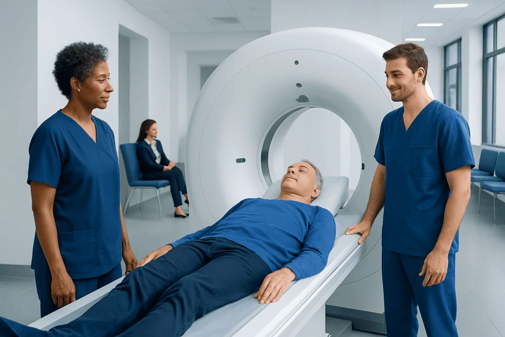
A cardiac PET scan assesses blood flow to the heart muscle at rest and during stress. It can help diagnose coronary artery disease and identify areas of reduced blood supply or damaged heart tissue.
Key Takeaways
- A cardiac PET scan assesses blood flow to the heart muscle at rest and during stress.
- It can help diagnose coronary artery disease and identify areas of reduced blood supply or damaged heart tissue.
- The test uses a small amount of radioactive tracer and is generally considered safe when medically indicated.
- Preparation may include avoiding caffeine and fasting for several hours before the scan.
- Results are interpreted together with symptoms, medical history, and other heart tests.
A cardiac PET scan is a specialized nuclear imaging test that shows how well blood flows to the heart muscle and how the heart is functioning. It is commonly used to evaluate suspected coronary artery disease, clarify unclear test results, and help guide treatment decisions.
Overview: what a cardiac PET scan shows
A cardiac PET scan is an advanced imaging test that helps doctors see how blood moves through the heart muscle and how well the heart tissue is working. PET stands for positron emission tomography. In heart care, it is most often used to look for areas that are not getting enough blood, to assess heart muscle viability, and to support decisions about treatment.
Unlike a routine chest X-ray or an electrocardiogram, a cardiac PET scan provides functional information. This means it can show whether the heart muscle receives enough oxygen-rich blood at rest and under stress. That makes it especially useful when symptoms suggest reduced blood flow but the diagnosis is still uncertain after initial evaluation.
The test is often used in people with suspected or known coronary artery disease, especially when a doctor needs more detail than other tests can provide. It may also be used after a heart attack, before certain procedures, or when other imaging studies have given inconclusive results. In some cases, it helps distinguish heart muscle that is scarred from heart muscle that is weakened but still potentially recoverable.
Why doctors recommend this test
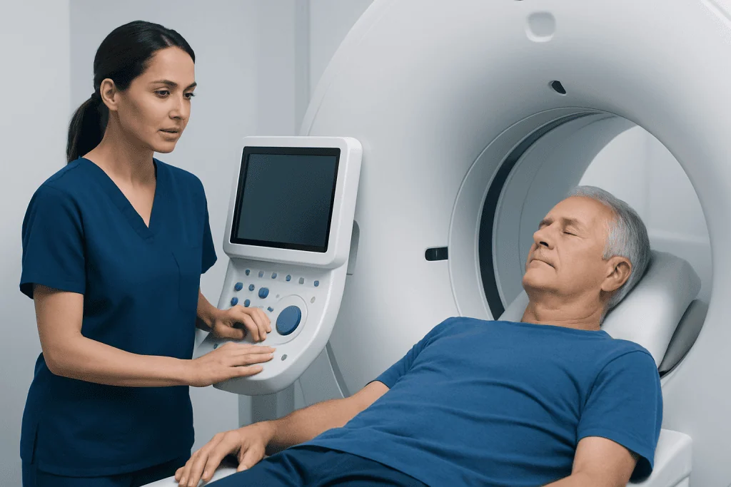
A doctor may recommend a cardiac PET scan for several practical reasons. One of the most common is to investigate symptoms such as chest discomfort, shortness of breath on exertion, or unexplained fatigue when a heart-related cause is being considered. The scan can help determine whether these symptoms are linked to reduced blood flow in the coronary arteries.
It may also be used to refine risk assessment in people with known heart disease. For example, if a patient has had a prior heart attack, coronary stent, or bypass surgery, PET imaging can help show how much heart muscle is still well supplied and whether further treatment might be beneficial. This can be especially important in complex cases or in people with diabetes, obesity, or previous inconclusive stress tests.
Cardiac PET is sometimes chosen because of its image quality and its ability to quantify blood flow. That may make it helpful in patients where other tests are less clear. It can be part of the broader evaluation of coronary artery disease or used alongside other cardiac imaging and consultation with heart specialists.
How the scan works and what to expect

During a cardiac PET scan, a small amount of radioactive tracer is injected into a vein. The tracer travels through the bloodstream and is taken up by the heart muscle. A special camera then detects signals from the tracer and creates detailed images showing blood flow and tissue activity.
Most cardiac PET scans include images taken at rest and during stress. The “stress” part may be produced with medication that temporarily increases blood flow in a way that simulates exercise. This helps doctors compare how the heart performs under different conditions. Blood pressure, heart rhythm, and symptoms are monitored throughout the procedure.
The scan itself is not usually painful, although the intravenous line and the stress medication may cause brief discomfort or sensations such as flushing, shortness of breath, or chest pressure. These effects are usually temporary and are closely supervised by trained staff. The full appointment may take a few hours, though the actual imaging time is shorter.
In some centers, a cardiac PET scan may be combined with CT for more anatomical detail. Depending on the question being asked, the care team may compare the results with an echocardiogram, a stress test, or a comprehensive cardiology check-up to build a clearer picture of heart health.
Preparation before a cardiac PET scan
Preparation instructions can vary slightly by center, so patients should always follow the specific guidance given by their care team. In many cases, fasting is required for several hours before the scan. Water is often allowed, but food and certain beverages may need to be avoided.
Caffeine is especially important to mention. Coffee, tea, cola, energy drinks, chocolate, and some headache or cold medicines can interfere with the stress portion of the test. Patients are commonly asked to avoid caffeine for at least a day before the appointment. Smoking may also affect results, so temporary avoidance may be recommended.
Some medicines may need to be paused or adjusted before the test, particularly those that affect heart rate or blood flow. This should only be done under medical instruction. Patients should tell the imaging team about all prescription drugs, over-the-counter medicines, supplements, allergies, pregnancy, breastfeeding, asthma, or prior reactions to stress agents or contrast-related tests.
Comfortable clothing is helpful, and it is wise to allow enough time for registration and monitoring. If the patient has diabetes, special preparation may be needed to balance fasting requirements with safe blood sugar control. Asking questions in advance often helps the day of testing feel more predictable and less stressful.
What the results can mean
The images from a cardiac PET scan are reviewed by a specialist, usually in the context of the patient’s symptoms, physical examination, and other test results. In broad terms, the scan can show normal blood flow, reduced blood flow during stress, reduced flow both at rest and during stress, or evidence of tissue injury. Each pattern suggests a different clinical picture.
If blood flow looks normal at rest and stress, significant coronary artery narrowing is less likely. If the scan shows reduced flow only during stress, that may suggest a blockage that limits blood supply when the heart has to work harder. If reduced flow is seen even at rest, it may indicate more severe disease or prior damage. A viability study can help determine whether weakened heart muscle may still improve after restoring blood flow.
These results do not stand alone. They may lead to further steps such as medication changes, lifestyle measures, additional imaging, or procedures to improve blood supply. Depending on the findings, doctors may discuss options that can include coronary angiography or treatment planning for coronary artery bypass grafting when clinically appropriate.
Benefits, limitations, and safety
One of the main benefits of cardiac PET is that it can provide highly informative images of heart blood flow and function. It can be particularly useful when previous stress tests are unclear or when a more precise estimate of blood flow is needed. In some patients, it helps avoid unnecessary procedures by showing when blood supply is actually adequate.
Like all medical tests, it has limitations. A cardiac PET scan does not replace a full medical evaluation, and it may not answer every question about heart rhythm, valve disease, or structural abnormalities. Results can also be influenced by preparation, body factors, underlying medical conditions, and the specific clinical reason for the test.
The amount of radiation used is generally low and carefully managed, but it is still an important consideration. The test is ordered when the expected benefit outweighs the risk. Reactions to the stress medication are usually short-lived, and serious complications are uncommon under proper supervision. Patients with pregnancy, severe lung disease, unstable symptoms, or certain medication concerns should discuss these issues with their doctor before the scan.
For some people, alternative tests may be more appropriate, such as stress echocardiography, cardiac MRI, CT coronary angiography, or invasive angiography. The best choice depends on the patient’s symptoms, history, and the exact question the doctor needs to answer.
After the test and next steps
After a cardiac PET scan, most patients can return to normal activities unless their doctor advises otherwise. Drinking fluids may be recommended to help clear the tracer from the body. If stress medication was used, any temporary sensations such as flushing or mild headache usually settle quickly.
The next step depends on the result. Some patients are reassured that blood flow appears normal and may continue with prevention-focused care. Others may need medication adjustments, closer follow-up, or further testing. If reduced blood flow is found, the doctor may discuss whether the goal is symptom relief, risk reduction, or evaluation for a procedure.
Because heart symptoms can have many causes, the interpretation is most useful when paired with a complete cardiovascular assessment. If ongoing symptoms suggest a broader heart issue, the care team may also consider related conditions such as heart failure or other forms of structural or ischemic heart disease.
Near the end of this process, some patients seek care at specialized centers that combine imaging, cardiology, and cardiac surgery expertise. Acibadem International’s multidisciplinary specialists and JCI-accredited hospitals diagnose and treat heart conditions for international patients when advanced evaluation is needed.
When to seek medical care
A cardiac PET scan is usually planned in a non-emergency setting, but symptoms should always guide the urgency of care. A person should seek prompt medical evaluation for chest pain, pressure, or tightness that is new, severe, worsening, or brought on by exertion. Shortness of breath, fainting, unexplained sweating, or pain spreading to the arm, back, neck, or jaw also deserve urgent attention.
After the scan, patients should contact their care team if they have persistent dizziness, worsening breathing difficulty, ongoing chest discomfort, or any symptoms that feel unusual or severe. It is also reasonable to ask for medical advice if preparation instructions were not followed, because this may affect interpretation and next steps.
Even when symptoms are mild, recurring chest discomfort, reduced exercise tolerance, or unexplained fatigue should not be ignored. Early assessment can help identify whether the cause is heart-related and whether further evaluation, monitoring, or treatment is appropriate.
Frequently asked questions
What is a cardiac PET scan used for?
A cardiac PET scan is used to evaluate blood flow to the heart muscle and assess how well the heart is functioning. It is commonly ordered to help diagnose coronary artery disease, investigate symptoms, or determine whether damaged-looking heart muscle may still be viable.
Is a cardiac PET scan the same as a regular stress test?
Not exactly. A cardiac PET scan is a type of stress imaging test, but it uses a radioactive tracer and specialized imaging to show heart blood flow in more detail. A standard exercise stress test may rely mainly on ECG changes, symptoms, and exercise performance.
How should someone prepare for a cardiac PET scan?
Preparation usually includes fasting for several hours and avoiding caffeine before the test. The exact instructions can vary, so patients should follow their center’s guidance carefully and ask about medications, diabetes management, and smoking restrictions.
How long does a cardiac PET scan take?
The full visit often takes a few hours, including preparation, tracer injection, monitoring, and both rest and stress imaging. The actual image acquisition time is shorter, but timing depends on the tracer used and the center’s protocol.
Is a cardiac PET scan safe?
For most people, a cardiac PET scan is considered safe when medically indicated and performed under supervision. It uses a small amount of radiation, and the stress medication can cause temporary symptoms, but serious complications are uncommon.
What happens if the results are abnormal?
An abnormal result may suggest reduced blood flow, prior heart damage, or heart muscle that could benefit from restored circulation. The doctor will interpret the findings together with symptoms and other tests, then discuss treatment options such as medications, more imaging, or procedures.
References
- American Heart Association
- Society of Nuclear Medicine and Molecular Imaging
- Radiological Society of North America
- National Heart, Lung, and Blood Institute
- American College of Cardiology
This article is for general information only and is not a substitute for professional medical advice. Please consult a qualified doctor about your individual situation.
Heart care in Turkey — expert evaluation and treatment
JCI-accredited · board-certified surgeons · reply within 24h
Add us as a Preferred Source to see more of our trusted health content across Google Search, AI Overviews and Discover.
Check your numbers in seconds
BMI, calories, due date, blood pressure and 30+ more clinical calculators — free, instant, doctor-reviewed ranges.