Cerebral Angiography for Brain Vessel Disorders: Preparation, Procedure, and Recovery
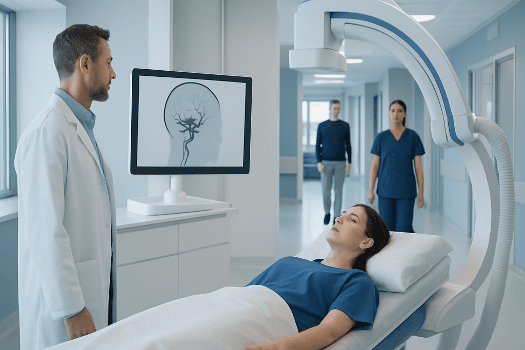
Cerebral angiography provides highly detailed images of blood vessels in the brain. The test is commonly used to investigate aneurysms, strokes, vessel narrowing, and arteriovenous malformations.
Key Takeaways
- Cerebral angiography provides highly detailed images of blood vessels in the brain.
- The test is commonly used to investigate aneurysms, strokes, vessel narrowing, and arteriovenous malformations.
- Preparation may include fasting, medication review, and blood tests, especially for kidney function and clotting.
- Most patients go home the same day or after a short observation period, depending on why the test was done.
- Recovery usually involves rest, fluids, and watching the catheter site for bleeding or swelling.
Cerebral angiography is a specialized imaging test used to examine the blood vessels of the brain in great detail. It helps doctors diagnose and plan treatment for conditions such as aneurysms, narrowing, malformations, and other brain vessel disorders.
Overview: What Cerebral Angiography Is
Cerebral angiography is a medical imaging procedure that allows doctors to see the blood vessels of the brain clearly and in real time. It is sometimes called a cerebral angiogram or brain angiography. During the test, a thin tube called a catheter is guided through a blood vessel, usually from the wrist or groin, and a contrast dye is injected so the vessels can be seen on X-ray imaging.
This test is considered one of the most detailed ways to examine brain circulation. It can show the shape, size, and flow pattern of arteries and veins, helping specialists find abnormalities that may not be fully visible on standard CT or MRI scans. In many cases, it is used after other imaging tests suggest a problem that needs closer evaluation.
Cerebral angiography may be done for diagnosis alone, or it may be combined with treatment during the same session. For example, if a specialist identifies a vessel problem that can be treated through the catheter, a minimally invasive procedure may sometimes follow. Because of this, cerebral angiography is an important tool in modern neurovascular care.
Why It Is Done and What Conditions It Can Show
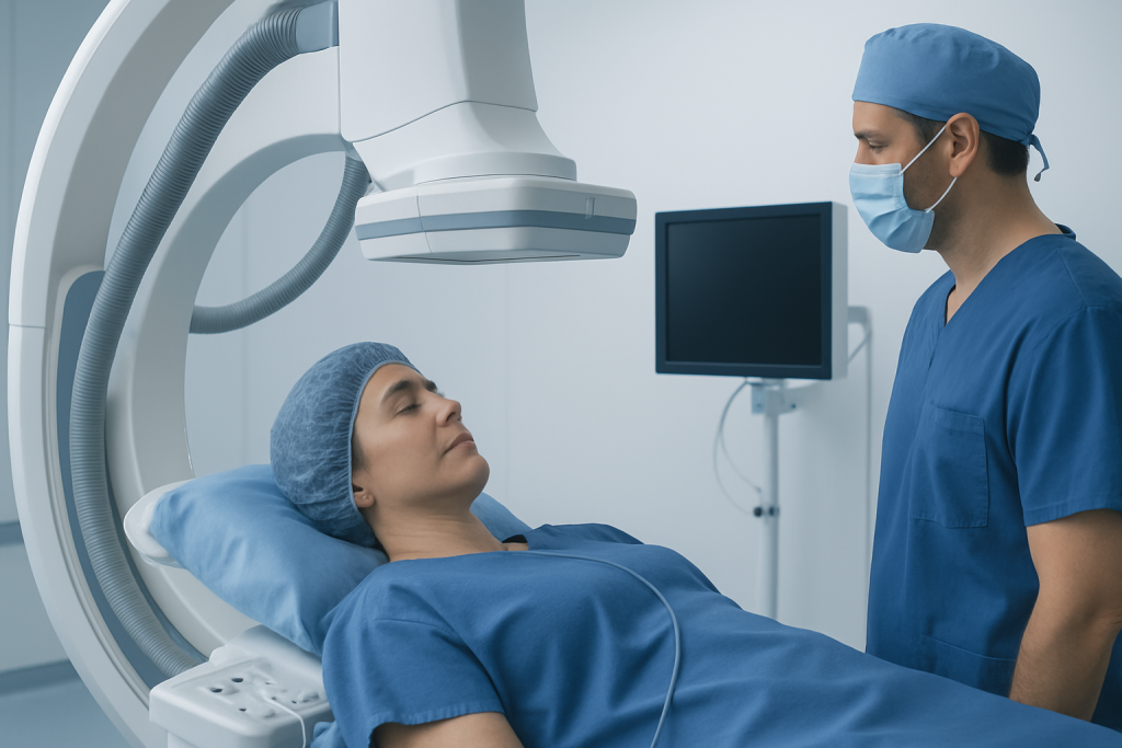
Doctors use cerebral angiography to investigate symptoms or imaging findings that suggest a disorder of the brain’s blood vessels. It is especially helpful when precise detail is needed before treatment planning. The test may be recommended for sudden severe headache, stroke-like symptoms, unexplained bleeding in or around the brain, or concern about abnormal vessels.
Conditions that may be identified or assessed with cerebral angiography include:
- Brain aneurysms
- Narrowing or blockage of brain arteries
- Arteriovenous malformations and fistulas
- Blood vessel spasm after bleeding
- Vasculitis affecting brain vessels
- Tumors with abnormal blood supply
- Sources of stroke or transient ischemic attack
It may also be used to assess conditions such as brain aneurysm or arteriovenous malformation. In some situations, the angiogram helps doctors decide whether a patient may benefit from minimally invasive treatment such as interventional neuroradiology procedures. This level of detail is valuable when treatment decisions depend on the exact anatomy of the affected vessel.
Preparation Before the Procedure

Preparation for cerebral angiography is usually straightforward, but it is important for safety. Before the procedure, the medical team reviews the patient’s symptoms, medical history, allergies, and current medicines. Special attention is given to blood thinners, diabetes medications, previous reactions to contrast dye, and any kidney problems.
Patients are often asked not to eat or drink for several hours beforehand. Blood tests may be done to check kidney function, clotting, and sometimes blood counts. If there is any chance of pregnancy, the team should be informed so that imaging risks can be carefully considered.
On the day of the test, patients usually change into a hospital gown and have an intravenous line placed for fluids or medicines. The doctor explains the procedure, benefits, and possible risks, and asks for informed consent. In many centers, a mild sedative is given to help the patient relax while still remaining able to follow instructions during the exam.
Patients should also arrange for someone to accompany them home if same-day discharge is planned. Following the care team’s instructions closely, including when to stop or continue medications, can help the procedure go more smoothly and reduce the chance of complications.
How the Procedure Is Performed
Cerebral angiography is usually performed in a specialized imaging suite by an interventional neuroradiologist, neurosurgeon, or another trained specialist. After the skin is cleaned, a local anesthetic numbs the area where the catheter will enter the body, often the wrist or groin. This helps minimize discomfort while the patient remains awake or lightly sedated.
The doctor then inserts the catheter into the blood vessel and carefully guides it toward the arteries supplying the brain. X-ray imaging is used continuously to track its position. Once the catheter is in place, contrast dye is injected, and a rapid series of images is taken to map blood flow through the brain vessels.
Patients may notice a brief warm feeling, a metallic taste, or a flushing sensation when the contrast is injected. These sensations are usually short-lived. The procedure itself may take from about 30 minutes to longer, depending on how complex the anatomy is and whether treatment is also being planned or performed.
After the images are complete, the catheter is removed and pressure or a closure device is used to reduce bleeding at the access site. If the angiography leads directly to a treatment plan, the team may discuss options such as aneurysm treatment or other catheter-based therapies once the findings are reviewed.
Risks, Safety, and Possible Side Effects
Cerebral angiography is generally safe when performed by experienced teams, but like any invasive procedure, it carries some risks. Most side effects are mild and temporary, such as bruising, tenderness, or minor bleeding where the catheter was inserted. Some patients feel tired afterward, especially if they received sedative medicine.
Less common but more important risks include allergic reaction to contrast dye, kidney strain from the contrast, blood vessel injury, infection, or blood clots. Because the test involves blood vessels supplying the brain, there is also a small risk of stroke or transient neurological symptoms. The medical team takes careful precautions to reduce these risks, including sterile technique, imaging guidance, and close monitoring.
Radiation exposure is part of the procedure, but the amount is kept as low as reasonably possible while still obtaining clear images. Patients with kidney disease, bleeding disorders, severe contrast allergy, or certain other health conditions may need additional precautions or an alternative imaging approach. Discussing any concerns in advance helps the team tailor the procedure safely to the individual patient.
Recovery After Cerebral Angiography
After cerebral angiography, the patient is moved to a recovery area for observation. Nurses monitor blood pressure, heart rate, neurological status, and the catheter site. If the procedure was performed through the groin, lying flat for a period may be necessary to reduce bleeding. Wrist access may allow earlier movement, depending on the device used and the doctor’s instructions.
Many patients go home the same day, while others stay longer if they have had treatment, have underlying medical concerns, or were admitted for symptoms such as stroke or bleeding. Mild soreness, bruising, or fatigue can happen for a day or two. Drinking fluids, unless otherwise advised, may help the body clear the contrast dye.
Patients are usually asked to avoid heavy lifting, strenuous exercise, and soaking the access site in water for a short period. The puncture site should be checked for increasing swelling, continued bleeding, worsening pain, or color changes in the affected limb. Clear written discharge instructions are typically provided before leaving the hospital.
If the angiogram is part of a broader care plan, the next step may include follow-up imaging, specialist review, or procedures such as stroke treatment when appropriate. Acibadem International’s multidisciplinary specialists and JCI-accredited hospitals diagnose and treat brain vessel disorders for international patients using coordinated imaging and neurovascular care.
When to Seek Medical Advice and What Follow-Up Involves
Patients should contact a doctor promptly if they develop significant bleeding from the catheter site, severe swelling, chest pain, shortness of breath, fever, increasing redness, or severe pain after going home. Urgent medical attention is also needed for new weakness, numbness, trouble speaking, confusion, vision changes, or a severe headache, as these may suggest a neurological complication.
Follow-up depends on why the angiography was done. Some patients receive results on the same day, while others need a detailed review with a neurologist, neurosurgeon, or interventional specialist. If an abnormality is found, the doctor explains whether monitoring, medication, surgery, or a catheter-based treatment is most appropriate.
Questions that are often helpful at follow-up include what the angiogram showed, whether additional testing is needed, what treatment choices are available, and what symptoms should prompt urgent care. Understanding the findings can help patients take an active role in decisions about their brain and vascular health.
Frequently asked questions
Is cerebral angiography painful?
Most patients do not describe cerebral angiography as painful, although some discomfort can occur. The skin is numbed with local anesthetic, and patients may feel pressure at the catheter site or a brief warm sensation when contrast is injected.
How long does a cerebral angiography procedure take?
The procedure often takes about 30 minutes to an hour, but it can be longer in complex cases. Extra time may be needed for preparation, recovery, and monitoring before discharge.
Will the patient be awake during cerebral angiography?
Many patients are awake but relaxed with mild sedation during the test. In some situations, deeper sedation or anesthesia may be considered depending on the patient's condition and the planned procedure.
What is the difference between cerebral angiography and CT or MR angiography?
CT angiography and MR angiography are noninvasive imaging tests, while cerebral angiography uses a catheter placed inside a blood vessel. Cerebral angiography usually provides more detailed and dynamic information and may also allow treatment planning during the same session.
How soon can normal activities resume after cerebral angiography?
Light daily activities can often be resumed within a day or two, depending on the access site and the reason for the test. Heavy lifting and strenuous exercise are usually delayed for a short time based on the doctor's instructions.
Who should tell the doctor about allergies or medicines before the test?
Every patient should inform the medical team about all medications, supplements, and allergies before cerebral angiography. This is especially important for blood thinners, diabetes medications, kidney disease, and any past contrast dye reactions.
References
- American Heart Association
- National Institute of Neurological Disorders and Stroke
- Radiological Society of North America
- NHS
- Society of NeuroInterventional Surgery
This article is for general information only and is not a substitute for professional medical advice. Please consult a qualified doctor about your individual situation.
Explore treatments in Turkey — costs, top hospitals & a free quote
JCI-accredited · board-certified surgeons · reply within 24h
Add us as a Preferred Source to see more of our trusted health content across Google Search, AI Overviews and Discover.