Coloboma: Diagnosis, Outlook, and Modern Treatment Approaches

Coloboma is present at birth and can affect one or several parts of the eye. Its impact ranges from mild cosmetic differences to significant vision impairment, depending on the structures involved.
Key Takeaways
- Coloboma is present at birth and can affect one or several parts of the eye.
- Its impact ranges from mild cosmetic differences to significant vision impairment, depending on the structures involved.
- Diagnosis relies on a detailed eye examination and sometimes imaging or genetic assessment.
- Treatment does not close the gap itself, but it can improve vision, reduce complications, and support eye health.
- Regular follow-up is important because some people with coloboma have a higher risk of retinal problems or other eye conditions.
Coloboma is a congenital eye condition in which part of the eye does not form completely before birth, leaving a gap in structures such as the iris, retina, choroid, or optic nerve. Outlook varies widely, but careful diagnosis, vision support, and targeted treatment can help many people protect vision and manage daily life well.
Overview: What coloboma means
Coloboma is a condition in which part of the eye does not develop fully during early pregnancy. The result is a gap or missing piece of tissue that may affect the iris, lens, retina, choroid, optic nerve, or eyelid. Because different eye structures serve different functions, the effects of coloboma can range from barely noticeable to more significant visual impairment.
Many people recognize coloboma by the appearance of an irregular or keyhole-shaped pupil when the iris is involved. However, not all colobomas are visible from the outside. Some affect the back of the eye, where they may only be found during a specialist eye examination. In these cases, symptoms may include reduced vision, blind spots, or sensitivity to light rather than a visible change in appearance.
Coloboma is usually present from birth and may occur in one eye or both. It can appear on its own or as part of a broader genetic or developmental syndrome. A person’s outlook depends mainly on which structures are involved, whether both eyes are affected, and whether related eye or health conditions are also present.
Types and how coloboma can affect vision
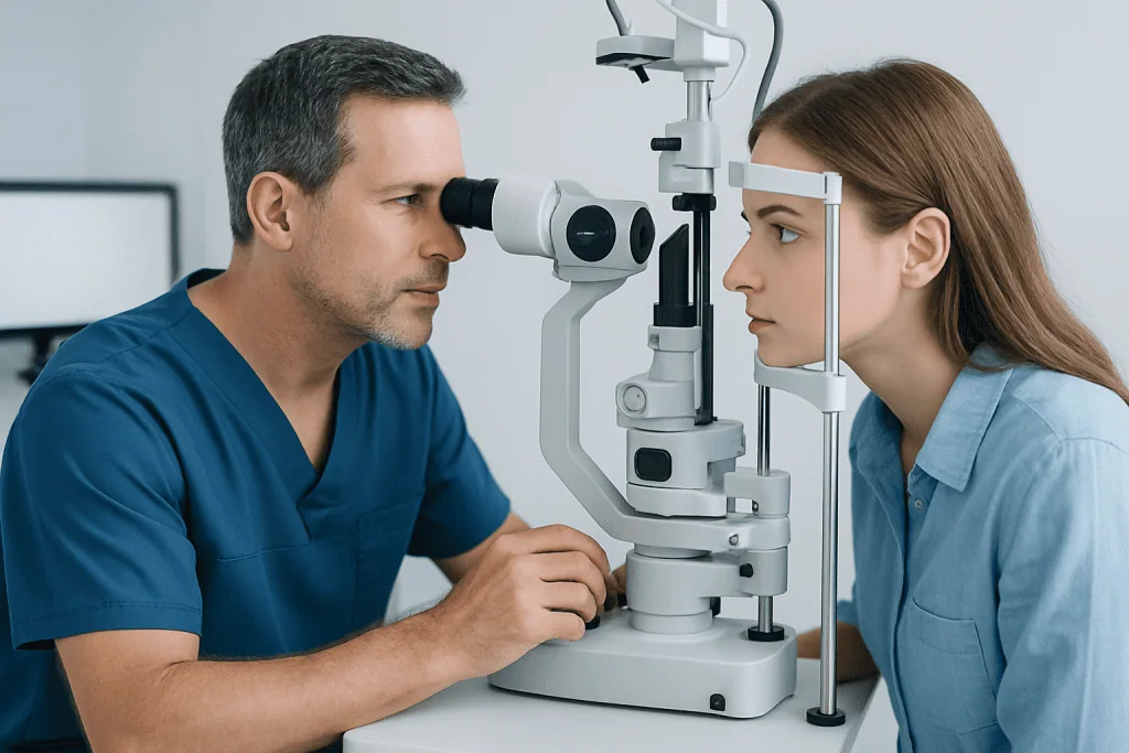
Doctors often describe coloboma by the eye structure involved. An iris coloboma affects the colored part of the eye and may change the shape of the pupil. A retinal or chorioretinal coloboma affects the light-sensing and supporting layers at the back of the eye. An optic nerve coloboma involves the nerve that carries visual information to the brain. Less commonly, the lens or eyelid can also be affected.
Vision changes depend on the location and size of the gap. An isolated iris coloboma may cause glare or light sensitivity but sometimes has little effect on sharp vision. By contrast, a coloboma involving the retina, macula, or optic nerve may reduce visual clarity, cause field defects, or impair depth perception. Some children develop lazy eye (amblyopia) if one eye sees much less clearly than the other.
Coloboma can also occur alongside other eye conditions. These may include microphthalmia, in which the eye is smaller than usual, cataract, nystagmus, refractive errors such as nearsightedness or farsightedness, or strabismus. When this happens, vision care focuses not only on the coloboma itself but also on treating the associated problems that influence long-term visual development and comfort.
Because the range of severity is broad, people with coloboma benefit from individualized assessment. Two people with the same general diagnosis may have very different visual needs, school or work accommodations, and follow-up plans.
Symptoms, signs, and possible complications
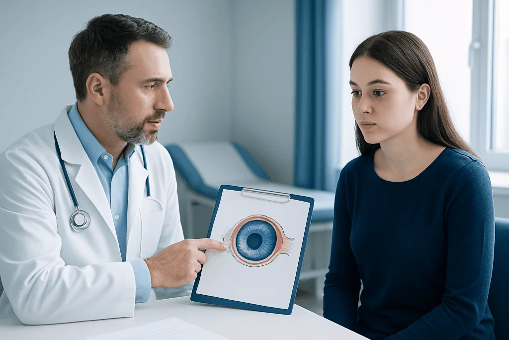
Symptoms vary according to the part of the eye involved. Some people have no obvious symptoms and are diagnosed during a routine eye examination. Others notice blurred vision, reduced vision in one eye, blind spots, glare, or increased light sensitivity. If the iris is affected, the pupil may appear elongated or keyhole-shaped.
In babies and young children, signs may be subtle. Parents may notice unusual eye appearance, wandering eyes, delayed visual attention, trouble following objects, or head turning to see more clearly. Since infants cannot describe visual symptoms, pediatric eye evaluation is especially important when any eye difference is present from birth.
Potential complications depend on the type of coloboma. A retinal coloboma may raise the risk of retinal detachment, which requires urgent care. Some people also have reduced depth perception, difficulties with reading or mobility in low light, or visual fatigue during school and screen-based tasks. Associated conditions such as cataract or strabismus can further affect vision.
- Common possible features include light sensitivity, reduced visual acuity, and visual field defects.
- Complications may include amblyopia, strabismus, cataract, nystagmus, or retinal detachment.
- Some individuals have syndromic coloboma with hearing, heart, growth, or developmental differences.
Causes and risk factors
Coloboma develops early in pregnancy when a temporary structure called the embryonic fissure does not close completely. This process is part of normal eye formation. If closure is incomplete, a gap remains in the affected eye tissue. This is why coloboma is considered a congenital condition, even if it is diagnosed later in childhood or adulthood.
In many cases, the exact reason is not fully known. Some cases are linked to genetic changes that affect eye development. Coloboma may occur on its own or as part of a syndrome involving other organs or body systems. Family history can be relevant, although many families have no prior known history.
Certain prenatal exposures or developmental disruptions have also been associated with congenital eye abnormalities in general, but this does not mean a parent caused the condition. In most situations, there is nothing a family knowingly did to create the problem. When coloboma is identified, clinicians may ask about pregnancy history, relatives with eye differences, and any associated health concerns to guide further evaluation.
If coloboma appears together with other congenital findings, doctors may consider genetic counseling or broader pediatric assessment. This helps clarify whether the eye finding is isolated or part of a larger diagnosis, which can influence follow-up and long-term care planning.
How coloboma is diagnosed
Diagnosis begins with a comprehensive eye examination. An ophthalmologist checks visual behavior or visual acuity, eye alignment, pupil shape, refraction, and the health of the front and back of the eye. Dilating the pupils allows a clearer view of the retina, choroid, and optic nerve, which is important because deeper colobomas may not be visible without this step.
Children often need age-appropriate testing over time, since visual function changes as they grow and can be difficult to measure accurately in infancy. Imaging may be used in selected cases to better define the anatomy of the eye or to monitor complications. If a doctor suspects associated systemic findings, hearing tests, heart evaluation, or genetic assessment may be recommended.
Diagnosis also includes identifying conditions that may affect sight more directly than the coloboma itself, such as refractive error, amblyopia, or cataract. This is one reason early assessment matters. Even when the gap in tissue cannot be reversed, other treatable vision problems may be detected and addressed promptly.
In some patients, advanced retinal evaluation may help monitor the back of the eye, especially when there is concern about retinal changes. Where appropriate, teams may use comprehensive eye check-up services and retina care to define the condition in detail and tailor follow-up.
Modern treatment approaches and long-term management
There is no single treatment that repairs every type of coloboma, because the condition represents missing or incompletely formed eye tissue. Management instead focuses on making vision as strong and comfortable as possible, protecting the eye, and treating associated conditions. The plan depends on the affected structure, the person’s age, and whether one or both eyes are involved.
Glasses or contact lenses may improve vision if refractive error is present. Children with unequal vision between the eyes may need amblyopia treatment, which can include patching or other strategies recommended by an eye specialist. If light sensitivity is bothersome in iris coloboma, tinted lenses or cosmetic contact lenses may help reduce glare and improve appearance.
Surgery is not needed for everyone, but it can be useful in selected situations. Procedures may be considered for associated cataract, strabismus, eyelid coloboma, or certain complications affecting the retina. Some patients with other structural problems may also need broader evaluation in services such as ophthalmology care or pediatric ophthalmology.
Regular follow-up is an important part of treatment. The goal is to monitor vision development in children, watch for retinal complications, update lenses when needed, and support educational or occupational adjustments. Near the end of the care pathway, some international patients may seek evaluation at centers such as Acibadem International, where multidisciplinary specialists in JCI-accredited hospitals assess and treat complex eye conditions.
Outlook, daily life, and self-care
The outlook for coloboma is highly individual. Some people have mild findings and live with little day-to-day limitation, while others have more significant visual impairment that requires ongoing support. Visual prognosis is generally better when the central retina and optic nerve are less affected and when associated conditions are recognized early.
Practical self-care often makes a meaningful difference. Good lighting, sun protection, regular eye appointments, and prescribed glasses or contact lenses can help reduce strain and improve function. Children may benefit from school accommodations, including seating adjustments, larger print, visual aids, or extra time for certain tasks.
People with retinal coloboma should follow their ophthalmologist’s advice on symptom monitoring because sudden flashes, new floaters, or a curtain-like shadow can signal retinal detachment. Those with syndromic coloboma may also need coordinated care with pediatricians, genetic specialists, hearing professionals, or neurologists depending on associated findings.
Families often find it reassuring to know that support can evolve over time. As a child grows, visual needs, learning demands, and treatment priorities may change. A structured follow-up plan helps make those adjustments in a timely way and supports the best possible quality of life.
When to seek medical care
Medical care should be sought promptly if a newborn or child has an unusual pupil shape, visible eyelid gap, wandering eye, poor visual attention, or any other difference in eye appearance. Early assessment is valuable even when the child seems otherwise well, because some forms of coloboma are not obvious without a full eye examination.
Adults and older children should arrange an eye evaluation if they notice unexplained blurred vision, glare, blind spots, or increasing light sensitivity. Urgent care is needed for warning signs that could suggest retinal detachment, including sudden flashes of light, many new floaters, or a dark shadow in part of the vision.
If coloboma occurs along with hearing problems, delayed development, heart concerns, or other congenital differences, broader medical review is also appropriate. In some cases, doctors may evaluate related conditions and determine whether a syndrome is present. Families looking for more information about associated ocular disorders may also encounter topics such as retinal detachment or strabismus during follow-up.
Frequently asked questions
Is coloboma a serious eye condition?
Coloboma can be mild or more significant depending on which part of the eye is affected. Some people have little change in vision, while others have reduced visual acuity, blind spots, or light sensitivity. A specialist examination helps define the level of concern and the need for follow-up.
Can coloboma be cured?
The missing tissue itself usually cannot be restored. However, many related problems can be managed, such as refractive errors, amblyopia, cataract, or glare. Treatment aims to support the best possible vision and reduce the risk of complications.
Does coloboma always affect both eyes?
No, coloboma may affect one eye or both eyes. It can also involve different structures in each eye. The pattern is important because it influences symptoms, treatment planning, and visual outlook.
Is coloboma inherited?
Sometimes it is linked to a genetic change, but not always. Many people with coloboma have no known family history. If the condition appears with other congenital findings or affects multiple family members, genetic counseling may be helpful.
Can a child with coloboma have normal vision?
Yes, some children with small or less centrally located colobomas have near-normal vision. Others may need glasses, patching, or long-term visual support. Early pediatric eye care is important because vision develops rapidly in childhood.
What symptoms need urgent attention in someone with coloboma?
Sudden flashes of light, many new floaters, or a curtain-like shadow over the vision should be assessed urgently. These symptoms can suggest retinal detachment, especially in some forms of retinal coloboma. Prompt treatment can help protect vision.
References
- American Academy of Ophthalmology
- National Eye Institute
- MedlinePlus
- National Organization for Rare Disorders
- American Association for Pediatric Ophthalmology and Strabismus
This article is for general information only and is not a substitute for professional medical advice. Please consult a qualified doctor about your individual situation.
Explore treatments in Turkey — costs, top hospitals & a free quote
JCI-accredited · board-certified surgeons · reply within 24h
Add us as a Preferred Source to see more of our trusted health content across Google Search, AI Overviews and Discover.