Cortical Cataract Images: An Evidence-Based Patient Guide

A cortical cataract affects the outer cortex of the eye’s natural lens and may look like spokes on a wheel during an eye examination. Glare, halos, reduced contrast and difficulty driving at night can occur even when distance vision seems fairly clear.
Key Takeaways
- A cortical cataract affects the outer cortex of the eye’s natural lens and may look like spokes on a wheel during an eye examination.
- Glare, halos, reduced contrast and difficulty driving at night can occur even when distance vision seems fairly clear.
- Cataracts usually develop gradually, but the rate of progression differs from person to person.
- Cataract surgery is the only treatment that removes a cataract; it is generally considered when vision affects daily activities.
- Regular eye examinations can identify cataracts and other eye conditions that may cause similar symptoms.
Cortical cataract images typically show whitish, wedge-shaped or spoke-like opacities beginning around the outer edge of the eye’s natural lens. These images can help explain the condition, but an eye examination—not a photograph alone—is needed to diagnose a cataract and decide whether treatment is needed.
Overview: what cortical cataract images show
Cortical cataract images are clinical photographs or slit-lamp examination views showing clouding in the cortex, the outer portion of the eye’s natural lens. A typical cortical cataract picture shows pale, triangular streaks that extend inward from the lens edge. Eye specialists may describe this spoke-like pattern as resembling the spokes of a wheel.
The lens normally focuses light onto the retina at the back of the eye. When areas of the lens become cloudy, light is scattered rather than focused clearly. This can reduce image quality, especially in bright sunlight, against headlights or when moving between light and dark environments.
A cortical sheet cataract is a less common descriptive term that may be used when cortical clouding appears more broad, layered or sheet-like rather than as distinct spokes. The wording used in a report can vary, but the ophthalmologist will assess the location, density and effect of lens changes on vision.
Images are useful for education and for documenting changes over time, but they cannot replace a complete examination. The appearance of the lens is considered alongside visual acuity, symptoms, eye pressure, retinal health and a person’s everyday visual needs.
What are the signs of a cortical cataract?
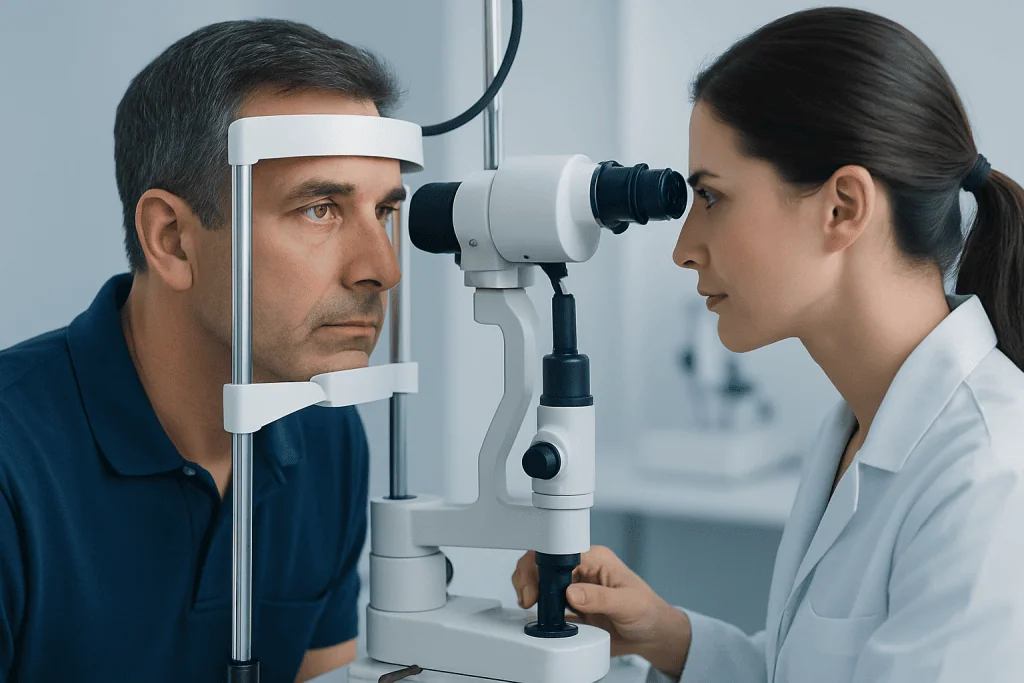
Symptoms depend on how central and dense the lens clouding is. Early cortical cataracts may cause few noticeable changes, particularly if the central lens remains relatively clear. Some people first notice that lighting conditions feel more difficult than they used to.
Common signs include glare from headlights or lamps, halos around lights, reduced contrast, blurred or hazy vision, fading or yellowing of colors, and difficulty seeing in bright sun. Night driving may become uncomfortable because scattered light can make oncoming headlights appear more intense.
Vision may fluctuate as the cataract changes, and a person may feel that glasses no longer provide the same clarity. Reading, recognizing faces, navigating stairs or judging curbs may become harder when contrast is low. These symptoms can also result from dry eye, refractive errors, glaucoma or retinal conditions, so assessment by an eye professional is important.
A cataract itself is usually painless. Sudden loss of vision, eye pain, marked redness, flashes, a curtain-like shadow or a sudden increase in floaters should not be assumed to be caused by a cataract and needs urgent medical evaluation.
Is cortical cataract serious?

A cortical cataract is a common lens change and is not usually an emergency. However, it can become serious in practical terms when it interferes with safe driving, work, reading, mobility or independence. The impact is based more on a person’s visual function and symptoms than on the name or appearance of the cataract alone.
Even a cataract that seems modest on an examination may cause troublesome glare if the cloudy areas affect how light enters the eye. Conversely, a cataract may look more advanced but cause limited symptoms in some people. Regular review helps an ophthalmologist monitor changes and discuss the right time for treatment.
Very advanced, untreated cataracts can make examination or treatment of other eye disorders more difficult. Rarely, long-standing lens changes may contribute to eye pressure problems. This is one reason to continue eye care rather than waiting until vision is severely limited.
People with diabetes, prior eye injury, long-term steroid exposure, high myopia, previous eye surgery or a family history of early cataracts may benefit from individualized follow-up. An ophthalmologist can also check for related conditions that influence the safest treatment plan.
Causes, risk factors and progression
Age-related changes are the most frequent cause of cortical cataracts. Over time, lens proteins and water distribution change, creating areas that scatter light. Cataracts can also develop earlier or progress differently because of diabetes, smoking, ultraviolet exposure, eye trauma, inflammation inside the eye, radiation exposure, certain medicines such as long-term corticosteroids, or previous eye surgery.
Protective habits support overall eye health but cannot reverse an existing cataract. These include not smoking, managing diabetes and other long-term conditions, eating a balanced diet, and wearing sunglasses that block ultraviolet radiation outdoors. People should not stop prescribed medication because of cataract concerns without speaking to the clinician who prescribed it.
How quickly do cortical cataracts progress? There is no single timetable. Some remain stable or change slowly over years, while others become more visually significant over months to a few years. Progression may be faster in the presence of diabetes, ongoing smoking, steroid use, eye injury or other ocular disease, but individual patterns remain variable.
Follow-up is usually guided by symptoms and examination findings. A change in glasses may improve vision temporarily when refractive changes are present, but it does not remove lens clouding. If everyday tasks are increasingly affected, an ophthalmology review can clarify whether surgery would be appropriate.
Diagnosis and the role of clinical images
An ophthalmologist or optometrist diagnoses cataracts through a detailed eye examination. This commonly includes a vision test, refraction to check whether glasses improve clarity, measurement of eye pressure, and examination of the lens with a slit lamp. Dilating eye drops may be used to allow a clearer view of the lens, retina and optic nerve.
A slit-lamp cortical cataract photo is taken using specialized magnification and lighting. It can show the distribution of cortical spokes, whether they approach the center of the lens, and whether other lens regions are affected. Cataracts may have cortical, nuclear and posterior subcapsular features at the same time.
Images may be kept in the medical record to support monitoring, but doctors do not rely on a single image to judge treatment. They also assess the person’s reported difficulty with vision, whether glasses help, and whether the cornea, retina and optic nerve are healthy enough to support expected visual improvement after surgery.
Before surgery, measurements of eye shape and corneal curvature help select an intraocular lens. Additional retinal imaging or testing may be recommended when symptoms or examination findings suggest macular degeneration, diabetic eye disease, glaucoma or another condition affecting vision.
What is the best treatment for cortical cataracts?
The best treatment depends on how much the cataract affects a person’s quality of life and whether vision can be improved adequately with glasses, lighting adjustments or other measures. There are no eye drops, supplements or exercises proven to clear a cataract. When symptoms are mild, observation and periodic eye examinations are often appropriate.
Cataract surgery is the only treatment that removes the cloudy natural lens. During modern surgery, the ophthalmologist removes the lens through a small incision, usually using ultrasound energy to break it into smaller pieces, and places a clear artificial intraocular lens in its position. The aim is to improve visual quality and reduce cataract-related symptoms, although the outcome also depends on other eye conditions.
Suitability for surgery is individualized. It may be considered when vision limits driving, work, hobbies, reading, self-care or confidence moving around. The decision also takes account of general health, eye anatomy, medications, the presence of retinal or optic nerve disease, and the person’s visual goals. Patients can learn more about cataract surgery during a specialist consultation.
Acibadem International’s multidisciplinary specialists and JCI-accredited hospitals assess and treat cataracts for international patients, with care plans based on eye findings and individual needs.
Cataract surgery: what happens, recovery, benefits and risks
Before the procedure, the surgical team reviews medical history, current medicines, allergies and eye measurements. Most cataract operations are performed as an outpatient procedure using anesthetic eye drops and, when appropriate, medication to help the person relax. The patient is awake but should not feel pain; the care team explains what to expect on the day.
In a typical procedure, a small opening is made at the edge of the cornea. The surgeon opens the thin capsule around the cloudy lens, breaks up and removes the lens material, then inserts a folded artificial lens implant into the remaining capsule. The small incision often seals without stitches. The other eye, if needed, is usually treated on a separate day.
Many people notice visual improvement within several days, although healing and adaptation continue for several weeks. Prescription eye drops are commonly used after surgery to support healing and reduce inflammation or infection risk. Patients are generally advised to avoid rubbing the eye, follow activity guidance, attend follow-up visits and contact the surgical team if symptoms worsen.
Benefits may include clearer vision, less glare and improved ability to perform everyday activities. Risks are uncommon but can include infection, inflammation, raised eye pressure, swelling of the retina, retinal detachment, lens-position problems or a later clouding of the lens capsule. Prompt follow-up helps identify and manage concerns early. An ophthalmologist can explain the expected benefits and relevant risks for the individual eye.
When to seek medical care
It is sensible to arrange a routine eye examination if vision becomes blurred, glare makes driving uncomfortable, glasses no longer seem effective, or colors and contrast appear less clear. A timely review is also helpful for people who have diabetes, use long-term steroid medicines, have had an eye injury, or have previously been told they have a cataract.
Urgent assessment is needed for sudden vision loss, severe eye pain, significant redness, nausea with eye pain, new flashes of light, many new floaters, or a dark curtain or shadow across vision. These are not typical cataract symptoms and may indicate another eye problem that needs prompt care.
People considering cataract surgery should discuss their work, driving needs, hobbies and health conditions with an ophthalmologist. This supports a shared decision about whether to monitor the cataract or proceed with treatment at a time that fits their visual needs and clinical findings.
Regular eye care remains important after cataract surgery because cataracts are only one possible cause of visual change. Continued monitoring can help protect long-term eye health and identify retinal, optic nerve or corneal conditions early.
Frequently asked questions
Can cortical cataract images diagnose a cataract?
Cortical cataract images can show characteristic spoke-like clouding in the lens, but they cannot provide a complete diagnosis on their own. An ophthalmologist uses a slit-lamp examination, vision testing and assessment of the retina and optic nerve to confirm the cause of visual symptoms.
What does a cortical cataract look like?
A cortical cataract often appears as whitish wedges or spokes starting near the edge of the lens and moving inward. The exact appearance varies, and some people have cortical changes together with other cataract types.
Is a cortical cataract the same as a nuclear cataract?
No. A cortical cataract affects the outer layer of the lens, while a nuclear cataract develops in the central part of the lens and often causes gradual yellowing or browning. Both can be present in the same eye and may affect vision differently.
Can glasses fix a cortical cataract?
Glasses may improve vision if a cataract has changed the eye’s prescription, especially in earlier stages. They do not remove the cloudy lens areas, so their benefit may become limited as the cataract progresses.
When is cataract surgery needed for a cortical cataract?
Surgery is generally considered when cataract-related vision problems affect important daily activities, safety or quality of life. There is no requirement to wait until the cataract is mature; the decision is individualized after an eye examination and discussion of goals.
How long does recovery take after cataract surgery?
Many people see improvement within a few days, but the eye commonly continues healing over several weeks. Recovery time can vary according to the eye’s condition, the type of lens implant and whether other eye diseases are present.
References
- American Academy of Ophthalmology
- National Eye Institute
- Mayo Clinic
- Royal College of Ophthalmologists
This article is for general information only and is not a substitute for professional medical advice. Please consult a qualified doctor about your individual situation.
Explore treatments in Turkey — costs, top hospitals & a free quote
JCI-accredited · board-certified surgeons · reply within 24h
Add us as a Preferred Source to see more of our trusted health content across Google Search, AI Overviews and Discover.