CT Scan of Epidural Hematoma: Preparation, Procedure and Results
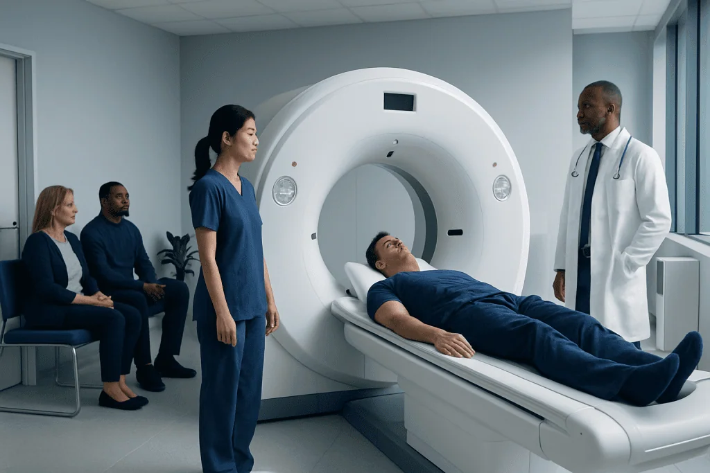
A non-contrast head CT is usually the first test used to identify a suspected epidural hematoma after head trauma. The scan is fast, painless and generally does not require injections, fasting or special preparation.
Key Takeaways
- A non-contrast head CT is usually the first test used to identify a suspected epidural hematoma after head trauma.
- The scan is fast, painless and generally does not require injections, fasting or special preparation.
- An epidural hematoma may appear as a lens-shaped collection of blood on CT images.
- CT findings are interpreted together with symptoms, neurological examination results and the mechanism of injury.
- New or worsening symptoms after a head injury need urgent medical assessment, even if the person initially seemed well.
A CT scan of epidural hematoma is the main rapid imaging test used after a head injury when doctors need to check for bleeding between the skull and the dura, the brain’s protective outer covering. It can show the location, size and pressure effects of a hematoma, helping the emergency team decide whether close monitoring or urgent neurosurgical treatment is needed.
Overview: Why CT Is Used for an Epidural Hematoma
A CT scan of epidural hematoma is a rapid brain imaging examination that helps doctors detect bleeding in the space between the inner skull and the dura mater, the tough outer membrane surrounding the brain. This type of bleeding most often develops after a blow to the head and can place pressure on nearby brain tissue if it enlarges.
Computed tomography (CT) uses X-rays and computer processing to create detailed cross-sectional images of the head. In an emergency setting, it is commonly the preferred first examination because it is widely available, takes only minutes to perform and is very effective at showing fresh blood and skull fractures.
An epidural hematoma can be serious, but the outlook is often better when it is recognised and treated promptly. CT results help the clinical team determine the urgency of care and whether the person may need observation, repeated imaging or neurosurgical treatment.
How a CT Scan Detects an Epidural Hematoma
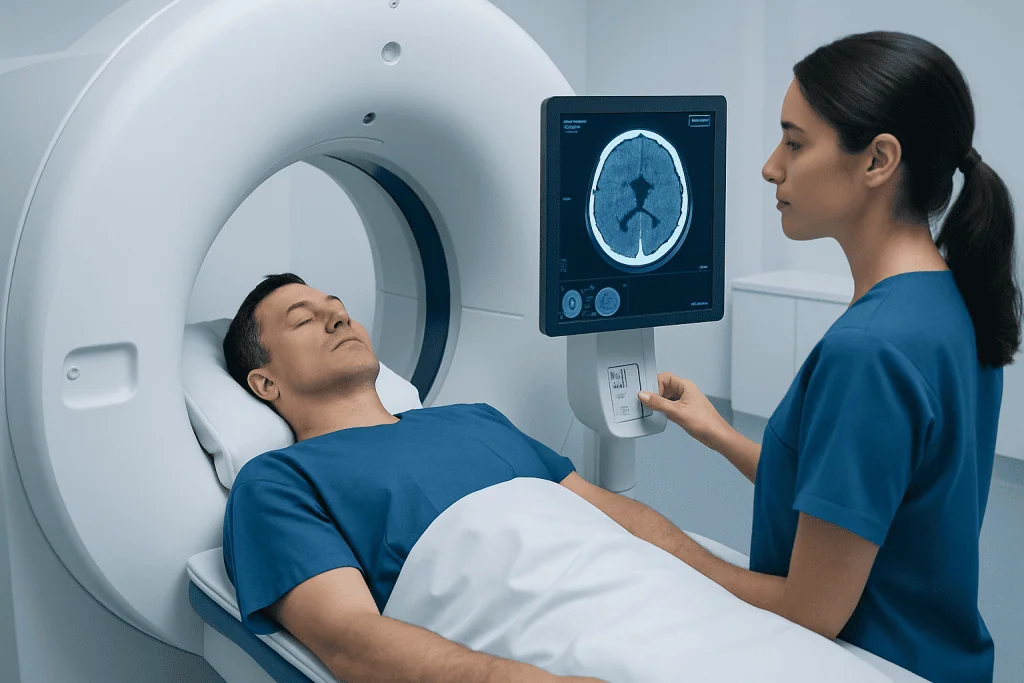
During a head CT, an X-ray scanner rotates around the head while the person lies still on a moving examination table. The computer converts the information gathered by the scanner into thin image slices that allow radiologists and emergency clinicians to examine the skull, brain tissue, fluid spaces and blood vessels indirectly.
Fresh blood usually looks denser, or brighter, than normal brain tissue on a non-contrast CT scan. An epidural hematoma often has a characteristic biconvex or lens-shaped appearance because the dura is closely attached to the skull at certain points. A CT may also reveal an associated skull fracture, brain swelling, shifting of brain structures or other types of traumatic bleeding.
CT is highly useful for urgent assessment, but the images are only one part of diagnosis. The team also considers consciousness level, pupil changes, weakness, vomiting, seizures, medications such as blood thinners and the details of the injury. Other injury patterns may be assessed at the same time, including subdural hematoma, which occurs in a different space around the brain.
Who May Need This Scan and How to Prepare

Doctors may recommend an urgent head CT after a significant head injury, particularly when there has been loss of consciousness, confusion, worsening headache, repeated vomiting, seizure, abnormal behaviour, weakness, unequal pupils or a skull fracture. Infants, older adults and people who take anticoagulant or antiplatelet medicines may need especially careful evaluation following head trauma.
Most CT scans for suspected epidural hematoma are performed without contrast dye. This means fasting is usually not required, and there is generally no injection. In emergency situations, the scan is arranged quickly and preparation is kept to a minimum so that imaging does not delay necessary care.
If possible, the patient or accompanying person should tell the team about recent injuries, previous brain surgery, pregnancy, allergies and all medicines being taken. Removable metal items near the head, such as hairpins, hearing aids, dentures or jewellery, may need to be removed because they can affect image quality. However, emergency staff will prioritise safety and stabilisation rather than delaying imaging for nonessential details.
- Bring medication information if it is readily available.
- Tell staff about blood-thinning medicines or bleeding disorders.
- Follow instructions about keeping still during the scan.
- Do not drive after a head injury unless a clinician has confirmed it is safe.
What Happens During the Procedure
Before the scan, a radiology professional confirms the person’s identity and explains the process as clearly as the situation allows. The patient lies on their back on a padded table, usually with the head supported to reduce movement. Emergency monitoring equipment may remain in place when needed.
The table moves through the circular opening of the CT scanner. The scan itself is painless and typically takes only a few minutes. The machine may make soft whirring sounds, and staff can communicate with the patient from an adjacent control area. Keeping the head still is important because movement can blur the images.
Most people remain awake for the examination. Children, very distressed patients or people who cannot stay still may require additional support, and in selected circumstances clinicians may consider sedation. If the patient is unconscious or medically unstable, the emergency and imaging teams coordinate care to complete the scan safely.
Once images are obtained, they are reviewed urgently by a radiologist and the treating team. When an epidural hematoma is suspected or confirmed, neurosurgical consultation may be arranged promptly. Depending on the findings, care may involve neurological observation, repeat CT imaging or a procedure to relieve pressure from the bleeding.
Understanding Results and the Recovery Timeline
CT results may be available quickly in an emergency department, although the exact timing depends on the clinical setting and the need for specialist review. A report generally describes whether bleeding is present, its size and location, whether there is a skull fracture, and whether the hematoma is causing pressure or displacement of brain structures.
A small epidural hematoma with no significant pressure effects may sometimes be managed with hospital observation and repeat scans. This decision depends on the scan findings, neurological status, symptoms, age, medications and the ability to monitor the person closely. A larger hematoma or one causing neurological deterioration may require urgent surgery to remove the blood and reduce pressure.
Recovery after the CT scan itself is immediate because a standard non-contrast examination has no recovery period. Recovery from the head injury, however, varies widely. Some people may return home after assessment and observation, while others require admission, surgery, rehabilitation or follow-up for symptoms related to traumatic brain injury.
Even when imaging is reassuring, patients should follow discharge instructions carefully. Rest, gradual return to usual activities and follow-up can be important. Persistent concussion-type symptoms, such as headache, dizziness, concentration difficulties or sleep changes, should be discussed with a clinician.
Benefits, Limitations and Possible Risks
The main benefit of a CT scan is speed. It can quickly identify an epidural hematoma and other urgent problems after head trauma, helping clinicians make time-sensitive treatment decisions. It can also show skull fractures and detect other intracranial bleeding that may affect care.
CT uses ionising radiation. The amount is kept as low as reasonably achievable while obtaining images that are medically useful. In a possible brain bleed, the benefit of promptly finding a potentially dangerous injury generally outweighs the small radiation risk. Clinicians take particular care when imaging children and pregnant patients, while still ensuring that urgent conditions are not missed.
A CT scan may not detect every subtle injury, especially some early or very small abnormalities. If symptoms and CT findings do not fully match, the treating team may recommend observation, repeat CT or magnetic resonance imaging (MRI) in selected situations. MRI is not usually the first test for suspected epidural hematoma because it generally takes longer and is less practical for many emergencies.
When to Seek Medical Care
Emergency medical care is needed after a head injury if a person becomes increasingly drowsy, confused, difficult to wake, develops a severe or worsening headache, vomits repeatedly, has a seizure, faints, has weakness or numbness, has trouble speaking or walking, or develops unequal pupils. These symptoms can occur with epidural hematoma and other serious brain injuries.
It is also important to seek urgent assessment after head trauma for people taking blood thinners, those with known bleeding disorders, older adults, young children and anyone who had a significant mechanism of injury. A person may occasionally seem relatively well shortly after an injury and then worsen later, so observation by a responsible adult may be advised after a clinician’s assessment.
People should not attempt to diagnose a head injury at home based on symptoms alone. If emergency symptoms are present, local emergency services should be contacted. Acibadem International’s multidisciplinary emergency, radiology and neurosurgery teams at JCI-accredited hospitals can assess and treat traumatic brain conditions for international patients when specialist care is required.
Frequently asked questions
What does an epidural hematoma look like on a CT scan?
On a non-contrast CT scan, an acute epidural hematoma commonly appears as a bright, lens-shaped or biconvex collection next to the skull. The scan may also show a nearby skull fracture and signs that the blood collection is pressing on the brain. A qualified radiologist and treating clinician interpret the images in the context of the injury and examination.
Is contrast dye needed for a CT scan of epidural hematoma?
Usually, no. A non-contrast CT scan is generally the first and most useful examination for identifying acute bleeding after head trauma. Contrast dye may be considered for other clinical questions, but it is not routinely required to diagnose an epidural hematoma.
How long does a head CT scan take?
The actual scanning portion commonly takes only a few minutes. More time may be needed for transfer, safety checks, positioning, monitoring and review of the images. In an emergency, the care team aims to complete and interpret the scan as quickly as safely possible.
Can a CT scan miss an epidural hematoma?
CT is very effective for detecting most acute epidural hematomas, particularly when performed promptly after injury. However, imaging findings can evolve, and a very small or early bleed may not always be apparent. If symptoms worsen or clinical concern remains high, doctors may repeat imaging or use another assessment method.
Can someone be awake with an epidural hematoma?
Yes. Some people are awake and may initially have mild symptoms after the injury. In certain cases, symptoms can worsen as bleeding expands and pressure increases, which is why concerning symptoms after a head injury require urgent assessment.
What happens if the CT scan confirms an epidural hematoma?
The next steps depend on the hematoma’s size, location, effect on the brain and the person’s neurological condition. Small, stable hematomas may be monitored closely with repeat examinations and scans. Larger hematomas or those causing pressure or neurological symptoms may require urgent neurosurgical drainage or removal.
References
- World Health Organization
- National Institute for Health and Care Excellence
- American College of Radiology
- Centers for Disease Control and Prevention
- Merck Manual Professional Edition
This article is for general information only and is not a substitute for professional medical advice. Please consult a qualified doctor about your individual situation.
Explore treatments in Turkey — costs, top hospitals & a free quote
JCI-accredited · board-certified surgeons · reply within 24h
Add us as a Preferred Source to see more of our trusted health content across Google Search, AI Overviews and Discover.