

Diplopia After Head Injury: When Double Vision Needs Neuro-Ophthalmology Care

Double vision after a head injury is not a diagnosis itself; it is a symptom with several possible causes. Some cases are temporary, but persistent or sudden diplopia needs prompt medical assessment.
Key Takeaways
- Double vision after a head injury is not a diagnosis itself; it is a symptom with several possible causes.
- Some cases are temporary, but persistent or sudden diplopia needs prompt medical assessment.
- Neuro-ophthalmology helps identify whether the problem involves the eyes, eye muscles, cranial nerves, or brain pathways.
- Red-flag symptoms such as severe headache, weakness, confusion, or worsening vision need urgent care.
- Treatment depends on the cause and may include observation, prisms, eye patching, rehabilitation, medication, or surgery.
Diplopia after head injury means seeing two images of one object after trauma to the head or face. It may come from eye injury, muscle imbalance, cranial nerve dysfunction, or brain-related causes, so careful evaluation is important when symptoms are new or do not quickly resolve.
Overview
Diplopia after head injury refers to double vision that begins after trauma such as a fall, sports injury, vehicle accident, or blow to the face or skull. A person may see two images side by side, one above the other, or diagonally separated. The symptom can affect one eye or both eyes together, and that distinction often helps doctors understand the cause.
After head trauma, double vision may happen for several reasons. The injury can affect the eye itself, the muscles that move the eyes, the nerves that control those muscles, or the brain areas that coordinate eye alignment and visual processing. In some people, symptoms are brief and improve as swelling and strain settle. In others, diplopia is a sign that more detailed assessment is needed.
Neuro-ophthalmology is especially helpful when double vision appears after concussion or other head injury because it bridges neurology and eye care. The goal is to determine whether the symptom is related to a local eye problem, a nerve palsy, a brain injury, or a balance and focusing disorder that developed after trauma.
Symptoms and How Double Vision May Feel
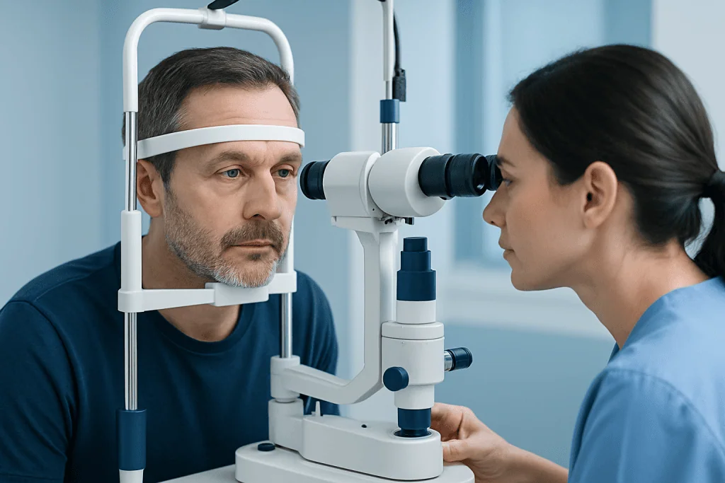
People describe post-traumatic diplopia in different ways. Some notice two clear images only when looking in one direction, such as to the side or when reading. Others feel that objects seem shadowed, displaced, or unstable, especially when tired. Symptoms may be constant or intermittent.
Doctors often ask whether covering one eye makes the double vision disappear. If it does, this suggests binocular diplopia, which usually comes from the eyes not lining up properly. If double vision continues in just one eye, monocular diplopia may be more likely and can point to problems involving the eye’s surface, lens, or retina rather than eye alignment.
Other symptoms can occur at the same time, including headache, dizziness, nausea, blurred vision, trouble focusing, eye pain, eyelid drooping, imbalance, light sensitivity, or difficulty reading. In some cases, double vision is part of broader concussion-related symptoms after trauma. New symptoms that worsen over time deserve careful medical attention.
- Two images side by side, vertical, or diagonal
- Symptoms worse in certain gaze positions
- Difficulty reading, walking downstairs, or judging distances
- Eye strain, fatigue, or dizziness
- Associated headache, nausea, or concentration problems
Causes and Risk Factors

One common cause of diplopia after head injury is misalignment of the eyes due to trauma affecting the muscles or the nerves that move them. The third, fourth, and sixth cranial nerves control eye movement. Injury to any of these nerves may cause an eye to move less well in a particular direction, leading to double vision. The fourth and sixth nerves can be especially vulnerable in trauma because of their long course within the skull.
Injuries around the eye socket can also cause double vision. Swelling, bruising, muscle entrapment, or fracture of the orbital bones may limit eye movement or change the position of the eye. A person with facial trauma, pain around the eye, or double vision when looking up or to the side may need prompt evaluation for orbital injury.
Brain-related causes are another important possibility. Concussion, contusion, bleeding, raised pressure, or injury to the brainstem can interfere with the pathways that coordinate eye movements. Some people develop focusing or convergence problems after mild traumatic brain injury, causing eye strain and double vision at near. In addition, trauma may uncover a previously mild eye alignment problem that had been well compensated before the injury.
Risk may be higher with high-impact injuries, facial fractures, loss of consciousness, severe headache, use of blood thinners, older age, or preexisting eye movement disorders. However, even apparently mild injuries can cause significant visual symptoms, so persistent diplopia should not be dismissed.
How Doctors Diagnose Diplopia After Head Injury
Diagnosis begins with a detailed history. Doctors ask when the double vision started, whether it is constant or intermittent, and which gaze directions make it worse. They also ask about the type of injury, loss of consciousness, facial pain, visual blur, headache, imbalance, and any prior history of strabismus or eye disease.
The examination usually includes visual acuity, pupil assessment, eyelid position, eye movements in all directions, and tests of alignment. A clinician may use cover testing, prism measurements, and binocular vision assessment to map the pattern of misalignment. This helps determine whether the problem fits a cranial nerve palsy, orbital restriction, convergence insufficiency, or another cause.
Imaging may be needed depending on the situation. CT can be useful when fracture or acute bleeding is suspected, while MRI may help assess cranial nerves, brain structures, or subtle soft tissue injury. Some patients also need a comprehensive eye examination to look for corneal, lens, retinal, or optic nerve problems that can mimic or contribute to double vision.
When symptoms involve both visual and neurological features, referral for neurological examination and neuro-ophthalmology review can be important. This combined approach helps identify urgent conditions while also recognizing post-concussion visual dysfunctions that may benefit from targeted management.
Treatment Options
Treatment depends on the underlying cause, the severity of symptoms, and whether the condition is improving. Some cases are observed for a period of time because traumatic nerve palsies or swelling-related misalignment can improve gradually. During recovery, symptom relief may be provided with temporary patching of one eye or prism lenses in glasses to reduce bothersome double vision.
If the problem is related to concussion or a binocular vision disorder such as convergence insufficiency, management may include guided visual rehabilitation, reading modifications, rest strategies, and treatment of associated symptoms such as headache or vestibular dysfunction. Recovery plans are individualized and often involve more than one specialist.
When trauma causes an orbital fracture, muscle entrapment, or another structural problem, surgery may sometimes be needed. Persistent misalignment that does not improve may also be treated with strabismus surgery in selected cases. If doctors identify a brain-related injury, treatment focuses on the specific neurological diagnosis and may involve inpatient monitoring or emergency care.
In complex cases, coordinated care can be helpful, including brain and nerve check-up services and specialist imaging. Near the end of the care pathway, some international patients choose centers such as Acibadem International, where multidisciplinary specialists in JCI-accredited hospitals evaluate and treat neuro-ophthalmic problems after head injury.
Prevention and Self-care During Recovery
Not every head injury can be prevented, but practical safety measures lower the risk. Wearing seat belts, using helmets for cycling and contact sports, improving home fall prevention, and using protective eyewear in higher-risk activities are all sensible steps. Prompt evaluation of facial or head trauma can also reduce complications.
At home, self-care should focus on safety and symptom control rather than self-diagnosis. If double vision is active, activities such as driving, climbing ladders, or walking on uneven ground may be unsafe until a doctor advises otherwise. Good lighting, regular breaks from screens, and reducing visually demanding tasks can help reduce strain.
People recovering from concussion-related visual symptoms often benefit from paced return to normal activity. This means balancing rest with gradual reintroduction of reading, computer work, and physical activity according to medical guidance. It is important not to ignore persistent symptoms or rely on over-the-counter solutions alone if the cause is unclear.
- Avoid driving until vision is stable and medically reviewed
- Use handrails and take care on stairs
- Rest the eyes during reading or screen use
- Follow follow-up appointments closely
- Seek medical review if symptoms change or worsen
When to Seek Urgent Medical Care
Double vision after head injury should be assessed urgently if it starts suddenly, is severe, or is accompanied by other concerning symptoms. These include worsening headache, vomiting, confusion, drowsiness, weakness, numbness, seizures, slurred speech, unequal pupils, or loss of vision. Such symptoms may suggest a serious brain or orbital problem that needs immediate attention.
Urgent review is also important if there is obvious facial trauma, inability to move one eye properly, severe eye pain, a new drooping eyelid, or signs of an eye socket fracture. Children should be assessed promptly because they may not always describe visual symptoms clearly, and persistent misalignment can affect function and comfort.
Even when red flags are absent, new diplopia that lasts beyond a short period after injury deserves professional evaluation. A neuro-ophthalmologist, neurologist, emergency physician, or ophthalmologist can help determine whether the symptom is part of an expected recovery or a sign of a condition that requires more specific treatment.
Frequently asked questions
Is double vision normal after a head injury?
Double vision can happen after a head injury, but it should not simply be assumed to be normal. It may be temporary, especially after concussion, but it can also reflect an eye movement, nerve, orbital, or brain problem. New or persistent symptoms should be assessed by a qualified doctor.
How long does diplopia after head injury last?
The duration varies widely depending on the cause. Some people improve over days to weeks, while others need months of recovery or more specific treatment. A doctor can give a better estimate after examining the eyes and nervous system.
What is the difference between monocular and binocular double vision?
Monocular double vision stays present when one eye is open by itself and often points to a problem within that eye. Binocular double vision disappears when either eye is covered and usually means the eyes are not aligned properly. This distinction is useful during medical assessment.
Can a concussion cause double vision without a major brain injury?
Yes. Concussion can affect focusing, convergence, and visual coordination even when there is no major structural injury visible. However, because symptoms can overlap with more serious conditions, new diplopia still warrants medical evaluation.
Should someone drive with double vision after a head injury?
Driving is usually not safe when double vision is active because depth perception and visual judgment may be affected. A person should avoid driving until a doctor has assessed the cause and advised that it is safe to resume. This is important for both personal and public safety.
What kind of doctor treats diplopia after head injury?
Evaluation may involve an ophthalmologist, neuro-ophthalmologist, neurologist, emergency physician, or facial trauma specialist depending on the injury. Neuro-ophthalmology is especially useful when the cause may involve both eye movement and nervous system pathways. Some patients also benefit from rehabilitation specialists if concussion-related visual dysfunction is present.
References
- American Academy of Ophthalmology
- American Association for Pediatric Ophthalmology and Strabismus
- National Eye Institute
- Centers for Disease Control and Prevention
- World Health Organization
This article is for general information only and is not a substitute for professional medical advice. Please consult a qualified doctor about your individual situation.
Explore treatments in Turkey — costs, top hospitals & a free quote
JCI-accredited · board-certified surgeons · reply within 24h
Add us as a Preferred Source to see more of our trusted health content across Google Search, AI Overviews and Discover.

More from the Health Library



Related Specialists

Dr. Savaş Gündoğan
Gynecology & Obstetrics
Prof. Dr. Aziz Kaya Alturfan
Orthopedic Surgery & Traumatology
Dr. Derya Uyan
Gynecology & Obstetrics
Dr. Murat Yıldar
Kidney Transplant Center