Dnet MRI: Preparation, Procedure and Results
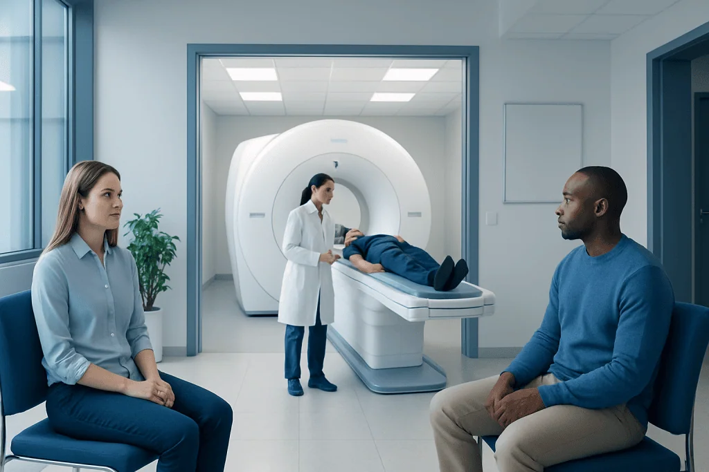
DNETs are uncommon, usually slow-growing brain tumors that often begin in childhood or young adulthood and may cause focal seizures. MRI is the main imaging test for evaluating a suspected DNET and for monitoring it over time.
Key Takeaways
- DNETs are uncommon, usually slow-growing brain tumors that often begin in childhood or young adulthood and may cause focal seizures.
- MRI is the main imaging test for evaluating a suspected DNET and for monitoring it over time.
- A typical DNET MRI appearance can suggest the diagnosis, but MRI alone cannot confirm whether any brain tumor is cancerous.
- Brain MRI preparation usually involves screening for metal implants, removing metal items and discussing contrast, kidney health, pregnancy and claustrophobia.
- When seizures or a lesion are present, care is individualized and may include seizure medicines, observation, neurosurgery or further diagnostic testing.
A DNET MRI is a brain magnetic resonance imaging scan used to identify and assess a dysembryoplastic neuroepithelial tumor (DNET), a rare tumor often associated with seizures. MRI provides important details about the lesion, but doctors interpret the results alongside symptoms, neurological examination findings and, when needed, tissue testing.
DNET MRI: what it shows and why it is used
A DNET MRI is a magnetic resonance imaging scan of the brain performed to look for and characterize a dysembryoplastic neuroepithelial tumor (DNET). DNET is a rare, generally low-grade tumor that develops from glial and neuronal cells. It commonly arises in the cerebral cortex, particularly the temporal lobe, and is often discovered during an evaluation for recurrent focal seizures.
MRI uses a strong magnetic field and radio waves rather than ionizing radiation. It produces detailed images of brain structures and helps the medical team assess the lesion’s location, size, internal features and relationship to nearby tissue. The scan may also help distinguish a possible DNET from other causes of seizures, including developmental changes, scarring and other low-grade tumors.
Although people may search for “dne MRI,” the accepted term is DNET MRI. Imaging is a central part of assessment, but it is not considered a stand-alone diagnosis. A neurologist, neuroradiologist and, where appropriate, neurosurgeon review MRI findings together with seizure history, electroencephalography (EEG) results and the person’s overall health.
How MRI works and who may need a DNET MRI
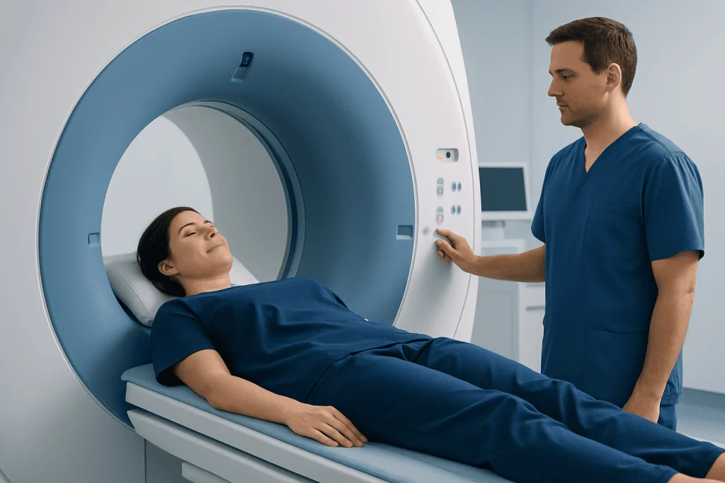
During MRI, the body lies inside a scanner containing a powerful magnet. Different MRI sequences show water, tissue structure, fluid and blood products in different ways. For a suspected DNET, the imaging protocol typically includes several detailed views of the brain. Some people also receive gadolinium-based contrast through a vein to show whether a lesion has areas of enhancement.
A doctor may request a brain MRI when a person has unexplained focal seizures, changes in awareness, unusual sensory episodes, persistent neurological symptoms or an abnormality seen on another scan. MRI may also be used after a DNET diagnosis to establish a baseline, support treatment planning or monitor a lesion that is being observed.
Most people can safely have an MRI, but screening is essential. Certain implanted devices, metal fragments, cochlear implants, older aneurysm clips or some stimulators may not be MRI-compatible. The imaging team should also know about pregnancy, kidney disease, prior contrast reactions, severe anxiety in enclosed spaces and any implanted medical device before scheduling the scan.
MRI procedure preparation and step-by-step process
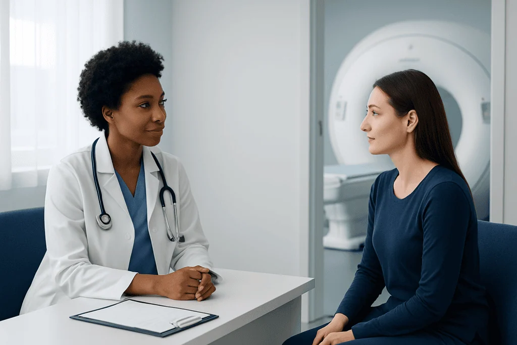
Brain MRI preparation instructions are usually straightforward. Patients should follow the imaging center’s specific directions about eating and drinking; many routine brain MRI scans do not require fasting, although different instructions may apply if sedation or contrast is planned. Current medication lists, prior imaging reports and information about implants should be brought or shared in advance.
For MRI pre-procedure instructions, patients should remove jewelry, watches, hairpins, hearing aids, removable dental appliances and clothing with metallic parts. Makeup or skin products may occasionally contain metallic particles, so the staff may advise avoiding them. It is important to tell the team about tattoos, permanent cosmetics, medication patches, insulin pumps or continuous glucose monitors, as some may require special precautions or temporary removal.
The procedure usually follows these steps:
- Staff complete a safety questionnaire and explain the scan.
- The patient lies on a padded table, and a head coil is placed around the head to improve image quality.
- Earplugs or headphones are provided because the scanner makes loud tapping sounds.
- The table moves into the scanner while the patient stays still for several image sequences.
- If contrast is needed, it is injected through an intravenous line during the examination.
A brain MRI commonly takes about 30 to 60 minutes, depending on the sequences required and whether contrast is used. The radiographer can communicate with the patient throughout the scan. For significant claustrophobia, children, or people who cannot remain still, a clinician may discuss options such as an open-style scanner where available or carefully supervised sedation.
What are the MRI findings of DNET?
The DNET MRI appearance is often a cortical or superficial lesion within a temporal lobe, though DNETs can occur elsewhere in the brain. They are commonly well-defined and may have a multicystic, “bubbly” or soap-bubble-like appearance. On T2-weighted and FLAIR MRI images, they often appear brighter than nearby normal brain tissue because of their high fluid content and tissue characteristics.
Many DNETs cause little swelling around the lesion and do not produce substantial pressure on surrounding structures. Contrast enhancement is absent or limited in many cases, although imaging appearances vary. Some lesions may be associated with nearby cortical developmental abnormalities, including focal cortical dysplasia, which can also contribute to seizures.
These features can make radiologists consider DNET, but they are not unique to it. Other low-grade tumors, glioneuronal tumors and non-tumor abnormalities can look similar. Follow-up imaging, advanced MRI techniques, specialist review and sometimes surgical pathology are used when the diagnosis remains uncertain or when treatment is being considered.
Can an MRI tell if a brain tumor is cancerous?
MRI can provide clues about whether a brain lesion may behave more like a low-grade or higher-grade tumor, but it cannot determine with certainty whether a tumor is cancerous. Features such as an irregular border, rapid change, extensive swelling, tissue destruction, bleeding or strong contrast enhancement can raise concern, while a stable, well-circumscribed lesion with typical DNET features may support a lower-grade process.
However, imaging findings overlap among different tumor types and grades. The most definitive way to identify a tumor’s cell type and grade is examination of tissue by a neuropathologist after biopsy or surgery. Whether tissue sampling is appropriate depends on the lesion’s location, symptoms, imaging pattern and the balance of likely benefit and risk.
For people with suspected DNET and difficult-to-control seizures, specialist assessment may include EEG, neuropsychological testing and detailed imaging to understand where seizures begin and how treatment could affect important functions. Brain tumor surgery may be considered when a lesion causes medication-resistant seizures, is growing, produces concerning features or needs tissue confirmation.
Treatment decisions, recovery and outlook
Not every DNET requires immediate intervention. For a lesion with a characteristic appearance, stable imaging and manageable symptoms, a specialist may recommend periodic MRI surveillance and treatment for seizures. Anti-seizure medicines can help some people, although seizures related to DNET may persist despite medication in others.
When surgery is recommended, the goal is usually to remove the lesion as safely and completely as possible while protecting normal brain function. Recovery varies with the lesion’s location, the extent of surgery, seizure history and individual health. After an uncomplicated MRI itself, there is no recovery period; most people can return to normal activities immediately. If sedation was used, an adult should accompany the patient home and driving should be avoided until the effects have fully worn off.
Potential MRI risks are low. The main concerns are metal-related safety issues, anxiety in the scanner and uncommon reactions to contrast. Gadolinium contrast is generally well tolerated, but the team takes additional care in people with severe kidney disease or previous contrast reactions. The benefit of MRI is that it gives a detailed, noninvasive view that supports careful and individualized decisions.
Acibadem International’s multidisciplinary neurology, neuroradiology and neurosurgery specialists at JCI-accredited hospitals assess brain lesions and seizure-related conditions for international patients, with treatment planning based on imaging and clinical needs.
How rare is a DNET tumor?
DNET is rare. It represents a small proportion of primary brain tumors and is most often recognized in children, teenagers and young adults, although diagnosis can occur at other ages. Because it is uncommon and can resemble other lesions on imaging, review by clinicians experienced in epilepsy and brain tumor imaging is valuable.
The exact number of people affected is difficult to state because DNET classification has evolved and some cases may historically have been grouped with other low-grade glioneuronal tumors. Its rarity does not automatically mean it is aggressive. In many people, the main health concern is seizure control rather than rapid tumor growth.
A diagnosis can understandably lead to questions about employment, school, driving and family planning. These issues should be discussed with the treating neurologist or neurosurgeon, especially when seizures are ongoing. Local regulations commonly affect driving eligibility after seizures, so individualized medical and legal advice is important.
What is the survival rate for patients with DNET tumors?
DNETs generally have a favorable outlook because they are typically slow-growing and classified as low-grade tumors. Long-term survival is usually excellent, particularly when the lesion can be fully removed when surgery is needed. However, a single survival rate is not the most useful measure for every individual because DNET is rare, published studies differ, and outcomes depend on diagnosis, location, seizure control, treatment and follow-up duration.
For many patients, the more immediate outcome question is whether seizures improve. Seizure outcomes after surgery are often favorable, especially when the tumor and the seizure-generating area can be safely addressed. Some people may continue to need anti-seizure medication for a period after surgery, and the care team decides on any medication changes gradually.
Regular follow-up remains important even with a reassuring outlook. Follow-up may include neurological review, seizure monitoring and repeat MRI at intervals chosen by the care team. New symptoms or changes on imaging do not necessarily mean a serious problem, but they should be assessed promptly.
When to seek medical care
Medical assessment is appropriate for recurrent episodes of altered awareness, unusual smells or tastes, involuntary movements, unexplained periods of confusion, new seizures or persistent neurological symptoms. A clinician can determine whether urgent testing, including a brain MRI, is needed. Anyone with a first seizure should seek prompt medical evaluation.
Emergency care is needed if a seizure lasts longer than five minutes, repeated seizures occur without full recovery between them, breathing is difficult, a serious injury occurs, or the person is pregnant or has diabetes. Emergency assessment is also important for sudden weakness, difficulty speaking, a severe new headache, loss of consciousness or a major change in mental status.
People who already have a known DNET should contact their treating team about increasing seizure frequency, new or worsening headaches, changes in vision, weakness, balance difficulties, memory changes or medication side effects. They should not stop anti-seizure medicine abruptly unless instructed by a qualified clinician.
Frequently asked questions
Does a DNET always cause seizures?
No. Seizures are a common reason DNET is found, but not every person with a DNET has seizures. Symptoms depend on the lesion’s location and its effect on nearby brain networks, and some lesions are identified incidentally during imaging for another reason.
Does a DNET MRI always require contrast?
No. Many DNET assessments can be informative using non-contrast MRI sequences, but contrast may be added to clarify lesion features or evaluate alternative diagnoses. The radiologist and referring clinician decide whether contrast is useful for the individual situation.
How should a person prepare for a brain MRI?
The person should complete the MRI safety screening carefully, remove metal objects and follow the center’s instructions about food, drink and medicines. They should tell the staff about implants, possible pregnancy, kidney problems, prior contrast reactions and claustrophobia before the appointment.
Is a DNET cancer?
DNET is usually considered a low-grade glioneuronal tumor and generally behaves less aggressively than malignant brain tumors. Still, it requires specialist evaluation because imaging alone may not establish the diagnosis and seizure symptoms may need treatment.
Can a DNET grow or come back after surgery?
Many DNETs remain stable or are successfully managed after removal, but imaging follow-up is still important. The chance of residual tumor, change over time or recurrence depends on whether complete removal was possible and on the final pathology findings.
What happens after a DNET MRI scan?
A radiologist reviews the images and sends a report to the requesting clinician, who explains what the findings mean in the context of symptoms and medical history. If a lesion is seen, next steps may include specialist referral, EEG, repeat MRI, observation or discussion of biopsy or surgery.
References
- World Health Organization Classification of Tumours Editorial Board
- National Cancer Institute
- American College of Radiology
- Epilepsy Foundation
- National Institute of Neurological Disorders and Stroke
This article is for general information only and is not a substitute for professional medical advice. Please consult a qualified doctor about your individual situation.
Oncology care in Turkey — second opinion and treatment plan
JCI-accredited · board-certified surgeons · reply within 24h
Add us as a Preferred Source to see more of our trusted health content across Google Search, AI Overviews and Discover.