Dural Arteriovenous Fistula Symptoms: Pulsatile Tinnitus, Headache, and When to See a Doctor

A dural arteriovenous fistula is an abnormal connection between arteries and veins in the covering of the brain or spinal cord. Common symptoms include pulsatile tinnitus, headache, vision changes, and neurologic symptoms, but some fistulas cause no symptoms at first.
Key Takeaways
- A dural arteriovenous fistula is an abnormal connection between arteries and veins in the covering of the brain or spinal cord.
- Common symptoms include pulsatile tinnitus, headache, vision changes, and neurologic symptoms, but some fistulas cause no symptoms at first.
- Imaging tests such as MRI, CT angiography, and cerebral angiography help confirm the diagnosis and show the exact blood flow pattern.
- Treatment may include endovascular embolization, surgery, radiosurgery, or careful monitoring depending on the fistula type and risk.
- Sudden severe headache, weakness, speech problems, seizures, or new confusion need urgent medical attention.
Dural arteriovenous fistula symptoms often include pulsatile tinnitus, headache, and sometimes vision or neurologic changes. Because these symptoms can overlap with more common conditions, proper medical evaluation is important to confirm the cause and decide whether treatment is needed.
Overview
A dural arteriovenous fistula, often called a dural AV fistula, is an abnormal connection between arteries and veins in the dura, the protective covering around the brain and spinal cord. In this condition, blood can flow directly from arteries into veins without the usual small capillaries in between. That abnormal flow may raise pressure in nearby veins and affect how blood drains from the brain or spinal cord.
Dural arteriovenous fistula symptoms vary widely. Some people notice a rhythmic whooshing sound in one ear, known as pulsatile tinnitus. Others may have headache, eye symptoms, dizziness, or neurologic changes. A few people have no clear symptoms and the fistula is found during testing for another problem.
The seriousness of a dural AV fistula depends less on its name and more on its location and venous drainage pattern. Some are relatively low risk and mainly cause bothersome symptoms. Others can increase the chance of bleeding or neurologic injury and need more urgent treatment. This is why specialist evaluation is important when symptoms suggest an abnormal blood vessel connection.
Symptoms
The most recognized dural arteriovenous fistula symptoms include pulsatile tinnitus, headache, and changes related to pressure in nearby veins. Pulsatile tinnitus is often described as a heartbeat-like sound, whooshing, or humming in one ear or occasionally both ears. It may become more noticeable at night, during exercise, or when lying down.
Headache may be mild or more persistent, and it can occur alone or together with pulsatile tinnitus. Some people develop visual symptoms such as blurred vision, double vision, eye redness, eye bulging, or a feeling of pressure behind the eye. These symptoms are more likely when the fistula affects veins near the eye socket or cavernous sinus.
Neurologic symptoms can happen if venous congestion affects brain or spinal cord function. Depending on the site, a person may notice weakness, numbness, balance problems, memory or concentration difficulties, speech changes, seizures, or progressive walking problems. In rare cases, a dural AV fistula can present with bleeding in or around the brain, causing a sudden severe headache or other emergency symptoms.
- Pulsatile tinnitus or a rhythmic sound in the ear
- Persistent or unexplained headache
- Blurred vision, double vision, red eye, or eye swelling
- Dizziness or imbalance
- Weakness, numbness, or trouble speaking
- Seizures or sudden neurologic change
Causes and Risk Factors

The exact cause of a dural AV fistula is not always clear. In some cases, it appears to develop after a change in normal venous drainage, such as clotting in a vein or venous sinus. This may trigger the growth of small abnormal vessel connections in the dura. In other people, the fistula appears without a clear trigger.
Possible risk factors include prior head trauma, surgery, infection, and venous sinus thrombosis. Hormonal influences, age-related vascular changes, or inflammatory processes may also play a role in some cases. Most dural AV fistulas are considered acquired rather than present from birth.
It is important to remember that having headache or pulsatile tinnitus does not automatically mean a person has a dural AV fistula. These symptoms are much more commonly caused by other conditions, such as migraine, ear disorders, anemia, high blood pressure, or vascular variations. Still, when symptoms are persistent, pulse-synchronous, or occur with neurologic or visual changes, a doctor may consider a vascular cause and investigate further.
Diagnosis
Diagnosis begins with a careful medical history and physical examination. The doctor will ask when the symptoms started, whether the sound in the ear matches the heartbeat, and whether there are accompanying headaches, vision problems, or neurologic symptoms. In some cases, a clinician may hear a bruit, an abnormal vascular sound, over the skull or around the ear with a stethoscope.
Imaging is central to confirming the diagnosis. MRI and MRA can show indirect signs of abnormal blood flow, while CT angiography may provide additional detail about nearby blood vessels and venous sinuses. These tests can strongly suggest a dural AV fistula, but they may not always define the exact anatomy or risk level.
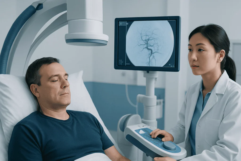
Cerebral angiography, also called digital subtraction angiography, is often the most definitive test. It shows which arteries feed the fistula, how blood drains, and whether there is dangerous cortical venous reflux. This information helps specialists classify the fistula and decide on the safest management plan. If symptoms overlap with other vascular conditions, doctors may also evaluate for problems such as brain aneurysm.
Because pulsatile tinnitus can come from several causes, evaluation may involve more than one specialty. Depending on the symptoms, a patient may be assessed by neurology, interventional neuroradiology, neurosurgery, or ENT. The goal is not only to find the abnormality, but also to understand whether it is low risk, high risk, or unrelated to the symptoms.
Treatment Options
Treatment depends on the fistula’s location, venous drainage pattern, symptom burden, and bleeding risk. Some low-risk fistulas can be monitored with regular follow-up if symptoms are mild and imaging does not show dangerous venous drainage. However, fistulas with cortical venous reflux, neurologic symptoms, or eye complications often need active treatment.
The most common treatment is an endovascular procedure, in which a specialist guides tiny catheters through blood vessels to the fistula and blocks the abnormal connection from inside. This approach is commonly called embolization and may use liquid embolic agents, coils, or other materials selected by the treating team. In many cases, it is the first-line option because it can directly target the abnormal flow while avoiding a large incision.
Some patients may need more than one treatment session or a combined approach. In selected cases, Gamma Knife radiosurgery or conventional surgery may be considered, especially when endovascular access is difficult or when residual abnormal vessels remain after treatment. If the anatomy is complex, treatment planning may involve teams experienced in brain and nerve surgery and advanced neurovascular care.
The best approach is individualized. Specialists weigh the benefits of closing the fistula against the potential risks of treatment, then explain the options in clear terms. Near the end of care planning, some international patients choose centers such as Acibadem International, where multidisciplinary specialists in JCI-accredited hospitals diagnose and treat complex neurovascular conditions.
Prevention and Self-Care
There is no guaranteed way to prevent a dural AV fistula, because many cases arise without a clearly preventable cause. Even so, general vascular health habits may support overall well-being. These include managing blood pressure, not smoking, staying physically active as appropriate, and following up on clotting or vascular conditions if a doctor has identified them.
Self-care focuses mainly on noticing symptoms early and avoiding delays in evaluation. A person with pulsatile tinnitus can keep a simple record of when it happens, whether it matches the heartbeat, and whether headaches, vision changes, or dizziness occur at the same time. This information can be helpful during a medical visit.
It is also wise not to assume persistent pulse-synchronous ear noise is only stress or wax buildup, especially if it is one-sided or new. While many cases of tinnitus are not dangerous, pulsatile tinnitus deserves assessment because it can occasionally signal a vascular problem. Self-treatment should never replace a professional evaluation when symptoms are unexplained or progressive.
When to See a Doctor
A doctor should evaluate new or persistent pulsatile tinnitus, especially if it occurs on one side, follows the heartbeat, or comes with headache, dizziness, or visual symptoms. Medical attention is also important when headaches change in pattern, become more frequent, or are accompanied by neurologic complaints. Early assessment can help identify whether the cause is a vascular issue, an ear-related condition, or another treatable problem.
Urgent care is needed for red-flag symptoms such as sudden severe headache, seizures, fainting, weakness on one side, difficulty speaking, confusion, or sudden vision loss. These symptoms can indicate bleeding or significant pressure changes affecting the brain and should not be ignored. Anyone with rapidly worsening eye redness, eye bulging, or double vision should also seek prompt evaluation.
Because several conditions can mimic each other, specialist review is often the safest next step when symptoms are concerning. In some cases, doctors may also compare the findings with other neurovascular conditions such as arteriovenous malformation. Timely diagnosis helps guide whether reassurance, monitoring, or treatment is the right path.
Frequently asked questions
What does pulsatile tinnitus from a dural AV fistula sound like?
People often describe it as a whooshing, humming, or heartbeat-like sound that matches the pulse. It may be more noticeable in quiet settings, when lying down, or after physical activity.
Is a dural arteriovenous fistula the same as a brain aneurysm?
No. A dural arteriovenous fistula is an abnormal connection between arteries and veins in the dura, while a brain aneurysm is a bulging weak area in an artery wall. Both involve blood vessels, but they are different conditions with different risks and treatments.
Can a dural AV fistula cause headaches without other symptoms?
Yes, it can. However, headache alone is very common and usually has a more common cause, so imaging and specialist assessment are used to determine whether a vascular abnormality is present.
Do all dural AV fistulas need treatment?
Not always. Some low-risk fistulas may be monitored, while others need treatment because of their venous drainage pattern, symptoms, or bleeding risk. The decision is based on detailed imaging and expert review.
How is a dural arteriovenous fistula diagnosed?
Doctors usually start with a history, examination, and imaging such as MRI, MRA, or CT angiography. Cerebral angiography is often the most definitive test because it maps the exact abnormal blood flow.
When should someone go to the emergency department?
Emergency evaluation is important for sudden severe headache, seizures, confusion, weakness, trouble speaking, or sudden vision loss. These symptoms can signal bleeding or significant neurologic involvement and need urgent care.
References
- National Institute of Neurological Disorders and Stroke
- American Heart Association
- Radiological Society of North America
- National Organization for Rare Disorders
- Merck Manual Consumer Version
This article is for general information only and is not a substitute for professional medical advice. Please consult a qualified doctor about your individual situation.
Joint and spine care in Turkey — expert assessment & treatment
JCI-accredited · board-certified surgeons · reply within 24h
Add us as a Preferred Source to see more of our trusted health content across Google Search, AI Overviews and Discover.
Check your numbers in seconds
BMI, calories, due date, blood pressure and 30+ more clinical calculators — free, instant, doctor-reviewed ranges.