Elbow Joint Anatomy: Which Bones Connect and What Stabilizes the Joint?

The elbow is formed by the humerus in the upper arm and the ulna and radius in the forearm. It functions as a combined hinge and pivot joint, allowing flexion, extension, pronation, and supination.
Key Takeaways
- The elbow is formed by the humerus in the upper arm and the ulna and radius in the forearm.
- It functions as a combined hinge and pivot joint, allowing flexion, extension, pronation, and supination.
- Joint stability comes from the fit of the bones, the joint capsule, key ligaments, and the muscles crossing the joint.
- Cartilage, synovial lining, and small fluid-filled structures help the elbow move smoothly.
- Pain, swelling, weakness, or loss of motion may signal injury or an underlying elbow condition.
Elbow joint anatomy centers on three bones—the humerus, ulna, and radius—that work together to allow bending, straightening, and forearm rotation. Its stability depends on bone shape, ligaments, cartilage, and the surrounding muscles and tendons.
Overview of Elbow Joint Anatomy
The elbow is a complex joint that connects the upper arm to the forearm. Although it is often described simply as a hinge, it is more accurate to think of it as a combination of joints working together. This design allows a person not only to bend and straighten the arm, but also to rotate the forearm so the palm can turn up or down.
Elbow joint anatomy includes three bones, several articulations, strong ligaments, smooth cartilage surfaces, and muscles that guide movement. These structures must work in balance to support everyday actions such as lifting, reaching, pushing, pulling, typing, and personal care. Because the elbow is used frequently, even small injuries or inflammation can affect comfort and function.
Understanding the anatomy can make it easier to see why elbow pain, stiffness, or instability develops. It also helps explain how doctors diagnose common problems and why treatment may focus on rest, bracing, exercise, imaging, medication, or sometimes surgery.
Which Bones Connect at the Elbow?
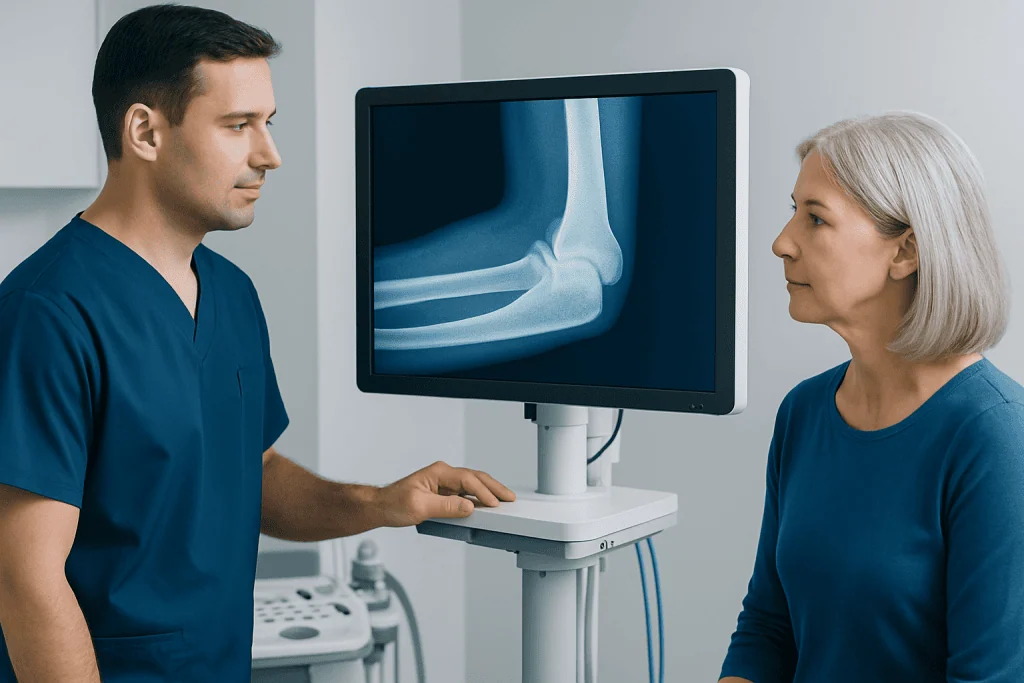
Three bones meet at the elbow: the humerus, the ulna, and the radius. The humerus is the long bone of the upper arm. The ulna and radius are the two long bones of the forearm. Together, they form the main framework of the elbow joint.
The lower end of the humerus has specially shaped surfaces that connect with both forearm bones. The ulna meets the humerus at the trochlea, creating the main hinge that allows bending and straightening. The radius meets the humerus at the capitellum, helping with both elbow motion and forearm rotation.
The elbow actually includes three related joints within one capsule:
- Humeroulnar joint: mainly responsible for flexion and extension.
- Humeroradial joint: supports movement and load transfer through the forearm.
- Proximal radioulnar joint: allows the radius to rotate around the ulna during pronation and supination.
A prominent part of the ulna called the olecranon forms the bony tip of the elbow. This structure acts like a lever for the triceps muscle, helping straighten the arm. The close fit between these bones contributes significantly to elbow stability, especially during daily use.
How the Elbow Moves
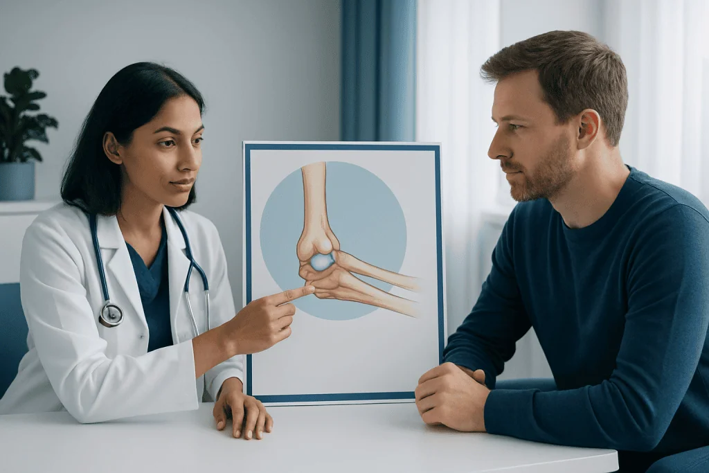
The elbow allows four main movements. Flexion bends the elbow and brings the hand toward the shoulder. Extension straightens the arm. Pronation rotates the forearm so the palm faces downward, while supination turns the palm upward. These movements are essential for practical tasks such as eating, dressing, opening doors, and using tools.
The humeroulnar joint works mainly like a hinge, providing a stable path for bending and straightening. At the same time, the radius rotates at the proximal radioulnar joint. This rotation is what gives the forearm its turning ability. The humeroradial joint helps coordinate these actions and transmit forces across the elbow.
Muscles around the elbow produce movement. The biceps and brachialis help bend the elbow, while the triceps helps extend it. Other muscles in the forearm control pronation and supination. Good coordination between these muscles is important not only for motion but also for dynamic joint stability during activity.
What Stabilizes the Elbow Joint?
Elbow stability depends on both static and dynamic stabilizers. Static stabilizers are structures that support the joint even when the muscles are relaxed. These include the shape of the bones, the joint capsule, and the major ligaments. Dynamic stabilizers are the muscles and tendons that cross the elbow and help keep the joint aligned during movement.
The two main ligament complexes are the medial collateral ligament, also called the ulnar collateral ligament, and the lateral collateral ligament complex. The medial collateral ligament resists forces that push the forearm outward relative to the upper arm, especially during throwing. The lateral collateral ligament complex helps prevent abnormal rotation and sideways instability. A separate ligament called the annular ligament wraps around the head of the radius and holds it in position while allowing rotation.
Soft tissues also contribute to smooth and safe motion. Articular cartilage covers the ends of the bones and reduces friction. The synovial membrane produces joint fluid for lubrication. Bursae, which are small fluid-filled sacs, reduce friction between moving tissues. If these structures become irritated, conditions such as bursitis may cause swelling and discomfort around the elbow.
When the stabilizing system is injured, the elbow may feel weak, painful, or unstable. In some cases, trauma can lead to elbow dislocation, while overuse can strain the ligaments and tendons over time.
Common Problems Related to Elbow Anatomy
Because many structures meet at the elbow, different problems can cause similar symptoms. Tendons can become irritated from repetitive gripping, lifting, or sports. Ligaments may be stretched or torn during falls, throwing, or contact injuries. Cartilage surfaces may wear down with age or after injury, and nerves around the elbow can also become compressed.
One of the best-known overuse problems is tennis elbow, which affects the tendons on the outer side of the elbow. Another common source of pain is inflammation of the inner side tendons, sometimes linked to repetitive wrist and forearm use. Children may experience different injuries because growing bones and growth plates behave differently from adult bone.
Fractures can involve the radial head, olecranon, or distal humerus. Dislocations may happen after a fall onto an outstretched hand. Inflammatory conditions, osteoarthritis, and crystal-related arthritis can also affect the joint. Because symptoms overlap, persistent pain, locking, significant swelling, or reduced motion should be assessed by a qualified clinician.
How Doctors Examine and Diagnose Elbow Conditions
Diagnosis starts with a medical history and physical examination. A doctor may ask when symptoms began, whether there was an injury, which motions are painful, and whether there is numbness, weakness, or joint locking. During the examination, they may inspect the elbow for swelling or deformity, feel for tender areas, and test range of motion and stability.
Imaging may be helpful depending on the suspected problem. X-rays are often used first to look for fractures, dislocation, arthritis, or alignment changes. Ultrasound can assess some tendon and soft tissue problems. Magnetic resonance imaging may be recommended when ligament, tendon, cartilage, or occult bone injury is suspected. In selected cases, doctors may use MRI or ultrasound imaging to clarify the diagnosis.
Not every elbow problem requires advanced testing. Many straightforward overuse injuries can be diagnosed clinically. However, prompt assessment is especially important after trauma, if the elbow looks misshapen, if movement is severely limited, or if there are signs of nerve or blood vessel involvement.
Treatment Options for Elbow Injuries and Instability
Treatment depends on which part of the elbow is affected and how severe the problem is. Many mild to moderate conditions improve with rest, activity adjustment, ice, short-term support, and guided exercise. Physical therapy may focus on restoring flexibility, strengthening the muscles around the elbow and shoulder, and improving movement patterns that reduce strain on the joint.
Medicines may sometimes be used to reduce pain or inflammation, but they should be taken under medical guidance. Bracing or splinting may help in selected injuries or after a dislocation has been reduced. If symptoms persist, clinicians may recommend targeted rehabilitation or additional evaluation with an orthopedic specialist.
Surgery is usually reserved for specific situations, such as unstable fractures, recurrent dislocation, major ligament tears, tendon ruptures, or conditions that do not improve with appropriate conservative care. Depending on the problem, treatment may involve arthroscopic surgery or an orthopedic surgical procedure to repair or reconstruct damaged structures.
For international patients needing evaluation or treatment, Acibadem International offers care through multidisciplinary specialists in JCI-accredited hospitals, with diagnosis and treatment planned according to the individual elbow condition.
Self-care, Prevention, and When to See a Doctor
Protecting the elbow starts with balanced use of the arm. Warming up before sports, strengthening the shoulder and forearm muscles, using correct technique, and avoiding sudden increases in training load can all help lower the risk of overuse injuries. At work or home, frequent breaks and ergonomic adjustments may reduce repetitive strain.
After minor strain, early self-care may include relative rest, avoiding painful activities, and gentle movement within a comfortable range. Prolonged immobilization without medical advice is usually not ideal, because stiffness can develop quickly in the elbow. A clinician or physiotherapist can advise on safe exercises and a gradual return to activity.
Medical attention is important if there is severe pain, visible deformity, major swelling, inability to move the elbow, numbness, hand weakness, or symptoms after a fall or direct trauma. A person should also seek care if discomfort lasts more than a short period, keeps returning, or interferes with daily activities. Early assessment can help protect joint function and guide the most appropriate treatment.
Frequently asked questions
What bones make up the elbow joint?
The elbow is formed by three bones: the humerus in the upper arm and the radius and ulna in the forearm. These bones meet in a way that allows both hinge-like motion and forearm rotation.
Is the elbow just a hinge joint?
Not exactly. The elbow behaves mainly like a hinge for bending and straightening, but it also includes a pivot component that allows the forearm to rotate. This is why the hand can turn palm up and palm down.
What structures keep the elbow stable?
Stability comes from the shape of the bones, the joint capsule, and important ligaments such as the medial collateral ligament, lateral collateral ligament complex, and annular ligament. The surrounding muscles and tendons also act as dynamic stabilizers during movement.
Why does elbow pain happen with overuse?
Repeated gripping, lifting, throwing, or twisting can place stress on the tendons and ligaments around the elbow. Over time, this may lead to irritation, small tissue injuries, or inflammation, especially if there is not enough rest or conditioning.
Can elbow anatomy explain clicking or stiffness?
Sometimes, yes. Stiffness can happen when the joint capsule tightens or after injury and inflammation. Clicking may come from tendons moving over bony areas or from joint changes, but persistent clicking with pain or locking should be checked by a doctor.
When should someone worry about an elbow injury?
An elbow injury should be assessed promptly if there is significant swelling, deformity, severe pain, inability to move the joint, numbness, or weakness in the hand. Medical review is also important if symptoms do not improve or keep returning.
References
- American Academy of Orthopaedic Surgeons
- National Institute of Arthritis and Musculoskeletal and Skin Diseases
- MedlinePlus
- Mayo Clinic
- OrthoInfo
This article is for general information only and is not a substitute for professional medical advice. Please consult a qualified doctor about your individual situation.
Joint and spine care in Turkey — expert assessment & treatment
JCI-accredited · board-certified surgeons · reply within 24h
Add us as a Preferred Source to see more of our trusted health content across Google Search, AI Overviews and Discover.