Encephalogram Test: An Evidence-Based Guide for Patients
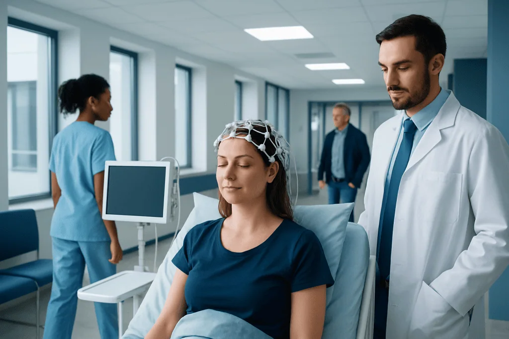
An encephalogram test is another name commonly used for an EEG, a test that measures brain wave patterns. The test does not send electricity into the brain; it only records natural electrical activity.
Key Takeaways
- An encephalogram test is another name commonly used for an EEG, a test that measures brain wave patterns.
- The test does not send electricity into the brain; it only records natural electrical activity.
- Doctors may use an EEG to investigate seizures, fainting-like episodes, confusion, sleep disorders, or changes in consciousness.
- Preparation may include clean hair and, in some cases, temporary sleep deprivation or medication instructions.
- EEG results are interpreted together with symptoms, medical history, and often brain imaging or other neurological tests.
An encephalogram test, usually called an electroencephalogram or EEG, records the brain’s electrical activity through small sensors placed on the scalp. It is a safe, noninvasive test that helps doctors evaluate seizures, episodes of lost awareness, sleep-related concerns, and some other neurological symptoms.
What is an encephalogram test?
An encephalogram test usually refers to an electroencephalogram, or EEG. This test records the brain’s natural electrical activity using small metal discs called electrodes placed on the scalp. It is widely used in neurology because it can show patterns that suggest how the brain is functioning at the time of the recording.
For patients, one of the most important points is that an EEG is noninvasive and painless. The electrodes do not put electricity into the body. Instead, they detect tiny electrical signals produced by brain cells. The test may be done in a hospital, outpatient clinic, sleep laboratory, or specialized neurophysiology unit.
An encephalogram test is not the same as a brain scan such as CT or MRI. Imaging tests show brain structure, while an EEG shows electrical activity over time. These tests often complement each other. For example, a doctor may combine an EEG with MRI scanning or CT imaging when evaluating unexplained neurological symptoms.
Why doctors order an EEG
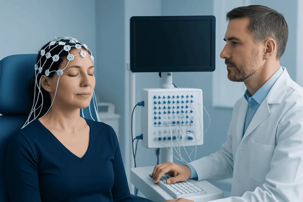
Doctors order an encephalogram test when they need more information about possible disturbances in brain activity. A common reason is to assess seizures or episodes that could be related to epilepsy. The test may also be useful after a person has had unexplained spells of staring, shaking, confusion, sudden falls, or temporary loss of awareness.
EEG can also support the evaluation of sleep-related problems, memory or attention changes, episodes of unusual behavior, and altered consciousness after head injury, infection, or metabolic illness. In some settings, it helps monitor brain function in critically ill patients or during surgery.
The test does not diagnose every neurological condition by itself. Instead, it adds one important piece to a larger clinical picture. A specialist may consider EEG findings together with the neurological examination and assessment for conditions such as epilepsy or brain tumors when symptoms suggest those possibilities.
How the test is done and what to expect
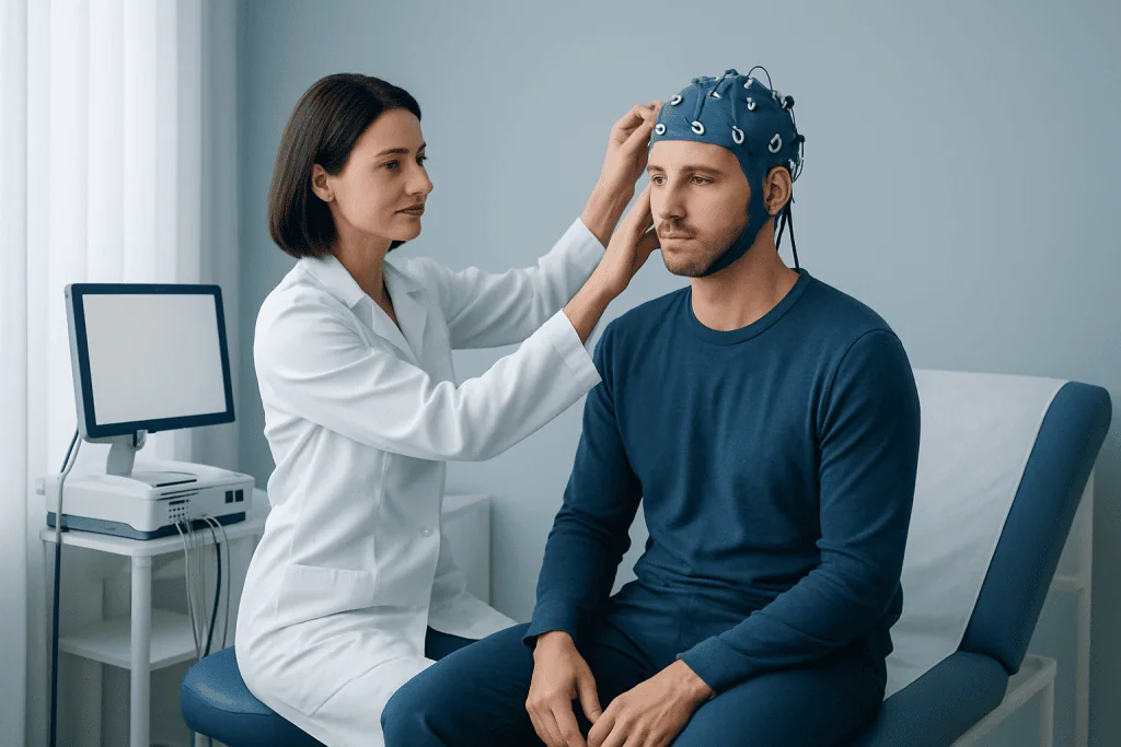
Before the recording begins, a technologist measures the head and places electrodes on specific areas of the scalp using a special adhesive or cap. The patient usually lies down or sits comfortably while the machine records the signals. The setup can take some time, but the recording itself is straightforward.
During the test, the patient is asked to relax and stay still because movement can affect the recording. Common routine steps may include opening and closing the eyes, deep breathing for a few minutes, and looking at flashing lights. These activities can help reveal certain brain wave changes that are not always visible at rest.
There are several types of EEG. A routine EEG usually takes less than an hour, while prolonged or ambulatory EEG can record brain activity over many hours or even days. Video EEG combines the electrical recording with continuous video to match symptoms or events with EEG changes. This can be especially helpful when doctors need to distinguish epileptic seizures from other episodes.
Most people can return to normal activities soon after the test, unless they were given medicine to help them sleep or are asked to follow other special instructions. If the EEG is part of a broader neurological evaluation, a doctor may also recommend neurology care and, when seizure symptoms are present, sometimes specialized epilepsy treatment in selected cases.
How to prepare for an encephalogram test
Preparation depends on the type of EEG being performed. In general, patients are asked to wash their hair the night before or the morning of the test and avoid hair sprays, gels, oils, or heavy styling products. Clean hair helps the electrodes attach properly and improves the quality of the recording.
Some patients are told to continue their usual medicines, while others may receive special instructions. Medication changes should never be made without direct advice from the prescribing doctor. For a sleep-deprived EEG, the patient may be asked to sleep less than usual before the appointment so the recording can capture drowsiness or sleep.
It can help to eat as usual unless told otherwise, because low blood sugar may affect how a person feels during testing. Patients may also want to bring a list of medications, a description of recent symptoms, and details about what happens before, during, and after any suspected episodes. This information can make the test more meaningful when the doctor interprets the results.
Understanding EEG results
EEG results are reviewed by a trained physician, often a neurologist or neurophysiologist. The report looks at the brain’s background rhythm and whether there are any unusual patterns, such as slowing or epileptiform discharges. These terms can sound technical, but they simply describe how the electrical activity appears compared with expected patterns for age and state of alertness.
A normal encephalogram test does not always rule out a seizure disorder or other neurological condition. Some people with epilepsy have a normal routine EEG between seizures, especially if the event does not happen during the recording. In these cases, a doctor may recommend repeat testing, sleep-deprived EEG, prolonged monitoring, or video EEG.
An abnormal EEG does not automatically mean a person has epilepsy either. Certain changes may be seen with sleep deprivation, medications, past brain injury, infections, metabolic disturbances, or structural brain conditions. This is why EEG findings are interpreted alongside symptoms, examination findings, blood tests, and imaging when needed.
Patients often find it helpful to ask practical questions after the test, such as whether the EEG was normal, whether any findings were specific or nonspecific, and what next steps are recommended. Clear follow-up is important because the result may influence diagnosis, safety advice, driving guidance, or treatment planning.
What conditions can an encephalogram test help assess?
An EEG is best known for helping assess seizures and suspected epilepsy, but its role is broader. It may support the evaluation of fainting-like spells, episodes of unresponsiveness, confusion, memory lapses, and unusual movements. In children and adults, it can help clarify whether brief events are likely to be epileptic or caused by another issue.
Sleep medicine specialists may use EEG as part of overnight testing because brain wave patterns help define sleep stages and identify certain sleep disorders. In emergency and intensive care settings, EEG can be used to monitor ongoing brain activity in a person who is very unwell, sedated, or not fully responsive.
EEG may also provide supporting information in suspected encephalopathy, after head trauma, or when a central nervous system infection or inflammatory problem is being considered. Even so, it is usually one part of a complete assessment rather than a stand-alone answer. The most accurate diagnosis comes from combining the test with a detailed history and examination.
- Possible seizure disorders
- Episodes of loss of awareness or staring
- Sleep-related concerns
- Confusion or altered mental status
- Monitoring after certain brain injuries or illnesses
Limits, safety, and self-care after the test
The encephalogram test is considered very safe. The electrodes only record activity and do not cause pain. If flashing lights or deep breathing are used, they are performed under supervision. In some people with photosensitive epilepsy, these steps can provoke a seizure, but this is exactly why trained staff monitor the patient closely in a controlled setting.
The main limitation of EEG is timing. If unusual brain activity does not happen during the recording, the test may miss it. A routine EEG is a snapshot, while some symptoms occur rarely. For this reason, a normal test does not always end the evaluation. Doctors may recommend longer monitoring if symptoms continue or if clinical suspicion remains high.
After the test, most people can wash their hair to remove the adhesive and resume normal routines. If they were sleep-deprived, they should be cautious about fatigue. If they have had a recent unexplained episode involving loss of consciousness, driving or operating machinery should only be resumed according to a doctor’s advice and local regulations.
Near the end of a patient’s workup, care may involve several specialists. Acibadem International’s multidisciplinary teams in JCI-accredited hospitals diagnose and treat neurological conditions for international patients when advanced evaluation is needed.
When to seek medical care
Medical review is important if a person has a first-time seizure, repeated episodes of blank staring, sudden unexplained falls, fainting with jerking movements, new confusion, or periods of lost awareness. These symptoms do not always mean epilepsy, but they deserve proper assessment.
Urgent care is needed if a seizure lasts more than a few minutes, if seizures repeat without recovery in between, if breathing seems difficult, or if an episode happens after head injury, during pregnancy, or in someone with diabetes or serious illness. Immediate evaluation is also important when weakness, trouble speaking, severe headache, fever, or persistent drowsiness occur with neurological symptoms.
For non-urgent concerns, patients should arrange an appointment if episodes are recurring, sleep problems are affecting daily life, or a doctor has recommended EEG as part of ongoing neurological care. Early assessment can help clarify the cause and guide safe, appropriate treatment.
Frequently asked questions
Is an encephalogram test the same as an EEG?
Yes. In everyday use, encephalogram test usually refers to an electroencephalogram, or EEG. It is a test that records the brain’s electrical activity through sensors placed on the scalp.
Does an encephalogram test hurt?
No, the test is generally painless. The electrodes sit on the scalp and only record signals; they do not deliver electric shocks. Some people only notice mild discomfort from the adhesive or from sitting still.
How long does an EEG take?
A routine EEG often takes less than an hour, although preparation can add extra time. Longer studies, such as ambulatory or video EEG, may continue for many hours or several days depending on the clinical need.
Can a normal EEG rule out epilepsy?
Not always. A routine EEG may be normal if abnormal brain activity does not occur during the recording. If symptoms strongly suggest seizures, a doctor may recommend repeat or prolonged EEG monitoring.
What should a patient do before the test?
Patients are often advised to arrive with clean hair and avoid styling products. They should follow any instructions about sleep, food, and medications carefully, and they should not stop medicines unless their doctor specifically tells them to.
Why might flashing lights or deep breathing be used during the test?
These are standard activation methods that may reveal patterns not seen during quiet rest. They are performed in a controlled setting and can help doctors identify certain seizure tendencies or other EEG changes.
References
- National Institute of Neurological Disorders and Stroke
- Epilepsy Foundation
- Mayo Clinic
- American Clinical Neurophysiology Society
- MedlinePlus
This article is for general information only and is not a substitute for professional medical advice. Please consult a qualified doctor about your individual situation.
Hair transplant in Turkey — costs, graft counts & a free assessment
JCI-accredited · board-certified surgeons · reply within 24h
Add us as a Preferred Source to see more of our trusted health content across Google Search, AI Overviews and Discover.