Eye Examination Procedure: An Evidence-Based Patient Guide

A comprehensive eye examination includes more than reading letters on a chart; it also assesses the front and back of the eyes. The visit may include vision testing, eye-pressure measurement, refraction, pupil dilation, and retinal examination.
Key Takeaways
- A comprehensive eye examination includes more than reading letters on a chart; it also assesses the front and back of the eyes.
- The visit may include vision testing, eye-pressure measurement, refraction, pupil dilation, and retinal examination.
- Dilation can temporarily blur near vision and increase light sensitivity, so travel planning is helpful.
- Regular eye examinations are important because glaucoma, diabetic eye disease, and some retinal conditions may develop without early symptoms.
- Sudden vision loss, severe eye pain, flashes, or a curtain-like shadow require urgent medical assessment.
An eye examination procedure is a structured assessment of vision, eye movement, and eye health performed by an eye-care professional. Most examinations are comfortable and noninvasive, and they can identify vision changes as well as early signs of eye conditions before symptoms are noticeable.
Overview: What Is an Eye Examination Procedure?
An eye examination procedure is a series of tests used to evaluate how well a person sees and whether the eyes are healthy. It may be carried out by an ophthalmologist, a medical doctor specializing in eye care and eye surgery, or by an optometrist, depending on the purpose of the visit and local care pathways. The examination is usually painless, although some tests may feel briefly uncomfortable because of bright lights or a puff of air.
A routine visit commonly checks visual acuity, refractive error, eye alignment, pupil responses, eye pressure, and the structures at the front and back of the eye. The findings can help update glasses or contact lens prescriptions and identify conditions that need monitoring or treatment, including cataracts, glaucoma, retinal disorders, and eye changes related to diabetes or high blood pressure.
The exact tests are tailored to the person’s age, symptoms, medical history, family history, and current medications. A comprehensive examination is different from a simple vision screening: screening can flag a possible concern, while a full examination helps confirm the cause and guide next steps.
Who May Need a Comprehensive Eye Examination?
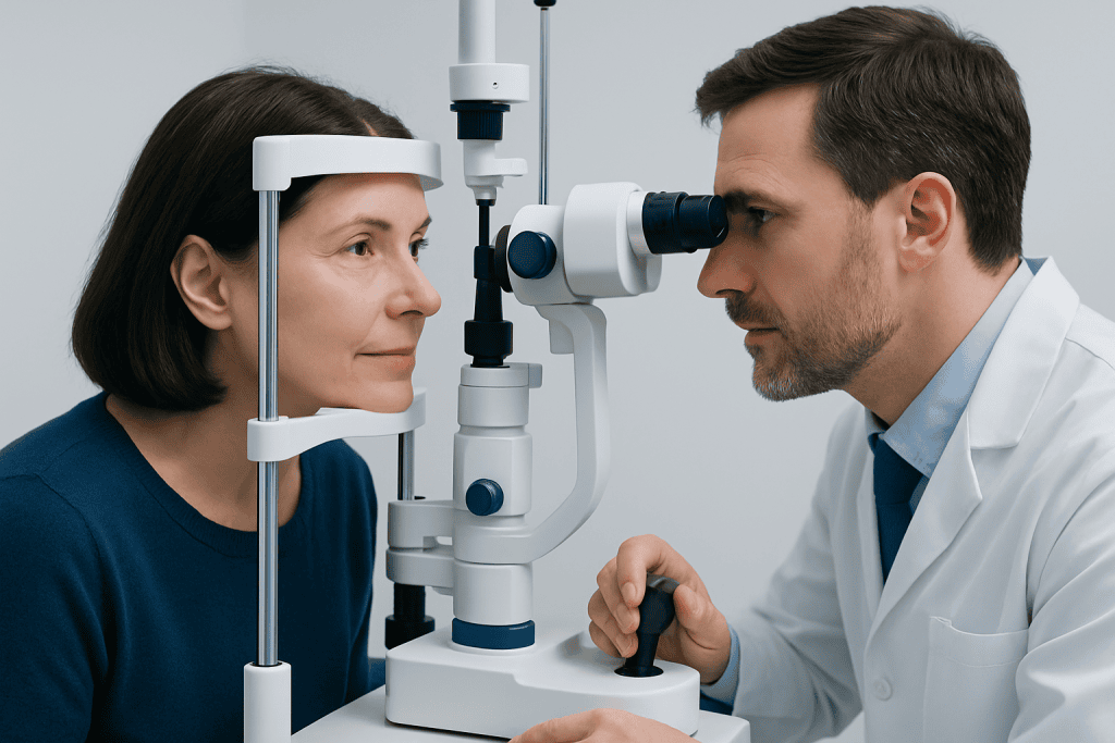
Eye examinations are appropriate for people of all ages. Children need age-appropriate vision assessments because uncorrected vision problems can affect learning and visual development. Adults may need examinations to assess changes in vision, eye discomfort, headaches linked to focusing difficulties, or the need for glasses, contact lenses, or workplace visual support.
Regular monitoring is particularly important for people with diabetes, high blood pressure, autoimmune disease, a history of eye injury or eye surgery, or a family history of glaucoma, retinal detachment, or inherited eye disease. People taking medicines that may affect vision or eye health may also be advised to have eye checks at intervals recommended by their clinician.
Frequency varies. A clinician can recommend an individual schedule based on age, risk factors, and previous findings. Someone with stable eye health may need checks less often than someone with established eye disease, while new symptoms may require an appointment sooner rather than waiting for a routine review.
How the Eye Examination Works
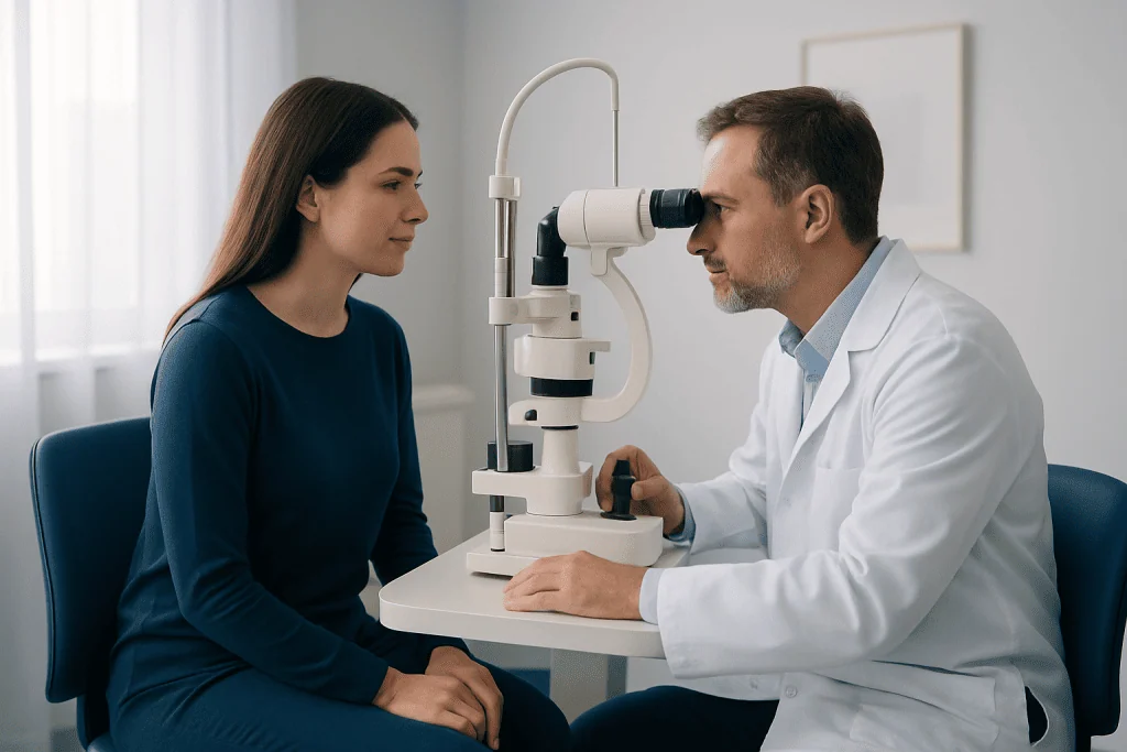
The eye examination procedure combines information from the patient’s history with measured findings. Before testing, the clinician may ask about blurred vision, double vision, pain, redness, floaters, flashes of light, headaches, injury, previous surgery, and any glasses or contact lenses used. Medical conditions and medicines are also relevant because many health issues can affect the eyes.
Testing generally progresses from basic vision measurements to a closer inspection of the eye. Specialized instruments allow the clinician to examine the cornea, lens, optic nerve, retina, and blood vessels without making an incision or touching many internal eye structures. Imaging or field testing may be added when there is a concern about the optic nerve, retina, or peripheral vision.
Some examinations use eye drops to widen the pupils. This provides a clearer view of the retina and optic nerve and is especially useful when assessing diabetes-related eye changes, retinal problems, and possible glaucoma. The effects are temporary, but they can influence comfort and activities for several hours.
Step by Step: Common Tests During the Visit
Most appointments begin with visual acuity testing. The person reads letters, symbols, or pictures at a measured distance, usually with each eye tested separately and then together. Near vision may also be checked. If vision is reduced, the clinician may perform refraction, using a phoropter or handheld lenses to determine whether glasses or contact lenses could improve clarity.
The clinician may then assess eye movements, alignment, and pupils. These tests can involve following a target, looking in different directions, or having a light shone briefly into each eye. Color vision or depth-perception tests may be used when clinically relevant. The front of the eye is often examined with a slit lamp, a microscope with a bright narrow beam of light that shows the eyelids, tear film, cornea, iris, and lens in detail.
Eye pressure may be measured because raised pressure can be associated with glaucoma. Methods include a gentle air puff or a device that lightly touches the numbed surface of the eye after anesthetic drops are applied. Pressure measurement alone does not diagnose glaucoma; it is interpreted alongside optic-nerve appearance, visual-field testing, and other findings.
If dilation is needed, drops are placed in each eye and take time to work. The clinician then uses lenses and a bright light to examine the retina and optic nerve. Additional tests may include optical coherence tomography, retinal photography, or visual-field testing. These investigations can support evaluation of conditions such as glaucoma and retinal disease.
Benefits, Limitations, and Possible Effects
The main benefit of an eye examination is early identification of correctable vision problems and eye disease. Many conditions can be managed more effectively when found early, sometimes before noticeable symptoms occur. An examination can also reveal eye findings that prompt assessment of broader health concerns, such as diabetes or high blood pressure.
An eye exam has limitations. A normal result reflects eye health at the time of the visit and does not eliminate the need to seek care for new symptoms later. Some conditions require repeat testing because changes may be subtle or develop gradually. Results are interpreted alongside symptoms, risk factors, imaging, and previous examination records when available.
Most tests have little or no risk. Bright lights may be temporarily uncomfortable, and eye drops can briefly sting. Dilating drops commonly cause blurred near vision and light sensitivity for several hours. Rarely, drops can trigger an acute rise in eye pressure in people predisposed to angle-closure glaucoma; eye pain, redness, nausea, headache, halos around lights, or worsening blurred vision after dilation should be assessed urgently.
After the Examination: Recovery and Self-Care
No recovery period is needed after most parts of an eye examination procedure. A person can usually return to work, school, and normal activities immediately. If contact lenses were removed for the assessment, the clinician can advise when they may be reinserted, particularly if drops, staining dyes, or additional tests were used.
After pupil dilation, near tasks such as reading or using a phone may be difficult, and sunlight may feel unusually bright. Wearing sunglasses outdoors can improve comfort. Some people can drive safely after dilation, while others cannot because vision is too blurred or light-sensitive; it is sensible to ask the clinic what to expect and arrange transport if needed.
It is helpful to bring current glasses, contact lens information, a medication list, and details of prior eye conditions or operations. Patients should ask when results will be available, whether a new prescription is recommended, and what follow-up interval is appropriate. If an eye condition is identified, the care team can discuss monitoring, medication, laser treatment, or surgery where appropriate, including comprehensive eye examination services.
When to Seek Medical Care
Routine examinations support long-term eye health, but some symptoms need prompt rather than scheduled assessment. A person should seek urgent medical care for sudden loss of vision, new severe eye pain, a painful red eye, a sudden increase in floaters, flashes of light, double vision, or a dark curtain or shadow across the field of vision. These symptoms can have several causes and should not be self-diagnosed.
Medical review is also appropriate for persistent blurred vision, ongoing redness or discharge, repeated headaches with visual symptoms, difficulty seeing at night, or a noticeable change in how the eyes look or move. Children should be assessed if they frequently squint, sit unusually close to screens, cover one eye, complain of headaches, or show concerns about visual development.
People with diabetes should follow their clinician’s recommendations for retinal screening, even when vision seems normal. Those with known glaucoma, retinal disease, or previous eye surgery should keep planned follow-up appointments. Acibadem International’s multidisciplinary specialists and JCI-accredited hospitals assess and treat eye conditions for international patients when specialist care is needed.
Frequently asked questions
How long does an eye examination procedure take?
The duration depends on the reason for the visit and the tests required. A straightforward examination may be relatively brief, while a comprehensive examination with pupil dilation, imaging, or visual-field testing can take longer. The clinic can advise what to expect when the appointment is booked.
Does an eye examination hurt?
Most eye examination tests are painless. Bright lights, air-puff pressure testing, and eye drops may cause brief discomfort or irritation, but they should not be painful. A person should tell the clinician if a test causes pain or significant distress.
Why are pupils dilated during an eye exam?
Dilation widens the pupil so the clinician can examine the retina and optic nerve more completely. It can help detect changes associated with diabetes, glaucoma, retinal disease, and other conditions. The drops may cause temporary blurred near vision and sensitivity to light.
Can an eye examination detect diabetes?
An eye examination can reveal retinal changes that may occur with diabetes, but it does not replace blood testing for diagnosis. If retinal findings suggest diabetes or another systemic condition, the eye clinician may recommend evaluation by a primary care clinician or relevant specialist. People with known diabetes need regular retinal monitoring as advised.
Should glasses be removed before the appointment?
People should usually bring and wear their usual glasses to the appointment unless the clinic gives different instructions. Bringing older prescriptions and contact lens details can also help the clinician understand changes in vision. Contact lens wearers may be asked to remove lenses for some tests.
How often should adults have an eye examination?
The ideal interval depends on age, symptoms, vision needs, medical conditions, family history, and previous eye findings. Adults with diabetes, glaucoma risk, established eye disease, or new symptoms may need more frequent assessment. An eye-care professional can recommend a personalized schedule.
References
- American Academy of Ophthalmology
- National Eye Institute
- Centers for Disease Control and Prevention
- World Health Organization
This article is for general information only and is not a substitute for professional medical advice. Please consult a qualified doctor about your individual situation.
Eye Examination in Turkey — costs, top hospitals & a free quote
JCI-accredited · board-certified surgeons · reply within 24h
Add us as a Preferred Source to see more of our trusted health content across Google Search, AI Overviews and Discover.
Check your numbers in seconds
BMI, calories, due date, blood pressure and 30+ more clinical calculators — free, instant, doctor-reviewed ranges.

Ophthalmology
Comprehensive eye care from laser vision correction to retina, cornea and cataract surgery.
75 specialists in this unit