Eye Freckle Treatment: How It Works, Results and What to Expect

Most eye freckles, also called nevi, are benign and are monitored rather than treated. Photographs and specialized eye imaging help clinicians compare a freckle over time.
Key Takeaways
- Most eye freckles, also called nevi, are benign and are monitored rather than treated.
- Photographs and specialized eye imaging help clinicians compare a freckle over time.
- Treatment may be considered if a lesion has features suggesting ocular melanoma or causes symptoms.
- A changing spot, blurred vision, flashes, floaters, or loss of side vision requires prompt eye assessment.
- Eye freckles do not usually fade quickly, and many remain stable throughout life.
Eye freckle treatment most often involves regular eye examinations and imaging rather than immediate removal. Most eye freckles are harmless, but an ophthalmologist should assess any lesion that is new, changing, elevated, or associated with symptoms.
Eye Freckle Treatment: What It Involves
An eye freckle is a small area of increased pigment in or on the eye. The medical term is nevus. Depending on its location, it may be a conjunctival nevus on the white of the eye, an iris nevus in the colored part of the eye, or a choroidal nevus at the back of the eye. Eye freckle treatment is usually careful observation, because most nevi are benign and do not affect vision.
The purpose of follow-up is not to remove every freckle. Instead, an ophthalmologist documents its size, shape, color, thickness, and location, then checks for meaningful change. If examination or imaging raises concern that a lesion could be an early eye cancer, referral to an ocular oncology specialist and treatment may be advised.
Management depends on the type of lesion and its individual features. A visible spot on the eye surface may be assessed differently from a freckle found during a dilated retinal examination. It is important not to assume that a pigmented spot is harmless without a professional eye examination.
People with a known eye nevus can also learn about related pigment changes through choroidal nevus information. This can help them understand why baseline photographs and scheduled reviews are useful.
How Eye Freckle Assessment and Treatment Work
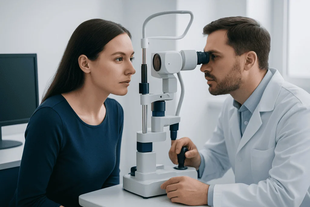
At the first visit, the ophthalmologist asks about eye symptoms, past eye conditions, family history, and sun exposure. The eye is examined with magnification and, when appropriate, dilating drops to allow a detailed view of the retina and choroid. A lesion that appears typical and stable may only require periodic review.
Digital photographs provide a reliable baseline for visible lesions. For freckles at the back of the eye, clinicians may use optical coherence tomography, ultrasound, fundus photography, autofluorescence imaging, or fluorescein angiography when indicated. These tests help assess thickness, fluid beneath the retina, pigment changes, and other characteristics that may guide follow-up.
If a lesion is suspicious for melanoma, treatment is directed at the suspected tumor rather than the pigmentation alone. Options may include plaque radiotherapy, proton beam radiotherapy, laser-based approaches in selected cases, surgery, or other individualized care. The best approach depends on the lesion’s size, location, growth pattern, and effect on nearby eye structures.
Specialist care for a suspected malignant lesion may include ocular melanoma treatment. The aim is to control the lesion while preserving vision and the eye whenever medically appropriate.
Who May Need Treatment Rather Than Monitoring?
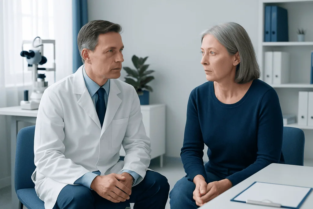
Most people with an eye freckle are candidates for observation, not a procedure. Monitoring is especially appropriate when the lesion has a stable appearance, has no concerning imaging features, and is not causing vision changes. Follow-up intervals are individualized and may be shorter at first to confirm stability.
Treatment or urgent specialist assessment may be considered when an eye freckle grows, becomes thicker, develops unusual features, causes fluid under the retina, or is linked with new visual symptoms. A lesion near important structures, such as the optic nerve or central retina, may also require closer surveillance because even benign changes can affect vision.
Conjunctival pigmented lesions may occasionally be removed or biopsied when they are enlarging, atypical, repeatedly irritated, or cosmetically concerning after medical assessment. Removal is not automatically recommended for cosmetic reasons, as surgery can leave scarring, alter eye appearance, or affect the surface of the eye.
Children and adults can both have eye nevi. However, a new pigmented lesion in adulthood, rapid change in a long-standing spot, or uncertainty about the diagnosis should be evaluated by an ophthalmologist rather than monitored at home.
What Happens During Eye Freckle Treatment?
There is no single eye freckle procedure because treatment depends on where the lesion is and why treatment is needed. For a suspicious lesion at the back of the eye, the care team first confirms the diagnosis through detailed examination and imaging. Biopsy is not needed for every lesion, but may be discussed when diagnostic uncertainty would change management.
Radiotherapy-based treatment is commonly planned by an ocular oncology team for confirmed or strongly suspected ocular melanoma. Planning scans and measurements are used to target the lesion as precisely as possible. Depending on the method, treatment may be delivered over one or several sessions, and follow-up imaging is used to assess response.
For selected surface lesions, a surgeon may remove the pigmented area under local or general anesthesia. The tissue may be examined in a laboratory to clarify the diagnosis. The procedure, expected recovery, and possible effect on eye appearance depend on the lesion’s size and location.
Before any intervention, the specialist explains expected benefits, alternatives, likely visual effects, and follow-up needs. A second opinion from an ocular oncology service can be appropriate when treatment for a suspicious lesion is being considered.
Recovery, Results, Benefits and Risks
Recovery after monitoring requires no physical healing, but it does require attending scheduled eye appointments. Many people feel reassured after baseline imaging confirms that their freckle has no high-risk features. The main benefit of monitoring is timely identification of an uncommon lesion that begins to change.
After surgery on the eye surface, temporary redness, grittiness, watering, or light sensitivity may occur. Healing time varies, and prescribed eye drops and activity guidance should be followed carefully. Contact lenses may need to be avoided until the eye has healed and the ophthalmologist confirms that they can be worn safely.
Recovery after treatment for ocular melanoma varies more widely. Vision may remain stable, improve, or decline depending on the lesion and the treatment area. Potential treatment-related effects can include dry eye, cataract, retinal swelling, bleeding, glaucoma, retinal damage, or changes in vision; the care team discusses risks based on the individual situation.
Benefits of treating a suspicious lesion include controlling a confirmed cancer or reducing the risk associated with a growing tumor. However, treatment is not beneficial for every stable benign nevus, which is why accurate diagnosis and ongoing review are central to good care.
What Percentage of People Have a Freckle in Their Eye?
Eye freckles are relatively common, particularly choroidal nevi, which occur in several percent of adults. Estimates vary by study population, age, ancestry, and the examination method used. Many are found incidentally during routine dilated eye examinations because they usually do not cause symptoms.
A visible freckle on the iris or conjunctiva may be noticed by the person or their family, while a choroidal nevus cannot generally be seen without an eye examination. Having an eye freckle does not mean a person has cancer or will develop cancer.
Because frequency estimates cannot predict an individual’s risk, the most useful step is a complete eye assessment. The ophthalmologist can identify the lesion type, establish a baseline, and recommend an appropriate monitoring schedule.
Will an Eye Freckle Go Away? How Long Does It Take for a Freckle to Fade?
Most eye freckles do not go away on their own. Like many skin moles, they may remain visible or detectable throughout life. Some lesions may show minor changes in pigment over time, but a stable eye nevus should not be expected to fade over a predictable period.
There is no safe home treatment, eye drop, supplement, or sun exposure strategy proven to make an eye freckle fade. Trying to self-treat a pigmented eye lesion can delay diagnosis and may irritate or injure the eye. Cosmetic removal should only be discussed with an experienced ophthalmologist after the lesion has been evaluated.
Ultraviolet protection is sensible for overall eye health. Wearing sunglasses that block UVA and UVB radiation and a brimmed hat outdoors may reduce exposure, although these measures cannot remove an existing nevus or guarantee that a lesion will not change.
Can an Eye Nevus Turn Into Melanoma? When to Seek Medical Care
Most eye nevi never become melanoma. However, a small proportion can grow or develop features that make ocular melanoma more likely. This is why an established baseline and periodic examinations are recommended. A specialist assesses risk using the lesion’s appearance, thickness, location, associated retinal fluid, and evidence of growth over time.
Medical care should be sought promptly for new blurred vision, flashing lights, new floaters, a shadow or missing area in vision, a noticeable enlarging dark spot, eye pain, or a change in the color or shape of a visible lesion. These symptoms can have many causes, and most are not due to melanoma, but they deserve timely assessment.
Routine eye care is also important when there are no symptoms, especially for people previously told they have an eye freckle. Acibadem International’s multidisciplinary specialists and JCI-accredited hospitals can assess and treat eye conditions for international patients when specialist evaluation is needed.
For concerns about pigmented lesions and their possible consequences, an ophthalmologist may coordinate care with ocular oncology, retinal, imaging, and radiation specialists. The appropriate pathway is based on the person’s examination findings, not on appearance alone.
Frequently asked questions
Is eye freckle treatment always necessary?
No. Most eye freckles are benign and need observation rather than treatment. An ophthalmologist may recommend photographs and imaging to confirm that the lesion remains stable over time.
How often should an eye freckle be checked?
The follow-up schedule depends on the lesion’s location and appearance. A specialist may request an earlier repeat examination after the first finding, then extend the interval if it remains stable.
Can sunlight cause an eye freckle?
The exact causes of eye nevi are not fully understood. Sun protection is recommended for general eye health, but it cannot remove an existing freckle or reliably prevent all pigmented eye lesions.
Does a choroidal nevus affect vision?
Most choroidal nevi do not affect vision and are discovered during an examination. Vision symptoms can occur if a lesion or related fluid affects the retina, particularly the central retina, and should be assessed promptly.
How is an eye freckle different from ocular melanoma?
An eye freckle is usually a benign collection of pigment cells, while ocular melanoma is a cancer that arises from pigment-producing cells. Examination and imaging help specialists distinguish a stable nevus from a lesion requiring further evaluation or treatment.
Can an eye doctor tell whether an eye freckle is dangerous?
An ophthalmologist can assess many risk features during a dilated examination and with imaging. In some cases, repeat images over time or referral to an ocular oncology specialist is needed to determine whether a lesion is stable or concerning.
References
- American Academy of Ophthalmology
- National Cancer Institute
- American Cancer Society
- National Eye Institute
- European Society of Ophthalmology
This article is for general information only and is not a substitute for professional medical advice. Please consult a qualified doctor about your individual situation.
Explore treatments in Turkey — costs, top hospitals & a free quote
JCI-accredited · board-certified surgeons · reply within 24h
Add us as a Preferred Source to see more of our trusted health content across Google Search, AI Overviews and Discover.