

Facial Pain, Visual Symptoms, and Numbness: When MRI of the Brain Is Recommended
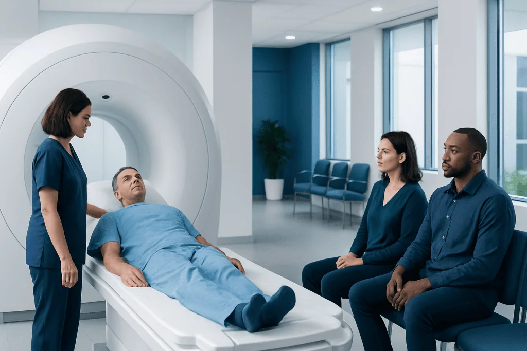
MRI of the brain provides detailed images of the brain, cranial nerves, and surrounding structures without using ionizing radiation. It may be recommended when facial pain, visual symptoms, or numbness are unexplained, persistent, worsening, or accompanied by other neurologic signs.
Key Takeaways
- MRI of the brain provides detailed images of the brain, cranial nerves, and surrounding structures without using ionizing radiation.
- It may be recommended when facial pain, visual symptoms, or numbness are unexplained, persistent, worsening, or accompanied by other neurologic signs.
- Brain MRI can help assess causes such as nerve compression, inflammation, demyelination, stroke, tumors, and some eye or sinus-related complications.
- Not every episode of facial discomfort or temporary visual change needs MRI; the decision depends on symptoms, examination findings, and medical history.
- Urgent medical evaluation is important if symptoms begin suddenly, are severe, or occur with weakness, speech difficulty, or loss of vision.
MRI of the brain is often recommended when facial pain, visual changes, or numbness could be related to the brain, cranial nerves, or nearby blood vessels. It helps doctors look for possible causes and decide whether symptoms need monitoring, medication, or more urgent treatment.
Overview: Why MRI May Be Recommended
Facial pain, visual symptoms, and numbness can come from many different causes. Sometimes the source is relatively local, such as the eyes, sinuses, teeth, jaw joint, or skin. In other cases, the symptoms may involve the brain, cranial nerves, or blood vessels that supply the head and face. When the pattern of symptoms suggests a neurologic cause, doctors may recommend an MRI of the brain.
MRI, or magnetic resonance imaging, creates highly detailed images of soft tissues. It is especially useful for viewing the brain, the optic pathways, and several important nerves that control facial sensation, facial movement, hearing, and eye function. Depending on the symptoms, the examination may include special sequences or contrast material to better evaluate inflammation, tumors, nerve compression, or changes related to blood flow.
The goal of a brain MRI is not simply to confirm a serious condition. Just as often, it helps rule out important causes and guide the next step in care. This can be reassuring for patients and useful for doctors deciding whether symptoms are more likely related to migraine, nerve irritation, infection, inflammation, or another condition.
Symptoms That May Lead to Brain MRI
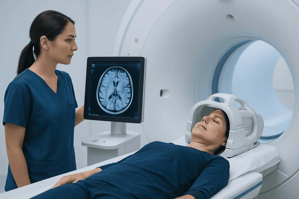
Doctors consider MRI when facial pain, visual symptoms, or numbness are unexplained or do not fit a simple pattern. Facial pain may feel sharp, electric, burning, pressure-like, or aching. It may affect one side of the face, the forehead, jaw, cheek, or around the eye. Visual symptoms can include blurred vision, double vision, reduced color vision, loss of part of the visual field, eye pain with movement, or brief or persistent vision loss. Numbness may involve the face, scalp, lips, tongue, or one side of the body.
The need for imaging becomes more likely when symptoms are persistent, recurrent, worsening, or associated with other neurologic changes. Examples include facial pain with facial weakness, numbness with imbalance, or visual symptoms with headache that feels different from usual. MRI may also be recommended when symptoms occur after a known neurologic event or when there is concern for conditions such as multiple sclerosis or a brain tumor.
Certain warning features deserve prompt assessment. These include sudden loss of vision, a new drooping face, trouble speaking, severe new headache, confusion, or weakness in an arm or leg. In those situations, imaging may be arranged urgently, and the most appropriate scan may depend on how quickly symptoms began and what doctors suspect.
- Persistent one-sided facial pain without a clear dental or sinus cause
- New numbness affecting the face or body
- Double vision or unexplained blurred vision
- Eye pain with reduced vision
- Symptoms that recur in episodes or steadily progress over time
Possible Causes MRI Can Help Evaluate

Brain MRI can help investigate several categories of conditions. One is nerve-related pain. For example, severe brief shocks of facial pain may suggest irritation of the trigeminal nerve, and MRI may be used to look for contact between the nerve and a blood vessel or to exclude less common structural causes. Symptoms around the eyes or face may also relate to inflammation of the optic nerve or other cranial nerves.
Another important group includes inflammatory and demyelinating conditions. If facial numbness or visual loss appears along with other neurologic episodes, doctors may consider disorders that affect the protective covering of nerves. MRI is a key test when evaluating this possibility because it can show characteristic areas of change within the brain and along the optic pathways.
Vascular conditions are another reason for imaging. MRI, sometimes combined with magnetic resonance angiography or venography, can help assess blood vessels when symptoms raise concern for stroke, reduced blood flow, vascular malformation, or pressure on a nerve caused by a nearby vessel. It may also be used as part of the evaluation for unexplained symptoms linked to stroke.
Less commonly, MRI may identify tumors, cysts, infection, sinus or orbital complications, or changes after trauma. Although these causes are not the most frequent explanation for facial pain or brief visual symptoms, they are important to consider when the examination or history suggests something beyond a routine headache, eye condition, or dental problem.
How Doctors Decide Whether MRI Is Needed
The decision to order MRI is based on the full clinical picture rather than one symptom alone. A doctor usually begins by asking detailed questions: when the symptoms started, whether they came on suddenly or gradually, how long they last, whether they affect one or both sides, and whether there are triggers such as chewing, touching the face, or moving the eyes. The pattern can offer clues about whether symptoms may be related to migraine, nerve compression, inflammation, infection, or a vascular event.
A physical and neurologic examination is equally important. Doctors may check vision, eye movements, pupil reactions, facial sensation, facial strength, hearing, balance, coordination, reflexes, and sensation elsewhere in the body. In some cases, an eye examination or dental or ENT review is also needed. MRI is most helpful when symptoms cannot be fully explained by these first evaluations or when exam findings suggest involvement of the central nervous system.
Not everyone with these symptoms needs MRI immediately. Short-lived, familiar migraine symptoms in a person with a stable pattern may not require urgent imaging. By contrast, a first episode of double vision, new facial numbness, pain around one eye with reduced vision, or progressive symptoms over days to weeks often justifies a closer look with brain MRI.
What a Brain MRI Can Show
A brain MRI can show detailed anatomy of the brain tissue, cranial nerves, eye sockets, and nearby structures, depending on the protocol used. It can reveal signs of inflammation, infection, old or new areas of ischemia, bleeding, tumors, demyelinating plaques, or pressure on nerves. When contrast is used, active inflammation and some tumors become easier to detect because they enhance differently from surrounding tissue.
Sometimes the MRI focuses closely on a specific region. For example, if trigeminal neuralgia is suspected, the scan may include thin slices through the pathway of the trigeminal nerve. If vision is the main concern, doctors may request sequences that better assess the optic nerves and the area where visual pathways travel through the brain. In selected cases, doctors may combine the exam with brain MRI protocols tailored to neurologic symptoms.
It is important to know that MRI does not answer every question. A normal scan can still occur in some conditions, especially early in the course of disease or when symptoms arise from causes outside the brain. For that reason, MRI results are interpreted together with the history, examination, blood tests, eye tests, and sometimes further studies such as vascular imaging or nerve testing.
What to Expect During the Scan
MRI is a noninvasive test that does not use X-rays. During the scan, the patient lies on a table that moves into the scanner. The machine makes loud tapping or thumping sounds, so ear protection is usually provided. It is important to lie still because movement can blur the images. The scan length varies, but many brain MRI examinations take between about 20 and 45 minutes depending on the details required.
Some patients receive contrast material through a vein. This is often used when doctors need a clearer view of inflammation, tumors, infection, or active lesions. Before contrast is given, the imaging team will ask about allergies, kidney problems, pregnancy, and any implanted medical devices. Patients should also mention claustrophobia, as support options may be available.
MRI is generally well tolerated, but it is not suitable for every person without screening. Metal implants, certain pacemakers, aneurysm clips, or retained metal fragments may require special precautions. The radiology team checks these issues carefully to make sure the examination is safe and appropriate.
Treatment After MRI Depends on the Cause
Treatment is guided by the reason for the symptoms, not by the scan alone. If MRI points to a nerve pain condition, treatment may include medications and specialist follow-up. If an inflammatory or demyelinating disorder is suspected, the patient may need further neurologic evaluation and a broader treatment plan. If there is concern for vascular disease, urgent management may be needed to protect brain and vision health.
When MRI identifies a structural cause, options may range from observation to targeted therapy or surgery. For example, some patients with severe nerve compression may be referred for procedures aimed at relieving pressure, while others may need treatment for a mass, infection, or orbital problem. Depending on the findings, doctors may recommend Gamma Knife radiosurgery or consultation with teams involved in neurosurgery.
If MRI is normal or shows no urgent concern, that result is still helpful. It can narrow the possibilities and support safer follow-up with a neurologist, neuro-ophthalmologist, ENT specialist, dentist, or pain specialist. Near the end of the diagnostic process, multidisciplinary centers such as Acibadem International may help coordinate imaging, specialist review, and treatment for international patients in JCI-accredited hospitals.
When to Seek Medical Attention
Medical review is advisable when facial pain, visual symptoms, or numbness are new, unexplained, recurrent, or affecting daily life. Even when symptoms seem mild, patterns such as one-sided numbness, facial pain triggered by light touch, or visual changes that keep returning should be discussed with a doctor. Early assessment can help identify whether the problem is neurologic, ophthalmic, dental, or related to another system.
Urgent care is especially important when symptoms begin suddenly or are accompanied by other concerning changes. Sudden loss of vision, double vision with severe headache, face drooping, speech difficulty, confusion, limb weakness, loss of balance, or severe numbness can signal a time-sensitive emergency. In these situations, a doctor will decide whether immediate imaging is needed and whether MRI or another scan is most appropriate.
Patients can prepare for an appointment by noting when symptoms started, what they feel like, how long they last, and any associated problems such as headache, eye pain, rash, fever, jaw pain, or recent infection. Bringing a list of medicines and past imaging can also help the clinical team decide whether a brain MRI is needed and how urgently it should be done.
Frequently asked questions
Does facial pain always mean a brain problem?
No. Facial pain often comes from dental issues, sinus disease, temporomandibular joint problems, migraine, or nerve irritation outside the brain. MRI is usually considered when the symptoms are unusual, persistent, severe, or associated with numbness, weakness, or visual changes.
Why would visual symptoms lead to an MRI of the brain?
Vision depends on the eyes, optic nerves, and brain pathways working together. If symptoms suggest the problem may involve the optic nerve, eye movement nerves, or the brain itself, MRI can help show inflammation, compression, demyelination, or other structural causes.
Can a normal brain MRI still happen if symptoms are real?
Yes. A normal MRI does not mean symptoms are imagined or unimportant. Some conditions do not show clear MRI changes, especially early on, and some causes arise outside the brain, so doctors interpret the scan together with the examination and other tests.
Is MRI better than CT for these symptoms?
It depends on the situation. MRI is usually better for detailed imaging of the brain, cranial nerves, and soft tissues, while CT may be used first in emergencies such as suspected bleeding or acute trauma. The choice is based on symptoms, timing, and the doctor’s clinical concern.
Will contrast always be needed for a brain MRI?
Not always. Contrast may be used when doctors need more information about inflammation, tumors, infection, or active lesions. In some cases, non-contrast MRI is enough, and the radiologist or referring doctor will decide which approach is most useful.
What symptoms should never be ignored?
Sudden loss of vision, new face drooping, trouble speaking, confusion, severe new headache, weakness, or sudden numbness need urgent medical attention. These symptoms can sometimes signal a stroke or another emergency where quick treatment matters.
References
- American College of Radiology
- National Institute of Neurological Disorders and Stroke
- National Eye Institute
- Radiological Society of North America
- World Health Organization
This article is for general information only and is not a substitute for professional medical advice. Please consult a qualified doctor about your individual situation.
Joint and spine care in Turkey — expert assessment & treatment
JCI-accredited · board-certified surgeons · reply within 24h
Add us as a Preferred Source to see more of our trusted health content across Google Search, AI Overviews and Discover.

More from the Health Library



Related Specialists

Prof. Dr. A. Taner Usta
Gynecology & Obstetrics
Prof. Dr. Ömer Faruk Taşer
Orthopedic Surgery & Traumatology
Asst. Prof. Dr. Tuğana Akbaş
Radiology
Dr. Özcan İnal
Internal Medicine