Foker Procedure: An Evidence-Based Patient Guide

The Foker procedure is mainly considered for long-gap esophageal atresia when a direct early connection is not possible. It aims to preserve the child’s own esophagus rather than replacing it with stomach or intestinal tissue.
Key Takeaways
- The Foker procedure is mainly considered for long-gap esophageal atresia when a direct early connection is not possible.
- It aims to preserve the child’s own esophagus rather than replacing it with stomach or intestinal tissue.
- Treatment occurs in a highly specialised pediatric surgical setting and may require a prolonged hospital stay.
- Children need close monitoring for complications such as narrowing at the repair site, reflux, feeding difficulties, and respiratory concerns.
- Long-term follow-up with pediatric surgery, gastroenterology, nutrition, and feeding specialists is important.
The Foker procedure is a specialised, staged approach for selected infants with long-gap esophageal atresia, a condition in which the upper and lower parts of the esophagus do not meet. It uses carefully controlled traction to encourage the child’s own esophageal ends to grow closer together before they are surgically joined.
Overview: What Is the Foker Procedure?
The Foker procedure is a surgical strategy used for some babies born with long-gap esophageal atresia. In esophageal atresia, the esophagus—the tube that carries food from the mouth to the stomach—ends in two separate sections instead of forming one continuous passage. When the distance between these sections is too large, joining them safely during an initial operation may not be possible.
In the Foker process, surgeons place traction sutures on the upper and lower esophageal ends. Gentle, sustained tension is applied over time to stimulate lengthening and bring the two ends closer together. Once there is enough length, the surgeon performs an anastomosis, meaning the ends are joined to create a continuous esophagus.
This approach is not appropriate for every child with esophageal atresia. It is a complex form of care that requires experienced pediatric surgeons, pediatric anesthesiologists, neonatal intensive care specialists, nurses, nutrition teams, and feeding therapists. The main goal is to retain the child’s own esophagus whenever this can be achieved safely.
How the Foker Process Works
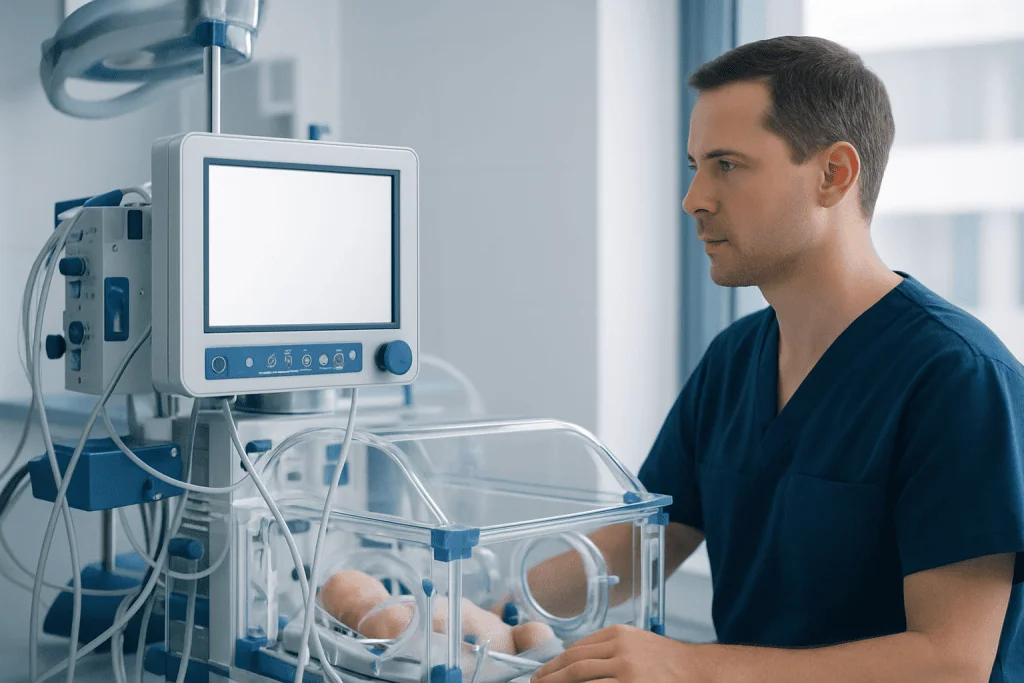
The esophagus has some capacity to lengthen when it is exposed to carefully controlled traction. The Foker procedure uses this biological response rather than immediately replacing the missing segment with another part of the digestive tract. The process is sometimes described as esophageal growth induction or traction-based lengthening.
During an operation, the surgeon identifies the upper and lower esophageal pouches and attaches sutures to each end. The sutures are brought out through the chest wall or managed through a planned traction system. Tension is adjusted gradually and monitored closely. In many centers, the baby remains sedated and receives intensive monitoring while traction is being applied, although specific practices vary by center and clinical situation.
The term Foker 2 procedure may be used informally to describe a later stage of treatment, such as the operation that connects the esophageal ends after traction. It is not always a standardized term, so families should ask the surgical team what is meant in their child’s individual treatment plan.
For a broader explanation of the underlying condition, parents can read about esophageal atresia and how it may affect feeding and early care.
Who May Be a Candidate?

The Foker procedure for esophageal atresia is generally considered when a child has long-gap esophageal atresia. This means the separated ends of the esophagus are too far apart for a safe primary repair shortly after birth. The exact definition of a long gap varies, and the decision is based on anatomy, the child’s overall health, and the surgical team’s assessment.
Before recommending an approach, clinicians assess the esophageal gap with imaging and operative findings. They also look for associated conditions, which can occur in some children with esophageal atresia. These may involve the heart, spine, kidneys, limbs, anus, or airway. A tracheoesophageal fistula, an abnormal connection between the esophagus and windpipe, may also be present and may need treatment as part of the overall surgical plan.
Suitability depends on more than gap length. The quality and position of the esophageal segments, respiratory stability, growth, nutritional needs, previous operations, and the expertise available at the treating center all matter. Some children may be better served by delayed primary repair, other traction-based methods, or esophageal replacement techniques. The care team should explain why a particular strategy is recommended and what alternatives are reasonable.
What Happens Before, During, and After Surgery?
Before surgery, newborns are cared for in a neonatal or pediatric intensive care setting. Feeding by mouth is usually not possible until there is a safe connection between the mouth and stomach. Nutrition may be provided through a vein initially and, when appropriate, through a feeding tube or gastrostomy tube placed directly into the stomach. Tests may include chest imaging, echocardiography, ultrasound, and assessment for related congenital differences.
The first operation involves examining the esophageal ends and placing traction sutures. The traction phase then takes place over days to weeks, depending on how the tissues respond. The infant is monitored for comfort, breathing, circulation, infection, and the position of the sutures. Repeat imaging or operative assessment may be used to determine when the ends can be joined.
Once adequate length has been achieved, the surgeon performs the final connection. A contrast swallow study is commonly performed later to check for leakage and to confirm that the repaired esophagus allows passage toward the stomach. Feeding is restarted cautiously, often with support from speech and feeding specialists. Some children require temporary tube feeding while they develop safe and effective swallowing.
Families considering this treatment can learn more about surgical care for esophageal atresia, including the role of individualized planning in complex repairs.
Recovery Timeline and Long-Term Follow-Up
Recovery after the Foker process varies considerably. The traction stage and final repair may require a hospital stay of several weeks or longer. Length of stay is influenced by the size of the initial gap, the child’s respiratory condition, the presence of associated anomalies, healing after the connection, feeding progress, and whether complications develop.
In the early period after repair, the team watches closely for leakage at the surgical connection, infection, breathing problems, and signs of narrowing. Nutrition is advanced only when it is safe to do so. Parents may receive training in tube feeding, medication administration, wound care, or recognizing symptoms that should be reported after discharge.
Long-term care is an essential part of treatment. Children may need regular reviews with pediatric surgery and gastroenterology, as well as nutrition, swallowing, speech, respiratory, and developmental specialists. Monitoring continues because swallowing difficulties, gastroesophageal reflux, recurrent chest infections, and narrowing of the repaired area can emerge over time.
Feeding support can be especially valuable as children transition from milk feeds to textured foods. Families may also benefit from care related to pediatric gastroenterology when reflux, nutrition, or swallowing concerns need ongoing assessment.
Benefits, Risks, and Possible Challenges
A potential benefit of the Foker procedure is that it may allow surgeons to preserve and connect the child’s native esophagus. Keeping the native esophagus can support more typical swallowing anatomy and may avoid some challenges associated with esophageal replacement. However, preserving the native esophagus does not guarantee that feeding or swallowing will be uncomplicated.
Like all major neonatal surgery, this procedure carries risks. These include bleeding, infection, injury to nearby structures, anesthesia-related complications, suture disruption, and leakage from the connection. The repaired area can also become narrow, known as an anastomotic stricture. If this occurs, a child may need endoscopic balloon dilation or another intervention to improve swallowing.
Other possible long-term issues include gastroesophageal reflux, delayed movement of food through the esophagus, food sticking, feeding aversion, poor growth, and respiratory symptoms related to aspiration or tracheomalacia. Some children require further procedures. The likelihood and severity of these concerns differ from child to child, and the treating team can provide the most relevant information based on the child’s anatomy and clinical course.
Families should feel comfortable asking about expected benefits, possible alternatives, the center’s experience with long-gap repair, and how complications are recognized and managed. Shared decision-making is particularly important because treatment often unfolds in stages.
Supporting Feeding, Development, and Family Well-Being
Feeding is often one of the most demanding parts of recovery. A child may take time to coordinate swallowing, tolerate textures, or build positive experiences around eating after prolonged hospital treatment. Pediatric feeding and speech-language specialists can help assess swallowing safety, develop feeding plans, and support gradual progress without unnecessary pressure.
Reflux management may involve feeding adjustments, positioning advice, and medicines when prescribed by the clinical team. Families should not make major changes to a child’s feeding plan without medical guidance, particularly if there is a history of aspiration, poor weight gain, or narrowing of the esophagus. Regular growth monitoring helps ensure nutritional needs are being met.
Parents and caregivers may experience significant stress during a prolonged neonatal admission and after discharge. It can be helpful to ask the hospital team about social work, psychological support, parent education, and practical arrangements for follow-up. Clear communication about the care plan and emergency contact routes can help families feel more prepared.
Acibadem International’s multidisciplinary specialists and JCI-accredited hospitals provide assessment and treatment planning for international families seeking care for complex pediatric surgical conditions.
When to Seek Medical Care
Parents should seek urgent medical assessment if a baby or child with repaired esophageal atresia has trouble breathing, blue or gray lips or skin, repeated choking during feeds, a fever with concerning illness, marked sleepiness, or signs of dehydration. Emergency care is also important if there is sudden inability to swallow, persistent vomiting, or suspected aspiration.
A prompt call to the child’s surgical or pediatric team is appropriate for worsening feeding refusal, coughing with feeds, recurrent vomiting, reduced wet diapers, poor weight gain, increasing distress during meals, or repeated respiratory infections. These symptoms do not always indicate a serious complication, but they should be evaluated because they may be linked to reflux, aspiration, or narrowing of the repaired esophagus.
Children who have undergone the Foker procedure need planned follow-up even when they appear well. Regular appointments allow the team to review growth, feeding, swallowing, reflux symptoms, respiratory health, and developmental progress, and to arrange tests or treatment when needed.
Frequently asked questions
What is the Foker procedure used for?
The Foker procedure is used for selected infants with long-gap esophageal atresia, where the two ends of the esophagus are too far apart to join safely at first. It applies controlled traction to encourage the esophageal segments to lengthen before they are connected.
Is the Foker procedure the same as esophageal replacement?
No. The Foker procedure aims to preserve the child’s own esophagus by bringing its separated ends together. Esophageal replacement uses tissue from another part of the body, such as the stomach or intestine, when using the native esophagus is not feasible or safe.
How long does the Foker process take?
The timing varies by child and by surgical center. The traction phase may take days to weeks, and the overall hospital recovery may be longer depending on healing, feeding progress, respiratory needs, and any complications.
Can a child eat normally after a Foker procedure?
Many children can gradually develop oral feeding, but progress is individual. Some experience reflux, swallowing difficulty, food aversion, or narrowing at the repair site and may need feeding therapy, dietary support, or further treatment.
What complications can occur after esophageal repair?
Possible complications include leakage, infection, narrowing where the esophagus was joined, reflux, and breathing or aspiration-related concerns. Ongoing follow-up helps clinicians identify and manage these issues early.
Will a child need more procedures after the Foker operation?
Some children need additional procedures, particularly if a stricture develops and requires dilation. The need for further treatment depends on the child’s healing, esophageal function, feeding ability, and associated health conditions.
References
- American Pediatric Surgical Association
- European Reference Network for Rare Inherited and Congenital Anomalies
- National Institute of Diabetes and Digestive and Kidney Diseases
- Children's Hospital of Philadelphia
- Society for Pediatric Radiology
This article is for general information only and is not a substitute for professional medical advice. Please consult a qualified doctor about your individual situation.
Persistent digestive symptoms? Get evaluated in Turkey
JCI-accredited · board-certified surgeons · reply within 24h
Add us as a Preferred Source to see more of our trusted health content across Google Search, AI Overviews and Discover.