Foot Xray — Explained by Medical Evidence, Not Myths
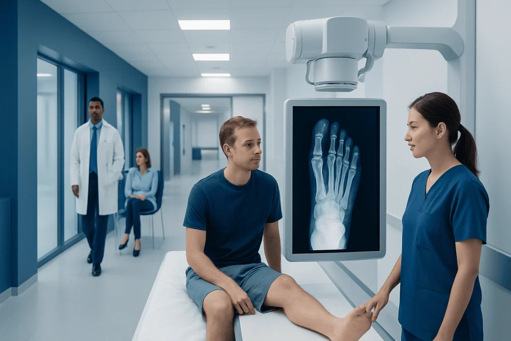
A foot xray uses a small amount of radiation to create images of the foot’s bones and joints. It is often used after injury or for ongoing pain, swelling, deformity, or suspected arthritis.
Key Takeaways
- A foot xray uses a small amount of radiation to create images of the foot’s bones and joints.
- It is often used after injury or for ongoing pain, swelling, deformity, or suspected arthritis.
- A normal foot xray does not rule out every cause of symptoms, especially soft-tissue injuries or very subtle stress fractures.
- The test is quick, usually requires no special preparation, and is interpreted together with symptoms and examination findings.
- Medical care is important for severe pain, inability to bear weight, visible deformity, or signs of infection.
A foot xray is a common imaging test used to look at the bones and joints of the foot. It can help doctors confirm fractures, alignment problems, arthritis changes, and some causes of pain or swelling, but it does not show soft tissues as clearly as ultrasound or MRI.
Overview: what a foot xray is and what it can tell doctors
A foot xray is a medical imaging test that uses a small amount of radiation to create pictures of the bones and joints in the foot. In everyday practice, it is one of the first tests doctors use when someone has foot pain after an injury, trouble walking, swelling, or a suspected bone or joint problem. It is fast, widely available, and especially useful for seeing whether a bone is broken or a joint is out of position.
The most important point is that a foot xray shows bone structure better than soft tissues. It can help identify fractures, dislocations, arthritis-related changes, bunions, bone spurs, and some deformities. However, tendons, ligaments, nerves, and many early soft-tissue problems are not seen well on xray alone, so another test may be needed if symptoms continue despite a normal result.
Doctors usually do not rely on myths such as “if it is broken, the person cannot walk” or “an xray always shows every injury immediately.” In reality, some people can still walk with a fracture, and some small or stress-related injuries may not be obvious on the first film. A foot xray is most useful when it is combined with a clinical examination and, if needed, follow-up imaging.
When a foot xray is commonly recommended
A foot xray may be recommended after a fall, twist, direct blow, sports injury, or other trauma to the foot. It is also commonly used when pain has lasted for days or weeks without a clear cause, especially if there is swelling, tenderness over a bone, bruising, or difficulty bearing weight. The goal is to check for a structural problem that could change treatment.
Doctors may also request a foot xray for long-term symptoms rather than a sudden injury. This includes concerns such as arthritis, changes in foot shape, persistent heel pain, bunions, suspected stress fractures, or conditions that affect the bones over time. In children, a foot xray may help assess injury, alignment, or growth-related issues when symptoms and examination suggest a bony cause.
Common reasons include:
- Suspected fracture or dislocation
- Ongoing foot pain or swelling
- Visible deformity or change in alignment
- Suspected arthritis or joint degeneration
- Possible stress injury from overuse
- Monitoring healing after treatment or surgery
In some situations, clinicians use decision tools based on where the pain is located and whether the person can bear weight. These tools can help avoid unnecessary imaging while still identifying people who are more likely to have a clinically important injury.
What a foot xray can and cannot show

A foot xray is particularly good at showing the number, shape, position, and alignment of bones. It can reveal clear fractures, small bone fragments, joint space narrowing from arthritis, deformities such as hallux valgus, and changes linked to chronic pressure or wear. It may also show signs that point toward conditions such as advanced infection in bone, though xray findings can lag behind early disease.
At the same time, xray has limits. It does not show cartilage, ligaments, tendons, and muscles in detail. This means a person can have a normal foot xray yet still have a painful ligament sprain, tendon injury, plantar fascia problem, or nerve-related condition. Very early stress fractures may also be missed, particularly in the first days after symptoms begin.
If a doctor suspects a problem that xray cannot explain well, other imaging may be more appropriate. For example, MRI scanning can help evaluate stress fractures, tendon tears, or ligament injuries, while ultrasonography may be useful for some soft-tissue and inflammatory conditions. If symptoms suggest a complex bone injury or preoperative planning is needed, computed tomography (CT) can provide more detailed bone images.
How the test is done and how to prepare
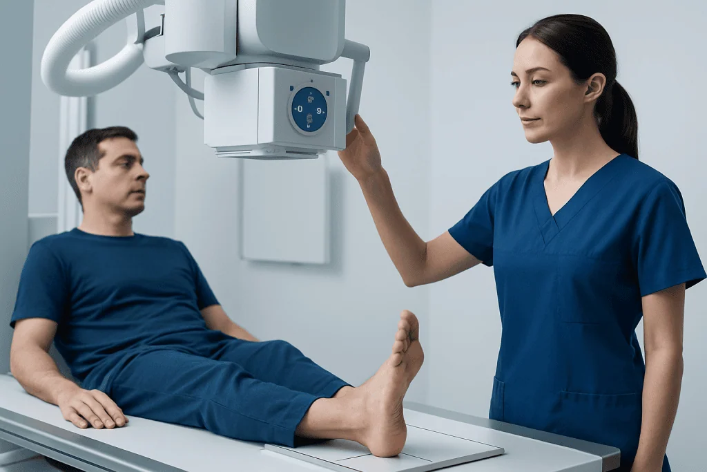
A foot xray is usually done in a radiology department, emergency department, or outpatient imaging unit. The person may be asked to remove shoes, socks, and any metal objects such as ankle jewelry. Depending on the reason for the test, images may be taken while sitting, lying down, or standing. Standing, or weight-bearing, views can be especially helpful when doctors want to assess alignment or how the foot functions under load.
The radiographer typically takes several images from different angles, often including top, side, and oblique views. Staying still for a few moments helps produce clear pictures. The test itself is painless, although moving an injured foot into position may cause temporary discomfort. The entire visit is usually brief.
Because xray uses ionizing radiation, it is important to tell the healthcare team about a current or possible pregnancy. For most people, the radiation dose from a foot xray is low. Medical teams use the lowest dose needed to obtain useful images and only request the test when it is clinically appropriate.
Understanding the results
A radiologist reads the images and looks for signs such as fractures, dislocations, joint changes, bone alignment problems, or other abnormalities. The report is then considered together with the medical history and physical examination. A clear explanation of symptoms is important, because the same xray finding may be significant in one person and incidental in another.
An abnormal result does not always mean surgery is needed. Many injuries seen on a foot xray can be treated with rest, activity modification, supportive footwear, a boot, cast, or other conservative care. If the images show a more complex break, unstable joint problem, or marked deformity, referral to an orthopedic or foot specialist may be recommended. This can be relevant for problems such as hallux valgus (bunion), where imaging helps assess severity and treatment planning.
A normal foot xray can also be helpful because it narrows the list of possible causes. Still, it does not completely rule out injury. If pain remains severe, worsens, or does not improve as expected, the doctor may repeat xray after some time or choose another imaging test. Clinical follow-up matters just as much as the first image.
Treatment after a foot xray depends on the cause
The xray itself is a diagnostic step, not a treatment. What happens next depends on what the images show and how severe the symptoms are. For many minor injuries, treatment may include protection of the foot, limiting weight-bearing, using ice and elevation, and taking doctor-approved pain relief. Supportive shoes or temporary immobilization can help the foot heal while reducing further strain.
If the xray suggests arthritis, treatment may focus on symptom control and preserving movement. This can include footwear changes, exercise guidance, weight management when appropriate, and medicines recommended by a physician. In some cases, a foot xray helps distinguish common wear-and-tear changes from inflammatory causes, though additional blood tests or imaging may still be needed.
When the images reveal a significant fracture, dislocation, or advanced structural problem, more specialized care may be required. This can include casting, reduction of a displaced injury, or evaluation for orthopedic surgery when conservative care is unlikely to restore alignment or function. International patients seeking coordinated assessment may also be seen by the multidisciplinary specialists at Acibadem International, where JCI-accredited hospitals diagnose and treat foot and ankle conditions.
When to seek medical care
Prompt medical assessment is important if foot pain starts after an injury and the person cannot bear weight, has marked swelling, significant bruising, numbness, or a visible deformity. These features can suggest a fracture, dislocation, or another injury that should not be ignored. Severe pain that feels out of proportion to the injury also deserves urgent attention.
Medical care is also important when there are signs of infection or reduced circulation. Redness that spreads, warmth, fever, drainage from a wound, or a foot that looks pale or bluish should be assessed quickly. People with diabetes, poor circulation, or reduced sensation should seek help earlier, because foot problems may become serious more easily.
Even without a major injury, a doctor should evaluate foot pain that persists, keeps returning, or interferes with normal walking and daily activities. A foot xray may or may not be the right first step, but ongoing symptoms should not simply be dismissed as strain if they are not improving. Early assessment can support more accurate diagnosis and safer treatment.
Frequently asked questions
What does a foot xray show?
A foot xray mainly shows the bones and joints of the foot. It can help detect fractures, dislocations, alignment problems, arthritis changes, bunions, and some bone-related causes of pain or swelling.
Can a foot xray miss an injury?
Yes. Some soft-tissue injuries, such as ligament or tendon problems, do not appear clearly on xray, and early stress fractures may not be visible right away. If symptoms continue despite a normal xray, a doctor may recommend repeat imaging or another test such as MRI or ultrasound.
Is a foot xray painful?
The xray itself is not painful. However, moving an injured foot into the right position for the images can cause temporary discomfort, especially after trauma.
How long does a foot xray take?
The imaging process is usually quick and often takes only a few minutes, although the full visit may be a little longer. Several views are commonly taken to help doctors see the foot from different angles.
Do people need to prepare for a foot xray?
Most people do not need special preparation. They may simply be asked to remove shoes, socks, and metal items, and should tell the healthcare team if they are or may be pregnant.
When is MRI or CT used instead of a foot xray?
MRI may be used when doctors suspect a stress fracture, tendon injury, ligament damage, or another soft-tissue problem that xray cannot show well. CT may be preferred for complex fractures or when more detailed bone imaging is needed.
References
- American College of Radiology
- Radiological Society of North America
- National Institute for Health and Care Excellence
- American Academy of Orthopaedic Surgeons
- MedlinePlus
This article is for general information only and is not a substitute for professional medical advice. Please consult a qualified doctor about your individual situation.
Joint and spine care in Turkey — expert assessment & treatment
JCI-accredited · board-certified surgeons · reply within 24h
Add us as a Preferred Source to see more of our trusted health content across Google Search, AI Overviews and Discover.

More from the Health Library

B Plague: An Evidence-Based Guide for Patients

Aphenphosmphobia: An Evidence-Based Guide for Patients

Inguinal Lymph Nodes — Explained by Medical Evidence, Not Myths

Primary Hypertension: An Evidence-Based Guide for Patients

Green Tea Benefits — Explained by Medical Evidence, Not Myths